Published on 19/03/2015 by admin
Filed under Dermatology
Last modified 22/04/2025
This article have been viewed 3077 times
Ginat W. Mirowski and Bethanee J. Schlosser
Evidence Levels: A Double-blind study B Clinical trial ≥ 20 subjects C Clinical trial < 20 subjects D Series ≥ 5 subjects E Anecdotal case reports
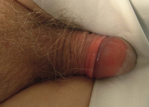
Balanitis (balanoposthitis, inflammation of the glans penis and occasionally the foreskin [prepuce]) occurs in both men and boys. Balanitis has many causes including poor hygiene, friction, infection, malignancy, and numerous dermatoses and may be multifactorial. The presence of foreskin (uncircumcised state) increases the risk of developing balanitis. Clinically, patients present with itching, irritation, and shiny erythema associated with an exudate or smegma. Urethral discharge is not present. Complications may include fissuring and pain, phimosis (inability to retract the foreskin due to agglutination/scarring), and stenosis or obstruction of the urethral meatus; surgical correction of sequelae may be necessary.
This chapter will focus on the treatment of non-specific balanitis, lichen sclerosus (LS, lichen sclerosus et atrophicus, balanitis xerotica obliterans, BXO) and Zoon’s (plasma cell) balanitis. Genital ulcers, genital warts, and urethral discharge will not be discussed.
Evaluation of a patient with balanitis should include chief complaint, history of present illness, past medical and surgical history, medications, allergies, and review of systems. Specific information should be sought regarding sexual habits (number, gender, and symptomatology of sexual partners) and alleviating or exacerbating factors. To identify potential allergens and/or irritants, the patient’s genital hygiene practices and the use of oral and topical agents (condoms, spermicides, sexual enhancing products, lubricants, etc.) should be sought. A complete mucocutaneous examination including extra-genital sites should be performed. The genital examination includes skin and soft tissue structures extending from the lower abdomen to the perianal skin/gluteal cleft. Examination findings should direct the acquisition of microbiologic studies (KOH preparation, bacterial, fungal and viral cultures), biopsy (hematoxylin and eosin, direct immunofluorescence), and serologic studies. Treatment of balanitis is dictated by results of these investigations.
Patients with balanitis should be instructed on appropriate local hygiene care including retraction of the foreskin prior to cleaning. The glans and shaft should be cleaned with plain water or normal saline twice daily and after sexual activity. Soap and topical products may be irritants or allergens and should be avoided. A bland emollient (plain white petrolatum or similar) applied twice daily will minimize friction and improve barrier function.
Medical therapy for balanitis is dictated by etiology. Circumcision is indicated in refractory cases. Urethral meatotomy or meatoplasty, glans resurfacing and other surgical procedures may be required for patients with significant anatomic distortion or compromised urinary function. Collaboration with urologic specialists is essential.
KOH microscopy for fungi
Tzanck smear or direct fluorescent antigen testing for herpes viruses
Swab culture for bacteria, viruses, and fungi
Biopsy for routine histopathology and direct immunofluorescence, if indicated
Fasting blood glucose
Urinalysis and urine glucose
Serologic tests for syphilis, herpes virus, and human immunodeficiency virus
Serologic tests for vesiculobullous diseases (systemic lupus erythematosus, pemphigus vulgaris, bullous pemphigoid, etc.)
Patch testing
Fornasa CV, Calabro A, Miglietta A, Tarantello M, Biasinutto C, Peserico A. Genitourin Med 1994; 70: 345–6.
Patients (n = 321) with balanitis were evaluated. Infection was diagnosed clinically in 185. Dermatologic conditions included traumatic/irritant contact dermatitis (n = 17), psoriasis (n = 11), lichen planus (n = 9), LS (n = 8), neoplasm (n = 8), Zoon’s (n = 3), and allergic contact dermatitis (n = 3). Of 51 individuals with mild balanitis who required further testing, etiologies included infection (n = 12, including Candida albicans, Chlamydia trachomatis, β-hemolytic streptococcus, gastrointestinal bacterial flora), irritant contact dermatitis (n = 9), mechanical trauma (n = 7), and allergic contact dermatitis (n = 6). No identifiable etiology was found in 17 patients.
Edwards S. Genitourin Med 1996; 72: 155–9.
Thorough review of infectious causes of balanitis with differential diagnosis considerations.
Edwards SK. Int J STD AIDS 2001; 12 (Suppl 3): 68–72.
Comprehensive review of the many causes of balanitis with specific recommendations for evaluation and management.
Teichman JM, Csea J, Thompson IM, Elston DM. Am Fam Physician 2010; 81: 167–74.
Practical approach to non-infectious inflammatory and neoplastic penile disorders with emphasis on clinical appearance, differential diagnosis, and management.
Birley HD, Walker MM, Luzzi GA, Bell R, Taylor-Robinson D, Byrne M, et al. Genitourin Med 1993; 69: 400–3.
Forty-three patients with recurrent balanitis were evaluated. Thirty-one patients diagnosed with irritant contact dermatitis had a greater lifetime incidence of atopy and more frequent genital hygiene habit; 90% responded to conservative treatment, use of emollient creams, and restriction of soap use.
Lisboa C, Santos A, Dias C, Azevedo F, Pina-Vaz C, Rodrigues A. J Eur Acad Dermatol Venereol 2010; 24: 820–6.
A prospective cross-sectional study of 478 men who attended a sexually transmitted diseases clinic revealed Candida balanitis in 18%, more than 40% of whom had a concomitant cause of balanitis. Candida albicans was the most common isolate. Candida colonization and infection were associated with age greater than 60 years and diabetes mellitus in males aged 40 years or older.
Pugliese JM, Morey AF, Peterson AC. J Urol 2007; 178: 2268–76.
Extensive review of the literature. Goals for treatment include symptomatic relief, prevention of scarring/anatomic distortion, and prevention of malignant transformation. An algorithmic approach to medical and surgical management of LS is provided with topical corticosteroids (CS) as first-line treatment.
Prevention of malignant transformation has not been assessed in the literature to date in the authors’ opinion.
Neill SM, Lewis FM, Tatnall FM, Cox NH. Br J Dermatol 2010; 163: 672–82.
Evidence-based recommendations for the evaluation and treatment of LS in men, women, and children. Topical CS are the treatment of choice. Testosterone is no longer recommended. Surgery is only recommended when scarring and destruction have occurred. Circumcision is highly effective; recurrences and koebnerization have been documented.
Dahlman-Ghozlan K, Hedblad MA, von Krogh G. J Am Acad Dermatol 1999; 40: 451–7.
Retrospective study of 22 men all of whom responded to clobetasol dipropionate 0.05% cream once or twice daily for 7 weeks with improvement in symptoms and urinary flow.
Kiss A, Csontai A, Pirót L, Nyirády P, Merksz M, Király L. J Urol 2001; 165: 219–20.
This double-blind, placebo-controlled, randomized study of 40 boys with clinical BXO showed that mometasone furoate 0.05% once daily improves BXO in the histologically early and intermediate stages of disease and may inhibit progression in the late stage.
Zavras N, Christianakis E, Mpourikas D, Ereikat K. J Pediatr Urol 2009; 5: 181–5.
A prospective study of 1185 boys with suspected phymosis using fluticasone propionate cream 0.05% (class 5 CS) twice daily for 4 to 8 weeks yielded successful resolution (full retraction of foreskin) in 91.1%.
Tang A, David N, Horton LW. Int J STD AIDS 2001; 12: 75–8.
Ten patients with plasma cell balanitis treated with a topical mixture of oxytetracycline 3%, nystatin 100 000 U/g, and clobetasone butyrate 0.05% (Trimovate) for 3 to 12 weeks had complete resolution. Four required re-treatment(s).
Kulkarni S, Berbagli G, Kirpekar D, Mirri F, Lazzeri M. Eur Urol 2009; 55: 945–54.
A total of 215 males (age range 11–85 years) with LS limited to the foreskin and/or external urethral meatus underwent circumcision or urethral reconstructive surgery. Thirty-four patients with foreskin-limited LS underwent circumcision with 100% success rate and no recurrences at mean follow-up of 65 months. Urethral involvement required more extensive surgical intervention with lower rates of success.
Kumar B, Sharma R, Rajagopalan M, Radotra BD. Genitourin Med 1995; 71: 32–4.
Twenty-seven patients with plasma cell balanitis were cured with circumcision. There were no recurrences at 3-year follow-up.
Kumar B, Narang T, Dass Radotra B, Gupta S. J Cutan Med Surg 2006; 10: 11–15.
A study of 112 males with plasma cell balanitis demonstrated complete resolution and no recurrence in the 85 who underwent circumcision. 22 of 27 showed healing at 2 to 3 months with topical therapy (CS, CS–antifungal, tacrolimus).
Hengge UR, Krause W, Hofmann H, Stadler R, Gross G, Meurer M, et al. Br J Dermatol 2006; 155: 1021–8.
Prospective, multicenter phase II study of tacrolimus 0.1% ointment in 84 patients (49 women, 32 men, three girls) with LS twice daily for 16 weeks demonstrated clearance of clinical disease in 43% and partial resolution in 34% at 24 weeks. Maximal response occurred at 10–24 weeks. No gender difference in response to therapy was noted.
Ebert AK, Rosch WH, Vogt T. Eur Urol 2008; 54: 932–7.
Twenty boys with biopsy-confirmed LS underwent circumcision followed by topical tacrolimus 0.1% ointment twice daily for 3 weeks. All completed treatment without adverse side effects and no evidence of clinical disease at follow-up. Topical tacrolimus without circumcision was not evaluated.
Roe E, Dalmau J, Peramiquel L, Perez M, Lopez-Lozano HE, Alomar A. J Eur Acad Dermatol Venereol 2007; 21: 284–5.
Three patients with Zoon’s balanitis refractory to topical CS, antifungals, and antibacterials responded favorably to tacrolimus 0.1% ointment twice daily within 3 to 4 weeks.
Santos-Juanes J, Sanchez del Rio J, Galache C, Soto J. Arch Dermatol 2004; 140: 1538–9.
Complete remission was reported in three patients with Zoon’s balanitis after using topical tacrolimus 0.1% cream or ointment twice daily for 3 to 5 weeks. Mild irritation was noted in one patient.
Georgala S, Gregoriou S, Georgala C, Papaioannou D, Befon A, Kalogeromitros D, et al. Dermatology 2007; 215: 209–12.
A randomized controlled study of 26 men with non-specific balanitis used pimecrolimus cream twice daily for 7 days. Seven of 11 men in the treatment group and one of 11 in the control group were free of all symptoms and lesions at day 14. As-needed use for 90 days showed good response.
Bardazzi F, Antonucci A, Savoia F, Balestri R. Int J Dermatol 2008; 47: 198–201.
Two cases of resistant Zoon’s balanitis were treated with topical pimecrolimus 1% cream twice daily for 2 months. One patient achieved complete clinical regression. One patient noted improvement with persistence of a hyperpigmented patch. Treatment was well-tolerated. Neither relapsed at nine to 10 months follow-up.
Marconi B, Campanati A, Simonetti O, Savelli A, Conocchiari L, Santinelli A, et al. Eur J Dermatol 2010; 20: 134–5.
Case report of Zoon’s balanitis successfully treated with imiquimod 5% cream three times weekly for 12 weeks.
Wollina U. J Cosmet Laser Ther 2010; 12: 120–3.
Ablative erbium:YAG laser treatment was used in 20 patients with Zoon’s balanitis with complete re-epithelialization within 2 to 3 weeks in all patients.
Aynaud O, Plantier F. Eur J Dermatol 2010; 20: 387–8.
Report of 4 cases of penile LS successfully treated with CO2 laser and review of the literature on CO2 laser treatment for genital LS.
Retamar RA, Kien MC, Chouela EN. Int J Dermatol 2003; 42: 305–7.
Discussion of CO2 laser treatment in five patients with Zoon’s balanitis with variable efficacy.
Treatment of Skin Disease Comprehensive Therapeutic Strategies 4e
WhatsApp us


 Hygiene
 Emollients
 Topical antifungals
 Topical corticosteroids
 Circumcision
 Topical tacrolimus
 Topical pimecrolimus
 Imiquimod
 CO2 laser
 Erbium : YAG laser