Published on 19/03/2015 by admin
Filed under Dermatology
Last modified 22/04/2025
This article have been viewed 2145 times
Tanya N. Basu, Chrystalla Macedo and Richard C.D. Staughton
Evidence Levels: A Double-blind study B Clinical trial ≥ 20 subjects C Clinical trial < 20 subjects D Series ≥ 5 subjects E Anecdotal case reports
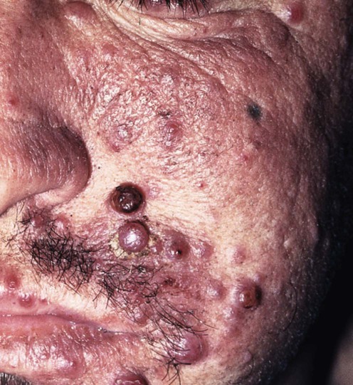
First described in 1983, bacillary angiomatosis (BA) is a vasculoproliferative disorder caused by the bacteria Bartonella henselae and Bartonella quintana (previously Rochalimaea spp.). It typically presents in profoundly immunocompromised patients (e.g., in advanced HIV infection, post transplant or during cytotoxic chemotherapy). Angiomatous papules, nodules, or plaques may arise in the skin or systemically in any organ including the bone, central nervous system, liver, where the condition is termed peliosis hepatis, and spleen (peliosis splenis).
Vascular proliferation may be due to an angiogenic factor produced by the Bartonella genus. B. henselae is transmitted through a cat scratch or bite; it also causes cat scratch disease. B. quintana is transmitted by the human body louse and also causes trench fever. Virtually identical localized cutaneous lesions to BA are seen in verruga peruana, which occurs in Peru due to the related Bartonella bacilliformis, transmitted by sandflies.
Prompt diagnosis of bacillary angiomatosis is essential to prevent dissemination, which can be fatal. Clinical suspicion should be aroused in the context of a low CD4 lymphocyte count (<100) or other immunosuppression, especially with a history of exposure to cats (harboring B. henselae) or body lice (which carry B. quintana). Cutaneous lesions can be superficial cherry-red round papules with an eroded surface, similar to pyogenic granulomas, or violaceous, lichenoid plaques or deep subcutaneous nodules. Single lesions have been reported in immunocompetent patients at inoculation sites whereas in the immunocompromised the entire body surface may be affected. Lesions can be mistaken for Kaposi sarcoma or in-transit metastatic amelanotic melanoma and other malignancies because of the highly vascular and erosive nature of the lesions. In advanced HIV infections, differentials include deep fungal infection, e.g., cryptococcosis and histoplasmosis. Patients with extracutaneous disease may or may not have skin signs and can present with vomiting, abdominal pain, and deranged liver function (peliosis hepatis) or pancytopenia and splenomegaly (peliosis splenis). Presentation can also include fever, lymphadenopathy, night sweats, endocarditis, and anemia.
Histology allows easy differentiation and shows a lobular proliferation of capillaries and venules, with swollen endothelial cells containing clumps of bacteria.
The response of bacillary angiomatosis to antibiotic treatment is usually dramatic, in contrast to the response of cat scratch disease. Our drugs of first choice are the macrolides (e.g., erythromycin 500 mg four times daily, azithromycin 500 mg daily, clarithromycin 500 mg twice daily); doxycycline 100 mg twice daily as an alternative; intravenous treatment may be needed in severe disease or with gastrointestinal intolerance. Their use is based on anecdotal experience in the absence of systematic trials. Current recommendations are that treatment should be continued for 3 months where there is skin disease only, and 4 months where there is bone/visceral involvement or peliosis hepatis. Should relapse occur on the above regimens, long-term prophylaxis with erythromycin or doxycycline may be indicated. In practice, however, the introduction of highly active anti-retroviral therapy (HAART) should reverse the immunocompromise state and thus alter the response to treatment, making long-term antibiotic less necessary (although BA has been reported during immune reconstitution during HAART). The patient should be evaluated for parenchymal and osseous disease prior to treatment and warned that a Jarisch–Herxheimer reaction may occur after the first few doses of antibiotic.
A wide variety of therapeutic agents are mentioned in the literature, but there is a lack of correlation between the in vitro and in vivo drug susceptibility of Bartonella spp., which reduces the usefulness of laboratory data. The picture is clouded further by the different response of Bartonella spp. to drugs in each of the diseases it causes.
Full blood count, liver function tests, and CD4 lymphocyte count
Biopsy and Warthin–Starry stains/electron microscopy
Prolonged culture of blood and biopsy tissue
Polymerase chain reaction of biopsy material
Serology – indirect fluorescence assay
Culture of the fastidious Gram negative rods of Bartonella spp. is extremely difficult, requiring special media and prolonged incubation of up to 45 days; it is invariably negative if antibiotics have been given. Skin biopsy is the essential diagnostic tool and shows characteristic appearances on histology and Warthin–Starry silver stains, which shows the organism, as can electron microscopy. Species confirmation can be obtained by PCR. Reliance on serology in the immunosuppressed is hazardous, but the Centers for Disease Control (CDC) definition of a positive test is an indirect fluorescence assay (IFA) titer of over 1 : 64.
Agan BK, Dolan MJ. Clin Lab Med 2002; 22: 937–62.
Culture methods have improved, but are still prolonged. Serologic testing for B. henselae has become the cornerstone for diagnosis in the immunocompetent patient. Ideal antigens for enzyme immunoassays have yet to be clearly identified. PCR currently offers the ability to establish the diagnosis when other tests fail.
Mohle-Boetani JC, Koehler JE, Berger TG, LeBoit PE, Kemper CA, Reingold AL, et al. Clin Infect Dis 1996; 22: 794–800.
Forty-two cases were compared to 84 matched controls and the distinguishing clinical characteristics were evaluated. Significant differences included the presence of anemia (hematocrit <0.36), raised alkaline phosphatase and aspartate aminotransferase levels, and a low CD4 lymphocyte count (median being 21/mm3 compared to 186/mm3 in controls). There was no difference in the white blood cell count, creatinine, bilirubin, and alanine aminotransferase levels. Clinical signs included fever, abdominal pain, and lymphadenopathy.
Gasquet S, Maurin M, Brouqui P, Lepidi H, Raoult D, et al. AIDS 1998; 12: 1793–803.
Diagnosis remains mainly based on histological appearance. On hematoxylin and eosin stains the appearance can be highly variable and so Warthin–Starry stains are essential to visualize the bacillus and confirm the diagnosis.
La Scola B, Raoult D. J Clin Microbiol 1999; 37: 1899–905.
In the large number of samples cultured, seven patients were diagnosed with bacillary angiomatosis. PCR was 100% sensitive in diagnosing these cases, in contrast to culture, which isolated Bartonella spp. from only three specimens. Serology was of no value, being positive in only one patient.
Jensen WA, Fall MZ, Rooney J, Kordick DL, Breitschwerdt EB. J Clin Microbiol 2000; 38: 1717–22.
The single step assay described provided a simple and rapid means of identifying Bartonella spp.
Wagner CL, Riess T, Linke D, Eberhardt C, Schäfer A, Reutter S, et al. Int J Med Microbiol 2008; 298: 579–90.
Two-step serodiagnosis, using a combination of an indirect immunofluorescence assay and adhesin A, improved identification of Bartonella henselae infections.
Koehler JE, Sanchez MA, Garrido CS, Whitfeld MJ, Chen FM, Berger TG, et al. N Engl J Med 1997; 337: 1876–83.
A case–control study of 49 patients (92% HIV positive) in whom macrolides, doxycycline, tetracycline, and rifampin were found to be effective. This was in contrast to patients treated with trimethoprim–sulfamethoxazole, ciprofloxacin, penicillins, and cephalosporins in whom Bartonella spp. could be isolated on PCR or culture.
Maurin M, Gasquet S, Ducco C, Raoult D. Antimicrob Agents Chemother 1995; 39: 2387–91.
The newer macrolides were highly effective in preventing bacterial growth with MIC 90s of 0.03 µg/mL for azithromycin and clarithromycin. Erythromycin, doxycycline, and rifampin all had MIC 90s of 0.25 µg/mL.
Koehler JE, Tappero JW. Clin Infect Dis 1993; 17: 612–14.
This review article refers to 50 patients whose lesions and symptoms responded to erythromycin or doxycycline therapy.
Zarraga M, Rosen L, Herschthal D. Am J Dermatopathol 2011; 33: 513–15.
Successful treatment with azithromycin.
Schlupen E-M, Schirren CG, Hoegl L, Schaller M, Volkenandt M. Br J Dermatol 1997; 136: 747–51.
An HIV-positive man presented with a 10-month history of bacillary angiomatosis on his ankle and was treated with erythromycin 500 mg four times daily. The swabs became negative on PCR at 12 weeks, at which point treatment was successfully stopped.
Rolain JM, Brouqui P, Koehler JE, Maguina C, Dolan MJ, Raoult D. Antimicrob Agents Chemother 2004; 48: 1921–33.
A good review article. Although erythromycin and azithromycin are the authors’ first-line treatments for bacillary angiomatosis, their use has been based on case series and case reports rather than on controlled clinical trials. Azithromycin has emerged as first-line treatment for cat scratch disease for which there are formal trial data.
Mukunda BN, West BC, Shekar R. Clin Infect Dis 1998; 27: 658.
A patient with AIDS presented with bacillary peliosis and was initially treated for a presumed Mycobacterium avium intracellulare complex infection with clarithromycin, ciprofloxacin, and rifabutin. He continued to be febrile and re-presented 15 days later with bacillary angiomatosis. This swiftly responded to doxycycline, which was continued for 6 weeks.
Schwartz RA, Nychay SG, Janniger CK, Lambert WC. Br J Dermatol 1997; 136: 60–5.
This article describes a variety of successful treatment regimens, including tetracycline and ciprofloxacin.
Although rifampin has activity in vitro, its efficacy when used alone has not yet been established and so it is recommended as a second-line drug in combination with either erythromycin or doxycycline for severely ill patients or where there is neurological involvement (doxycycline has good CNS penetration so doxycycline/rifampicin combination is recommended for CNS involvement). Rifampicin is, however, effective in the treatment of verruga peruana (B. bacilliformis) and cat scratch disease.
Riley LE, Tuomala RE. Obstet Gynecol 1992; 79: 818–19.
A pregnant patient was treated with a third-generation cephalosporin, ceftizoxime. However, there are inadequate data to recommend its use at present.
Musso D, Drancourt M, Raoult D. J Antimicrob Chemother 1995; 36: 101–8.
Aminoglycosides display in vitro bactericidal activity against Bartonella spp. and as such warrant further clinical investigation. For culture positive Bartonella endocarditis, doxycycline for 6 weeks plus intravenous gentamicin for the first 14 days are recommended.
Moulin C, Kanitakis J, Ranchin B, Chauvet C. Gillet Y, Morelon E, Euvrard S. Transpl Infect Dis 2012; 14: 403–9.
Cases were successfully treated with erythromycin, clarithromycin, and ciprofloxacin (quinolones are not currently recommended as they give inconsistent clinical results).
Treatment of Skin Disease Comprehensive Therapeutic Strategies 4e
WhatsApp us


 Erythromycin
 Azithromycin
 Clarithromycin
 Doxycycline
 Tetracycline
 Rifampin
 Gentamicin
 Third- and fourth-generation cephalosporins