Autonomic Nervous System and Cardiac Arrhythmias

Published on 21/06/2015 by admin

Filed under Cardiovascular

Last modified 22/04/2025

Print this page

rate 1 star rate 2 star rate 3 star rate 4 star rate 5 star
Your rating: none, Average: 0 (0 votes)

This article have been viewed 2699 times

Chapter 5 Autonomic Nervous System and Cardiac Arrhythmias

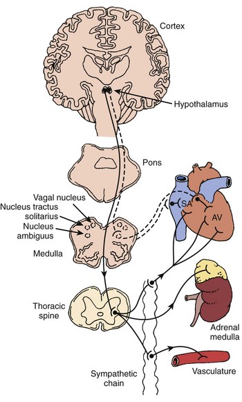
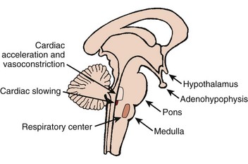
The autonomic nervous system (ANS) comprises the portion of the central nervous system that provides moment-to-moment regulation of the function of the cardiovascular system as well as that of all other organ systems. The ANS continuously monitors afferent neural signals from vascular beds and organ systems and coordinates efferent neural traffic to modify the responses of heart and blood vessels to ever-changing physiological and metabolic requirements. In this context, the sympathetic and parasympathetic components of the ANS are the dominant players (Figures 5-1 and 5-2).1 However, ANS cardiovascular control also incorporates actions of cardiac and extracardiac neurohumoral agents, intracardiac reflex arcs, and the contributions of certain less well-understood agents such as vasoactive intestinal peptide (VIP), neuropeptide Y, transmitters released by the so-called purinergic nerve endings, serotonin, inflammatory cytokines, vasopressin, and nitric oxide.2 Further, with respect to cardiovascular control, the ANS collaborates with the hypothalamic-pituitary-adrenal (HPA) axis. For its part, the HPA-axis, governed from the hypothalamus, participates by prompting the release of glucocorticoids, mainly cortisol and, to a lesser extent, mineralocorticoids. The HPA theater of operation therefore includes inflammatory, immune, metabolic, and pressor effects.3 Both systems (ANS and HPA) are involved in stress responses.2,3

It is not unexpected that any disturbance of ANS function, given its wide-ranging impact, may lead to clinically important consequences. In terms of cardiac electrophysiology and arrhythmias, common clinical conditions in which ANS effects are evident include acute myocardial ischemia, heart failure, and neurally mediated reflex syncope (particularly the vasovagal faint). Furthermore, it is now widely acknowledged that the nervous system has the capacity to injure the heart acutely (e.g., stress-induced cardiomyopathy); serious acute cerebral disorders such as subarachnoid hemorrhage, intracerebral bleeds, infections, and seizures may induce electrocardiographic changes, myocardial damage, arrhythmias, and even sudden death.37 Perhaps the most publicized direct cardiac effects of presumed autonomic “storms” are the immediate, apparently stress-triggered, increases in the number of cardiovascular events; these include acute myocardial infarctions, sudden cardiac deaths, and presumed stress-induced cardiomyopathy (Table 5-1).35

Table 5-1 Epidemiologic Associations: Stress and Increased Cardiovascular Event Rates

MI, Myocardial infarction; SCD, sudden cardiac death; ICD, implantable cardioverter-defibrillator.

References

a Stalnikowicz R, Tsafrir A: Acute psychosocial stress and cardiovascular events, Am J Emerg Med 20:488–491, 2002.

b Brown DL: Disparate effects of the 1989 Loma Prieta and 1994 Northridge earthquakes on hospital admissions for acute myocardial infarction: Importance of superimposed triggers, Am Heart J 137:830–836, 1999.

c Watanabe H, Kodama M, Okura Y, et al: Impact of earthquakes on Takotsubo cardiomyopathy. JAMA 294:305–306, 2005.

d Zhang XQ, Chen M, Yang Q, et al: Effect of the Wenchuan earthquake in China on hemodynamically unstable ventricular tachyarrhythmia in hospitalized patients, Am J Cardiol 103(7):994–997, 2009.

e Wilbert-Lampen U, Leistner D, Greven S, et al: Cardiovascular events during World Cup soccer. N Engl J Med 358:475–483, 2008.

f Katz E, Metzker J-T, Marazzi A, Kappenberger L: Increased sudden cardiac deaths in Switzerland during the 2002 FIFA World Cup, Int J Cardiol 107:132–133, 2006.

g Brotman DJ, Golden SH, Wittstein IS: The cardiovascular toll of stress, Lancet 376: 1089–1100, 2007.

h Meisel SR, Kutz I, Dayan KI, et al: Effect of Iraqi missile war on incidence of acute myocardial infarction and sudden death in Israeli civilians, Lancet 338:660–661, 1991.

i Tofler GH, Muller JE: Triggering of acute cardiovascular disease and potential preventive strategies, Circulation 114:1863–1872, 2006.

This chapter provides a brief overview of current concepts regarding the impact of autonomic innervation as they pertain to cardiac arrhythmias, conduction system disturbances, and related disorders.

Anatomic Nervous System and Cardiac Conduction System Physiology

Sinus Node, Atrioventricular Node, and His-Purkinje System

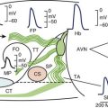
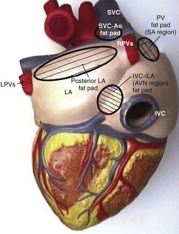
The sinus node (SN) and the atrioventricular (AV) node appear to be represented by separate cells within the nucleus ambiguus. However, it is uncertain whether the nodes are coordinated centrally; in fact, it seems increasingly likely that local circuits, often positioned within the epicardial fat pads of the heart, participate in the coordination of these structures (Figure 5-3).4,8

Atrioventricular Node and Cardiac Conduction System

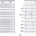
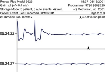
As a rule, AV nodal dromotropic responsiveness in the resting patient is under relatively balanced sympathetic and parasympathetic neural influence. However, this situation is readily altered by physiological events (e.g., exercise, sleep), the impact of disease states, drug effects, or during cardiac electrophysiology procedures when certain atrial regions are stimulated. Any tendency toward parasympathetic predominance markedly enhances the decremental properties of the AV node; in the extreme, this can be associated with transient complete AV nodal block (Figure 5-4). The latter is, in fact, a relatively common finding in sleeping patients and in very fit resting subjects. The relationship between ANS control of SN rate and AV conduction properties appears to foster both the maintenance of 1 : 1 AV conduction and a relatively optimal AV conduction interval.

The His bundle and bundle branches comprise cells with larger surface areas, more negative resting membrane potentials, and faster (sodium [Na+]-dependent) action potentials than those of the AV node. Furthermore, cells that make up the cardiac conduction system have abundant intercellular connections and are physically arranged in such a way as to promote longitudinal conduction. Consequently, decremental conduction is essentially absent, except in the setting of relatively severe conduction system disease. Sympathetic nerve endings are generally better represented in the distal aspects of the specialized conduction system than are parasympathetic nerves. However, it has become evident that parasympathetic influence penetrates farther than had previously been thought.

Ventricular Myocardium

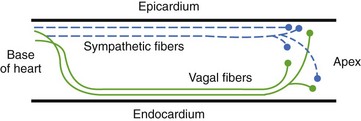
Ventricular sympathetics tend to lie within the subepicardial layer and follow the large coronary vessels as they spread out over the myocardium.9,10 The parasympathetics, in contrast, tend to penetrate the myocardium after crossing the AV groove and thereafter are subendocardial in location (Figure 5-5). The parasympathetic vagal efferents to the mycardium terminate not on the muscle cells themselves but on intracardiac ganglia. Evidence suggests that these ganglia not only form relay stations but also subserve certain local integrative functions, including the intracardiac reflex activity discussed earlier.

image

Figure 5-5 Diagram depicting the epicardial and endocardial locations of sympathetic and parasympathetic ventricular nerves, respectively.

(Modified from Zipes DP, Inoue H: Autonomic neural control of cardiac excitable properties. In Kulbertus HE, Franck G, editors: Neurocardiology, Mount Kisco, NY, 1988, Futura Publishing.)

Heightened adrenergic activation in the ventricular myocardium may be arrhythmogenic by causing enhanced pacemaker activity as well as by increasing the frequency and rate of automaticity. In addition, elevated adrenergic tone is known to increase the likelihood of the generation of early after-depolarizations (EADs) and delayed after-depolarizations (DADs).

Parasympathetic effects, in contrast, are thought to operate mainly as an antiadrenergic action in the setting of increased adrenergic tone. Consideration is being given to vagal nerve stimulation as an antiarrhythmic treatment strategy. The outcome of this activity may be diminished production of adrenergically induced EADs and DADs and an apparently anti-inflammatory action (diminished cytokine release and enhanced glucocorticoid release).

Autonomic Nervous System and Specific Bradyarrhythmias and Cardiac Conduction System Disturbances

Sinus Node Dysfunction

SN dysfunction (sick sinus syndrome) encompasses abnormalities of SN impulse generation, disturbances of impulse emergence into the atrium, abnormal impulse transmission within the atria, increased susceptibility to atrial tachycardias (particularly atrial fibrillation), chronotropic incompetence, and inappropriate sinus tachycardia. Clinical manifestations vary from seemingly asymptomatic electrocardiogram (ECG) findings to a wide range of complaints, including syncope, shortness of breath, palpitations, fatigue, and premature mental incapacity.

The causes of SN dysfunction are numerous but may be conveniently categorized as conditions that alter the SN, the sinoatrial structure or function directly (so-called intrinsic SN disease) or those that operate indirectly to impair sinoatrial function (i.e., extrinsic factors such as autonomic disturbances or drug effects). Ageing-associated idiopathic degenerative changes, fibrotic changes, or both are probably the findings most closely associated with “intrinsic” SN dysfunction. In regard to “extrinsic” SN dysfunction, drugs are the most important non-ANS contributors. β-adrenergic blockers, calcium channel blockers, membrane-active antiarrhythmics, and, to a lesser extent, digitalis are the most frequently implicated. Each of these may alter SN function as a result of direct pharmacologic effects (e.g., flecainide, d-sotalol), or indirectly via the ANS (e.g., β-adrenergic blockers) or both (e.g., quinidine, disopyramide, propafenone, amiodarone, digitalis). In terms of clinical outcomes, cardioactive drugs may initiate or aggravate sinus bradyarrhythmias or induce chronotropic incompetence.

Apart from drug-induced autonomic disturbances, the ANS may also contribute to apparent extrinsic disturbances of SN function. Sinus bradycardia, sinus pauses, sinoatrial exit block, and slow ventricular responses in atrial fibrillation may occur in the setting of parasympathetic predominance despite apparently normal underlying intrinsic SN or atrial function. In some cases, bradyarrhythmias are, in fact, extreme forms of sinus arrhythmia. Perhaps the best example of this is the physically fit individual in whom parasympathetic predominance at both the SN and the AV node levels may be present on a chronic basis. In such cases, sinus pauses and various degrees of AV block have been reported during sleep or at rest. Generally, these are asymptomatic and of little clinical consequence. Nonetheless, their occurrence (often detected inadvertently) may cause alarm. Carotid sinus syndrome and related conditions, in which excessive hypervagatonia is transient, are other instances in which intrinsic conduction system function is usually relatively normal and yet manifests clinically important ANS-induced disturbances. Fortunately, even in the setting of an apparently prolonged asystolic event, spontaneous restoration of the cardiac rhythm occurs in, by far, the vast majority of cases.

The syndrome of persistent or inappropriate sinus tachycardia provides another example of a clinical circumstance in which the ANS appears to play a primary role in arrhythmogenesis. The basis for the tachycardia is believed to be abnormal enhanced automaticity within the SN or nearby atrial regions. The cause of inappropriate sinus tachycardia in many cases, excluding those that turn out to be ectopic atrial tachycardias arising in the vicinity of the SN, remains unknown. Diminished parasympathetic control of SN function has been suggested; given the frequent association with recent radiofrequency ablation of cardiac structures (or in former times to surgical ablation of accessory connections), a disturbance of intracardiac vagal reflexes has also been proposed. However, one relatively recent report investigated the prevalence and the functional effects of circulating antiautonomic receptor antibodies in patients with inappropriate sinus tachycardia. Findings suggested a link between inappropriate sinus tachycardia and circulating anti–β-adrenergic receptor antibodies that induce a persistent increment in cyclic adenosine monophosphate (cAMP) production.

The coexistence of periods of bradyarrhythmia and bouts of atrial fibrillation or, less commonly, other paroxysmal primary atrial tachycardias in the same patient is a common manifestation of SN dysfunction (the so-called bradycardia-tachycardia syndrome). In bradycardia-tachycardia syndrome, symptoms may be the result of episodes of rapid heartbeats, the bradycardic component, or both. In this case, ANS influences are rarely entirely to blame. Similarly, true chronotropic incompetence is not usually attributable to ANS effects alone. As a rule, patients with parasympathetic predominance may exhibit low resting heart rates but ultimately manifest normal chronotropic responses to physical exertion. True chronotropic incompetence (i.e., inability of the heart to adjust its rate appropriately in response to metabolic need) implies intrinsic SN dysfunction, an undesirable effect of concomitant drug treatment, or both. In this regard, although conventional exercise testing is not generally useful in identifying most forms of SN dysfunction, such testing may be helpful in differentiating patients with resting sinus bradycardia but essentially normal exercise heart rate responses (e.g., physically trained individuals with presumably higher levels of parasympathetic influence on SN automaticity) from patients with intrinsically inadequate chronotropic responses.

Evaluation of SN responses to pharmacologic interventions and neural reflexes (e.g., carotid sinus massage, Valsalva maneuver, heart rate response to upright tilt, or induced hypotension [e.g., by administration of nitroglycerin]) is an important element in the diagnostic assessment of SN function. For example, pharmacologic interventions may assess SN response to β-adrenergic blockade, β-adrenergic stimulation, or parasympathetic muscarinic blockade (i.e., atropine infusion). The most important of these tests is estimation of intrinsic heart rate (IHR, SN rate in the absence of neural control) by pharmacologic autonomic blockade with combined administration of a β-adrenergic blocker and atropine. A normal IHR in a patient with apparent sinus pauses or marked SN bradycardia suggests extrinsic SN dysfunction.

Atrioventricular Conduction Disturbance

First- and second-degree type 1 AV block are most often the result of conduction disturbances at the level of the AV node (i.e., prolonged AH interval) and are frequently attributable to ANS influences. This is especially the case when there is no evidence of underlying cardiac disease, when the QRS morphology is normal, and when the individual is young, physically fit, or both. Of course, drug-induced AV block must also be excluded.

ANS-mediated higher degrees of AV block may also be observed (see Figure 5-4). These episodes of paroxysmal AV block are generally benign from a mortality perspective, although they may be associated with dizziness and syncope (e.g., vasovagal faint) and risk of physical injury. Sustained third-degree AV block is, however, not usually attributable to ANS effects. In adults, acquired complete heart block is almost always associated with structural heart disease.

In the setting of acute anterior myocardial infarction, transient or fixed complete AV block is reported to occur in 5% of cases and is typically infranodal. The ultimate poor prognosis in these patients is related to the magnitude of ventricular damage. By contrast, complete AV block occurs in 10% to 15% of patients after inferior wall myocardial infarction. In these instances, however, the block may often progress through stages beginning with PR interval prolongation, type 1 second-degree AV block, or both; the site of the block is most often within the AV node. The mechanisms eliciting this form of AV block are multiple, including nodal ischemia, adenosine release, and enhanced parasympathetic tone. Often the block can be reversed (at least temporarily) by atropine administration, which supports the importance of the parasympathetic autonomic etiology.

Drug effects are a common cause of AV nodal conduction disturbances. A variety of cardioactive drugs affect the AV node: by direct cellular action, indirectly as a result of their actions on the autonomic nervous system, or both. For example, cardiac glycosides are widely known to affect the AV node by ANS-mediated effects; first- or second-degree type 1 AV block occurs as a result of glycoside-induced enhanced vagal tone at the AV node. β-Adrenergic blockers cause AV nodal conduction slowing, and occasionally block, by diminishing sympathetic neural effects on the AV junction, or both. When certain antiarrhythmic drugs are prescribed, the important ANS effects that they have need to be taken into consideration. Both quinidine and disopyramide manifest prominent vagolytic actions that tend to counterbalance their direct negative dromotropic effects. This vagolytic effect can lead to apparently paradoxical increases of ventricular rate when these drugs are used to treat patients with certain primary atrial tachycardias, especially atrial flutter.

Autonomic Nervous System and Specific Tachyarrhythmias

ANS activity may be implicated to some extent in all tachyarrhythmias. For instance, sympathetic, parasympathetic, and purinergic neural inputs at the AV node may, in large, part determine whether AV node re-entry or AV re-entry supraventricular tachyarrhythmias can be triggered or sustained in patients with known substrates to these arrhythmias. In essence, the ability of a premature atrial or ventricular beat to dissociate conduction pathways and thereby permit re-entry may vary from moment to moment, depending on neural influences.

Atrial Fibrillation

The ANS may play a role both in setting the stage for and in triggering certain forms of atrial fibrillation, or flutter (AF). In addition, the relative balance of ANS input to the cardiac conduction system is a crucial determinant of the ventricular response in AF. Little is known regarding the possibility of ANS elements also participating in the termination of AF. The possibility that reduction of susceptibility to AF, or even AF termination, may be facilitated by ANS manipulations has received relatively little attention until recently. ANS manipulation by catheter ablation is now a topic of interest.

Supraventricular Tachycardias (Other than Atrial Fibrillation)

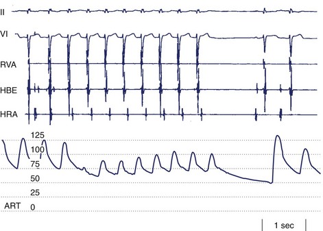
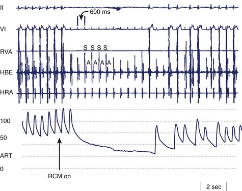
Supraventricular tachycardias are typically categorized into whether or not the tachycardia is dependent on AV node conduction. The first group is exemplified by AV nodal re-entry tachycardia (AVNRT) and AV re-entrant tachycardia (AVRT) using accessory AV connections. The AV nodal independent tachycardias include atrial re-entrant arrhythmias as well as those that are thought to be automatic or triggered in origin. In either case, the ventricular rate is determined by the ANS effect on the AV conduction system (particularly the AV node, see earlier). In certain cases, ANS effects may also play a role in terminating tachyarrhythmias, either spontaneously (Figure 5-6) or during medical interventions such as carotid sinus massage.

Ventricular Tachycardia

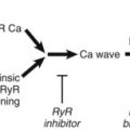
The importance of the ANS in determining susceptibility to ventricular tachyarrhythmias in certain disease states is well established. Acute ischemic heart disease is the best example.9,10 However, ANS influences may also be instrumental in triggering tachycardia events in patients with well-established long-standing substrates, such as those with pre-existing fibrotic areas as a consequence of prior myocardial infarction or remote cardiac surgery (e.g., childhood repairs). ANS participation in the triggering of arrhythmias is also almost certainly pertinent in other chronic states in which the arrhythmia substrate is present all the time and yet rhythm disturbances occur only sporadically. Among the best examples of the latter scenario are the abnormal ventricular repolarization syndromes (e.g., long QT syndrome [LQTS], Brugada syndrome).

Parasympathetic Neural Influences

Experimental and clinical evidence supports the view that enhanced parasympathetic tone diminishes the risk of arrhythmia in the setting of acute ischemia. In this regard, both neurally mediated heart rate–slowing effects and direct parasympathetic agonist effects contribute to the overall benefit. Furthermore, direct vagal nerve stimulation, by virtue of its anti-sympathetic effects as well as its parasympathetic actions, may become particularly valuable.

Studies of baroreceptor sensitivity (BRS) (a measure of vagal influence on the heart) offer important insights into the potentially protective role played by the parasympathetic nervous system in patients with ischemic heart disease. In a prospective trial of relatively low-risk patients, at 2-year follow-up, BRS values were seen to be lower in those individuals who failed to survive, and the effect appeared to be independent of left ventricular function as assessed by ejection fraction measurement. In this regard, a multicenter trial, Autonomic Tone and Reflexes after Acute Myocardial Infarction (ATRAMI), provided convincing additional evidence.13 Patients were monitored for 21 ± 8 months. Cardiac mortality was higher (9% vs. 2%; 10% vs. 2%) among individuals with low BRS (<3 ms/mm Hg) or low standard deviation of normal NN intervals (SDNN) (<70 ms) than those with normal BRS or SDNN (>6.1 ms/mm Hg, >105 ms). Combining both indices resulted in recognition of even greater risk. Once again, the effect appeared to be independent of ejection fraction.

The observations related to BRS led to the evaluation of heart rate variation (heart rate variability [HRV]) as an ANS-related means to stratify risk of lethal arrhythmias in patients with ischemic heart disease. The findings suggested that diminished HRV is associated with a much greater mortality risk in post–myocardial infarction patients. Other techniques using markers of ANS status as markers of arrhythmia risk include heart rate turbulence (HRT) and deceleration capacity (DC).

In summary, markers of reduced parasympathetic cardiac control appear to be indicative of increased risk, whereas enhanced parasympathetic control appears to be associated with reduced risk of lethal arrhythmia in patients with ischemic heart disease (at least in the post–myocardial infarction group). Consequently, enhancing parasympathetic predominance through exercise has become part of the overall approach to reducing mortality risk in patients with ischemic heart disease.

Long QT Syndrome, Brugada Syndrome, and Other Channelopathies

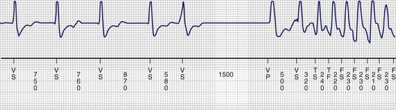
Disturbances of ventricular repolarization have been the subject of considerable interest in recent years, partly because of the recognition that they are a common cause of potentially life-threatening arrhythmias (Figure 5-7) but even more as a result of rapid progress in better understanding of why they occur.14 Currently, it is believed that underlying susceptibility to arrhythmia (primarily torsades de pointes ventricular tachycardia) is based on one or more genetically determined disturbances of the structure of cardiac membrane ionic channels, and/or disturbances of their function, or trafficking to the cell membrane.

The initial recognition of these conditions was based on overt ECG manifestations (e.g., long QT interval, typical Brugada pattern). However, it is now suspected that many more individuals manifest a less overt form of channelopathy; in such cases, the ECG signature may become apparent only after exposure to a trigger, particularly certain drugs or electrolyte disturbances, and, in some cases, acute neurologic injury (e.g., subarachnoid bleed). These latter circumstances are considered acquired, although a concealed congenital predisposition may well be present.

Genotype and Phenotype: Differences in Response to the Autonomic Nervous System in Long QT Syndrome

With the identification of specific genotypes for LQTS, it has become apparent that there are clinically significant differences in the expression of their phenotypes. Particularly striking is the observation that patients with LQT1 are especially prone to lethal cardiac arrest during exercise, whereas sudden death during exercise appears to be distinctly unusual in patients with LQT2 and LQT3. In contrast, patients with LQT3 and, to a somewhat lesser extent, those with LQT2 are more prone to lethal events during sleep or at rest.

Jervell and Lange-Nielsen, generally considered to be the earliest to report on LQTS, had noted that one of their subjects exhibited an increase in the QT interval in response to adrenaline and exercise. More recently, catecholamines such as isoproterenol or epinephrine have been used in various protocols trying to determine if an increase in the QT interval could be a tool in the diagnosis of LQTS. It has since become clear that the response to catecholamines cannot be generalized to all genotypes. During epinephrine infusion, the absolute QT interval increased in patients with LQT1 and decreased in those with LQT2 and LQT3. Similarly, the QTc increased during exercise stress testing in patients with LQT1 and decreased in those with LQT2. In effect, patients with LQT1 fail to appropriately shorten their QT interval in response to adrenergic stimulation that occurs during exercise or other types of stress.

More than one factor may make patients with LQT1 especially vulnerable to sudden death during exercise. The slow deactivation kinetics of the normal Iks channel produces an increase in the Iks current at fast heart rates; this shortens ventricular repolarization, which would be protective against torsades de pointes at fast rates, as has been demonstrated in guinea pig ventricular myocytes. A similar contribution of Iks appears to be present in human ventricular muscle in the setting of sympathetic stimulation. The loss of function of Iks that defines LQT1 renders these patients at risk for sudden death during exercise, whereas the presence of a normally functioning Iks channel in LQT2 and LQT3 appears to be protective against lethal cardiac events during exercise. Conversely, patients with LQT3 appear to be particularly vulnerable to sudden death at slow heart rates and more protected at fast heart rates.

Autonomic Responses and Treatment Considerations in Long QT Syndrome

Standard treatment for patients with LQTS has consisted of four principal options, singly or in combination: (1) β-adrenergic blockers, (2) pacing, (3) implantable defibrillators, and (4) LCSD. β-adrenergic blockers have reduced mortality, even though they slow the heart rate, presumably by reducing after-depolarizations and triggered activity postulated to underlie torsades initiation. Cardiac pacing prevents long cycle lengths that prolong the QT interval and lead to torsades. Implantable defibrillators are indicated for protection from sudden cardiac death in patients with prior cardiac arrest, ventricular tachycardia, or syncope. Before the development and acceptance of implantable cardioverter-defibrillators (ICDs), LCSD was considered a last-line option for patients refractory to β-blockers and pacing. LCSD is now considered an option for those with refractory symptoms and may be useful in patients with frequent ICD discharges.

Understanding the molecular basis of LQTS has opened the possibility of therapies targeted at the specific molecular mechanisms underlying an individual form of channelopathy. Even as these are being developed and tested, the traditional treatment options can be reassessed. A particular type of LQTS may respond well to one form of therapy, while other traditional options may be less effective or even undesirable. Patients with LQT1, having an impaired ability to shorten QT at faster heart rates and having a higher risk of death during periods of sympathetic hyperactivity such as exercise, might be expected to respond well to β-blockade, which limits heart rate increase and decreases after-depolarizations. In one clinical study, 81% of patients with LQT1 were able to avoid syncope, cardiac arrest, or sudden death with β-blocker therapy. In contrast, the symptom-free rate was 59% and 50% in patients with LQT2 and LQT3, respectively.

According to the International Long-QT Syndrome Registry, patients with LQT3 have a higher incident of lethal cardiac events than those with LQT1 or LQT2.14 Patients with LQT3 are at a particularly high risk of a cardiac event during sleep when EADs may result from a slow heart rate rather than from sympathetic activation. In these patients, β-blockers may be relatively unfavorable, whereas pacing may be particularly desirable. Besides pacing, patients with LQT3 may benefit from LCSD, which will diminish norepinephrine release without slowing the heart rate.

ANTIANGINAL AGENTS

PSYCHOACTIVE AGENTS TRICYCLIC ANTIDEPRESSANTS ANTIBIOTICS/ANTI-INFECTIVES NONSEDATING ANTIHISTAMINES OTHERS

Autonomic Nervous System and Syncope

Syncope is best viewed as a syndrome characterized by transient loss of consciousness, usually associated with concomitant loss of postural tone and spontaneous recovery. (Table 5-3).15 Mechanistically, syncope is most often the result of transient disturbances of cerebral blood flow. In this regard, maintenance of cerebral blood flow is normally facilitated by several factors, all of which are, to some extent, significantly influenced by the ANS. Certain of these factors include (1) cardiac output, (2) baroreceptor-induced adjustments of heart rate and systemic vascular resistance, (3) cerebrovascular autoregulation (which is, in part, contributed to by the status of systemic arterial pressure as well as by local metabolic factors, particularly pCO2), and (3) regulation of vascular volume by the kidneys and by hormonal influences.

Table 5-3 Classification of the Principal Causes of Syncope

NEURALLY MEDIATED REFLEX SYNCOPE

ORTHOSTATIC SYNCOPE CARDIAC ARRHYTHMIAS AS PRIMARY CAUSE OF SYNCOPE STRUCTURAL CARDIOVASCULAR OR CARDIOPULMONARY DISEASE CEREBROVASCULAR

Neurally Mediated Reflex Syncope

Of the many causes of syncope, ANS effects are of greatest importance in the various forms of neurally mediated syncope; the vasovagal faint and carotid sinus syndrome are the most common among these. Other conditions in this group (e.g., postmicturition syncope, cough syncope, swallow syncope) are relatively uncommon. However, ANS effects are crucial contributors to syncope associated with orthostatic stress and are also believed to play an important contributory role in certain tachyarrhythmias and cases of valvular heart disease.

In the so-called vasovagal faint, and especially faints associated with stress or emotional upset, primary central nervous system stimuli are believed to be responsible for the trigger signals.4 However, receptors in any of the organ systems may contribute. For instance, mechanoreceptors and, to some extent, chemoreceptors located in the atrial myocardium and in the ventricular myocardium may participate in certain neurally mediated events by initiating afferent neural signals when subjected to increased wall tension or changes in the chemical environment (e.g., myocardial ischemia). Similarly, mechanoreceptors and chemoreceptors in the central great vessels and lungs may contribute, which accounts for the reported occurrence of vasovagal faints in heart transplant recipients. The basis for apparent variations in susceptibility to vasovagal syncope among seemingly otherwise well individuals and the factors causing a faint to occur at a certain point in time still remain unknown.

Bradycardia in neurally mediated reflex syncope is primarily the result of increased efferent parasympathetic tone mediated via the vagus nerve. It may manifest as asystole, sinus bradycardia, or even paroxysmal AV block. If the bradyarrhythmia is sufficiently severe, it may be the principal cause of the faint (i.e., cardioinhibitory syncope). However, most patients also exhibit a vasodepressor picture comprising inappropriate ANS-induced vasodilatation (Figure 5-8). The mechanism of the vasodilatation is believed to be mainly the result of abrupt peripheral sympathetic neural withdrawal, although potential contributions of excess β-adrenergic tone caused by frequently associated elevated circulating epinephrine levels or altered epinephrine-norepinephrine balance are certainly considerations.

Orthostatic Syncope

The ANS participates importantly in the ubiquitous presyncopal or syncopal symptoms associated with abrupt postural changes. For the most part, these symptoms result from actual or relative central vascular volume depletion caused by inadequate or delayed peripheral vascular compensation in the presence of a change in gravitational stress (e.g., moving to upright posture). The outcome is posture-related symptomatic hypotension. Iatrogenic factors such as excessive diuresis or overly aggressive use of antihypertensive agents are important contributors.

Primary ANS disturbances are relatively rare but increasingly recognized causes of abnormal vascular control leading to syncope. Parkinsonism is perhaps the most commonly encountered neurologic disease in which ANS disturbances are associated with orthostatic hypotension as a prominent feature. ANS dysfunction may also occur in association with multiple system involvement (i.e., formerly called Shy-Drager syndrome). However, symptoms of orthostatic hypotension also occur in the absence of other apparent neurologic disturbances, and subtle forms may be easily overlooked. Furthermore, ANS diseases in which orthostatic hypotension is secondary in nature are far more common than those in which it is primary in nature. Examples include neuropathies of alcoholic or diabetic origin, dysautonomias occurring in conjunction with certain inflammatory conditions (e.g., Guillain-Barré syndrome), or paraneoplastic syndromes.

Noncardiovascular Conditions

Noncardiovascular causes result in syncope mimics (Table 5-4) rather than true syncope. However, temporal lobe seizures may induce neurally mediated reflex bradycardia and hypotension (i.e., a vasovagal faint). Furthermore, certain central nervous system syncope mimics may cause worrisome ECG changes and even myocardial damage, as discussed earlier.

Table 5-4 Conditions That Mimic Syncope

METABOLIC/ENDOCRINE DISTURBANCES

PSYCHIATRIC DISORDERS CENTRAL NERVOUS SYSTEM SUBSTRATES

* Potential nervous system–mediated cardiac damage and sudden death.

Metabolic or endocrine disturbances do not often cause true syncope. Acute hyperventilation provoked by or associated with panic attacks or anxiety attacks, and thus perhaps ANS related, is the most important exception. In these cases, abrupt reduction of pCO2 levels have been suggested to result in sufficient cerebral vasoconstriction to cause syncope. However, the evidence for hyperventilation causing frank syncope is weak at best.

The role of the ANS in the so-called chronic fatigue syndrome has been the source of some controversy following publication of findings suggesting an overlap with tilt-induced hypotension–bradycardia. It is most likely that ANS effects do play a role, but the magnitude of the impact is probably quite variable, and the evidence supporting a close connection between chronic fatigue syndrome and neurally mediated reflex syncope is far from convincing at this stage.

References

1 Randall WC, Wurster RD. Peripheral innervation of the heart. In: Levy MN, Schwartz PJ, editors. Vagal control of the heart: Experimental basis and clinical implications. Armonk, NY: Futura Publishing, 1994.

2 Herring N, Paterson DJ. Neuromodulators of peripheral cardiac sympatho-vagal balance. Exp Physiol. 2009;94:46-53.

3 Brotman DJ, Goldman SH, Wittstein IS. The cardiovascular toll of stress. Lancet. 2007;370:1089-1100.

4 Samuels MA. The brain-heart connection. Circulation. 2007;116:77-84.

5 Shishehbor MH, Alves C, Rajajgopal V. Inflammation: Implications for understanding the heart-brain connection. Clev Clinic J Med. 2007;74(Supp 1):S37-S41.

6 Tomson T, Nashef L, Ryvlin P. Sudden unexpected death in epilepsy: Current knowledge and future directions. Lancet Neurol. 2008;7:1021-1031.

7 Oppenheimer S. Cortical control of the heart. Clev Clinic J Med. 2007;74(Supp 1):S27-S29.

8 Armour JA. Potential clinical relevance of the “little brain” on the mammalian heart. Exp Physiol. 2008;93:165-176.

9 Pauza DH, Skripka V, Pauziene N, Stropus R. Morphology, distribution, and variability of the epicardiac neural ganglionated subplexuses in the human heart. Anat Rec. 2000;259(4):353-382.

10 Zipes DP, Rubart M. Neural modulation of arrhythmias and sudden death. Heart Rhythm. 2006;3:108-113.

11 Vaseghi M, Shivkumar K. The role of the autonomic nervous system in sudden cardiac death. Prog Cardiovasc Dis. 2008;50(6):404-419.

12 Saffitz JE. Sympathetic neural activity and the pathogenesis of sudden cardiac death. Heart Rhythm. 2008;5:140-141.

13 La Rovere MT, Bigger JTJr, Marcus FI, Mortara A, Schwartz PJ. Baroreflex sensitivity and heart rate variability in prediction of total cardiac mortality after myocardial infarction for the ATRAMI Investigators. Lancet. 1998;351(9101):487-494.

14 Zareba W, Moss AJ, Schwartz PJ, et al. Influence of genotype on the clinical course of the long QT syndrome. International Long QT Registry Research Group. N Engl J Med. 1998;339:960-965.

15 Brignole M, Alboni P, Benditt D, et al. Task force on syncope, European Society of Cardiology: Guidelines on management (diagnosis and treatment) of syncope—update 2004. Europace. 2004;6:467-537.