Atypical nevi

Published on 19/03/2015 by admin

Filed under Dermatology

Last modified 19/03/2015

Print this page

rate 1 star rate 2 star rate 3 star rate 4 star rate 5 star
Your rating: none, Average: 0 (0 votes)

This article have been viewed 1473 times

Atypical nevi

Julia Newton-Bishop

Evidence Levels:  A Double-blind study  B Clinical trial ≥ 20 subjects  C Clinical trial < 20 subjects  D Series ≥ 5 subjects  E Anecdotal case reports

image

The term ‘atypical nevi’ refers to clinically diagnosed lesions, defined as nevi that are more than 5 mm in diameter with an irregular or diffuse edge and variable color. Biologically such nevi are believed to be melanocytic neoplasms that result from more protracted proliferation (leading to a stromal reaction) than do banal benign melanocytic nevi. Histologically, atypical nevi are characterized by elongated rete ridges, bridging of melanocytes between rete ridges, a predominance of single melanocytes over nested melanocytes, and a dermal inflammatory reaction with papillary dermal fibroplasia. Although these histological changes are characteristic there may be a lack of correlation between the clinical and histological features, which has lead to controversy which has largely been unhelpful. Suffice it to say that the entity remains an important one, that it is clinically diagnosed but there are characteristic histological correlates which are variable in degree.

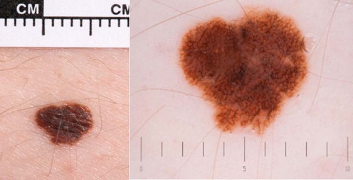
I use the term ‘atypical nevi’ to mean nevi that others might call dysplastic nevi.

Atypical nevi may be considered more of a marker of patients at higher risk of melanoma than as frequent precursors of melanoma. The indication for excision is to exclude melanoma, not to make a diagnosis of an atypical nevus. There is no role for prophylactic excision of atypical nevi with a banal dermoscopic appearance except perhaps when a single atypical nevus appears in an individual who is older than the usual age for such nevi (over 50 years). It is important to note that two-thirds of melanomas do not arise from previous nevi even in the atypical mole syndrome, so that removing all atypical nevi does not prevent melanoma. It is mandatory to perform total body skin examinations in patients at risk for melanoma, looking for the ‘ugly duckling nevus,’ which stands out as different from that patient’s typical ‘signature nevi.’ The mnemonic ABCDE has been used as a clinical aid (asymmetry, border irregularity, color variegation, diameter >6 mm, and evolution or change in a lesion), but in practice most experts make the diagnosis based upon a global clinical examination. This is similar to a child who recognizes her written name without understanding the meaning of the individual letters.

Some authorities advocate grading atypical nevi as mild, moderate, or severe atypical nevi (NIH Consensus Conference. Diagnosis and treatment of early melanoma. JAMA 1992; 268: 1314–1319). Others have argued that such grading has poor reproducibility, and therefore they do not grade the severity of the atypia. The majority of dermatopathologists prefer to grade the architectural and the cytologic atypia separately. It is not uncommon for severely atypical nevi to cause diagnostic difficulty. Histopathologists may report that such a lesion is of unknown malignant potential and recommend that the lesion best be treated as melanoma with a wide local excision as for melanoma.

It is common to have a single atypical nevus. In a mildly atypical nevus, with a bland dermoscopic appearance, the risk of malignant change under the age of 50 years is very small. Such nevi therefore should not be excised. It is then important to educate the patient how to monitor the lesion and to give that patient information booklets with photographs of atypical nevi and melanoma so that the patients knows what to look for. Merely asking the patient to ‘keep an eye out for change’ is insufficient.

Where the atypical nevus shows more markedly atypical features, and especially in older individuals, then the lesion should probably be excised. In such cases, taking a photograph and reviewing is rarely helpful as one usually feels no less comfortable to leave alone on review than at first visit; the hypothesis is that, if an atypical nevus is single, the patient’s risk can be removed by an excision of the lesion. If an atypical nevus causes concern then it should be excised in its entirety rather than sampled incisionally. Although data have been published to suggest that there is a low rate of clinical recurrence after biopsy of benign moderately ‘dysplastic’ nevi, sampling is risky as sample error may lead to examination of a less atypical portion of the tumor. For the patient and the clinician, complete excision is a safer approach as melanocyte pathology is difficult to interpret and the pathologist could make an error in this grey area – it is better to have excised the lesion completely in the face of ambiguity. Incisional biopsy may furthermore stimulate proliferation of melanocytes to lead to a clinically and histologically concerning lesion, known as a pseudo-melanoma; if there is enough clinical concern to sample such a nevus, an experienced clinician will be sampling a lesion with some clinically worrisome features thereby supporting the argument that a complete excision is desirable.

Patients with increased numbers of banal nevi and/or multiple clinically atypical nevi are said to have the atypical mole syndrome and require different management. These patients have a melanoma risk that cannot be removed by excision of nevi. The key components of good treatment are:

image Taking a detailed family history to determine if cases of melanoma have occurred in the family. Risk estimation is strongly modified by family history (see www.genomel.org)

image Education about monitoring of nevi

image Follow-up/supervision in clinic, for a period whose length is determined by risk estimation based upon family or personal history of melanoma, the competency of the patient in self examination and the clinical phenotype

image Excision of atypical nevi where it is necessary to exclude melanoma

image Education about ensuring sufficient sun protection without becoming vitamin D depleted. Sunburn avoidance is crucial in that sunburn is established to be associated with melanoma risk in multiple studies. Sunbathing, independently of sunburn may also increase risk so should be avoided in those with atypical moles.

Management strategy

The strategy is essentially to excise clinically atypical nevi if there is a reasonable suspicion of malignancy whilst avoiding excessive numbers of procedures. The history of the lesion, the appearance to the naked eye, and the dermoscopic appearance are all important. Clinically atypical nevi, which are behaving in an unusual fashion, should prompt a decision to excise such lesions. Examples include a new atypical nevus over the age of 50 or a lesion which looks like an atypical nevus which has grown rapidly in the previous 6 months. Dermoscopy has been shown to increase diagnostic accuracy. Although anecdotal reports in the literature have utilized topical tretinoin, imquimod, laser surgery, or cryosurgery, these modalities cannot be advocated for treating atypical nevi.

Treatment strategies for atypical nevi: