Atopic dermatitis

Published on 19/03/2015 by admin

Filed under Dermatology

Last modified 22/04/2025

Print this page

rate 1 star rate 2 star rate 3 star rate 4 star rate 5 star
Your rating: none, Average: 0 (0 votes)

This article have been viewed 1935 times

Atopic dermatitis

Shehla Admani and Lawrence F. Eichenfield

Evidence Levels:  A Double-blind study  B Clinical trial ≥ 20 subjects  C Clinical trial < 20 subjects  D Series ≥ 5 subjects  E Anecdotal case reports

image

Atopic dermatitis (AD) is a chronic, relapsing, intensely pruritic dermatosis that develops most commonly during early infancy and childhood and is, in most cases, associated with a personal or family history of atopy (allergic rhinitis, asthma, or eczema). It is frequently associated with abnormalities in skin barrier function and immune dysregulation. Skin involvement ranges from acute weeping and crusted areas of eczema to papular lesions or lichenified plaques. There is no single defining clinical feature or laboratory test and the diagnosis is based on a constellation of clinical findings.

Management strategy

Successful AD therapy considers the patient’s age and needs, the extent and localization of AD at presentation, and the overall disease course. Other factors to consider include previous response to treatment, disease persistence, frequency of flares, and susceptibility to and past history of infection (especially due to Staphylococcus aureus and herpes simplex). The goals of management are to educate patients and caregivers about the disease, promote excellent skin care, reduce the degree and frequency of flares, monitor medication quality/quantity of use, and, if possible, modify the overall disease course and the atopic march.

Interventional education

Comprehensive multispecialty ‘eczema clinics’ are beginning to test the utility of intensive education as a distinct component of long-term AD management. This model, with its longer appointments, focused educational curricula, patient support networks, and the ability to elicit patient/family feedback, parallels strategies shown to be effective for managing asthma, diabetes, and other chronic diseases. Supporters hope to empower patients and caregivers and improve both clinical and quality of life outcomes. Long-term comparative evaluation is required to examine the cost-effectiveness and suitability of these educational programs. Other learning modalities beyond in-clinic disease education may be useful to assist with disease management, including hand-outs, internet-based written material and instructional videos (e.g., www.eczemacenter.org; www.nationaleczema.org; www.eczema.org).

Skin care

Excellent skin care remains a cornerstone of management. Emollients improve the xerosis and skin barrier dysfunction associated with AD, and may improve pruritus and spare prescription anti-inflammatory medication use. With little evidence to recommend the use of one emollient/moisturizer over another, patient and caregiver preference should be considered with product selection based on the premise that ‘an emollient that is applied works better than one that remains on a shelf.’ Very occlusive ointments may not be tolerated during the summer months or in humid climates because of interference with the function of eccrine sweat ducts and the induction of folliculitis; in these situations, a cream may be a more practical choice. It is best to avoid preparations that contain topical sensitizers such as fragrance, neomycin, benzocaine, etc. Emollients/moisturizers should be applied after any topical pharmacologic therapies to allow active medications to reach the skin with full effect.

The value of bathing and the frequency with which it should be undertaken remain controversial, though most experts believe that daily bathing with application of emollients to follow is well tolerated and may be beneficial. The chief benefits of bathing include cleansing, debridement of infected eczema, improved penetration of topical therapies, and skin hydration (when emollients are used to ‘lock in’ moisture). Potential drawbacks of bathing are drying of the skin and disruption of the stratum corneum barrier during water evaporation when emollients are not used. Recent data regarding transepidermal water loss suggest that the frequency of application of emollients/moisturizers may be more important than timing the application to coincide strictly with bathing (the traditional ‘soak and seal’ approach); this finding remains to be confirmed in larger, more controlled studies.

Fillagrin is a structural protein found in the stratum corneum. Loss of function mutations in the fillagrin gene can lead to increased transepidermal water loss and an increased risk of developing AD as well as asthma, allergies, and herpes virus infection. Several barrier repair products have been approved by the US Food and Drug Administration (FDA) as ‘510(k) medical devices.’ These products contain ingredients that attempt to replace or correct deficiencies of epidermal lipids, improve skin hydration, reduce skin barrier dysfunction, and relieve the pruritus, burning, and pain associated with AD. Several over-the-counter products similarly target barrier function abnormalities. There have been limited comparative effectiveness studies to assess the relative efficacy and safety of these prescription devices, specially formulated emollients, and traditional emollients/moisturizers in helping to manage AD.

Wet wraps are a useful tool in the intensive treatment of severe AD and/or disease that is refractory to standard topical therapies. They may increase skin hydration, serve as an effective mechanical barrier to scratching, and act as an occlusive layer that promotes penetration of topical corticosteroids into the skin thereby increasing the amount of medication delivered to the most severely affected areas. Temporary systemic bioactivity of the corticosteroids is a concern, as is the potential to induce hypothermia. When wet wraps are overused or used incorrectly, maceration of the skin may occur. Because of these concerns wet wraps should only be used under close supervision of a physician.

Topical therapies

Topical corticosteroids remain the first-line therapy for inflammation and pruritus associated with AD unresponsive to good skin care and moisturizers. Variation in corticosteroid-prescribing habits (e.g., quantity, frequency, and duration of therapy) is common even among dermatologists. Some clinicians start treatment with high-potency topical corticosteroid preparations in order to induce remission, followed by a relatively quick tapering-down of potency as the dermatitis improves. Alternatively, some clinicians use short bursts of high-potency corticosteroids followed by moisturizer alone until relapse occurs. Another treatment regimen advocates more prolonged treatment with less potent steroid preparations. Drug-specific FDA indications and an expanding body of clinical trial data should help guide clinicians when educating and instructing patients on topical corticosteroid usage.

The topical calcineurin inhibitors (TCIs) tacrolimus and pimecrolimus have the important advantage of not being associated with skin atrophy. Current indications for TCI use are as ‘second-line therapy for the short-term and non-continuous chronic treatment’ of mild to moderate AD (pimecrolimus) or moderate to severe AD (tacrolimus) in non-immunocompromised adults and children ‘who have failed to respond adequately to other topical prescription treatments for atopic dermatitis, or when those treatments are not advisable.’ Patients who are especially likely to benefit include those in whom the clinical course of AD is marked by steroid tachyphylaxis (versus simple non-compliance), disease persistence, and/or frequent flares, which would otherwise result in an almost continuous need for topical corticosteroid treatment. TCIs may also be specifically indicated in sensitive thin skin areas, such as around the eye, face, neck, and genital area where local safety and systemic absorption are of special concern.

The safety and efficacy of tacrolimus and pimecrolimus have been studied in multiple short- (6 week) and long-term (longer than 2 years) clinical trials. Data from these trials demonstrate that pimecrolimus reduces the number and severity of flares, extends the period between major flares, and reduces pruritus and other cutaneous signs associated with AD. Likewise, long-term, intermittent (once daily, two or three times weekly) maintenance use of tacrolimus ointment in patients with stabilized AD has been shown to significantly increase the period between disease exacerbations and the total number of disease-free days compared to vehicle. The incidence of side effects in these studies was generally low and most commonly included transient application-site stinging. The currently available data suggest that the use of TCIs is not associated with systemic immunosuppression or an increased risk of skin cancer; nor does it appear to affect the delayed-type hypersensitivity response. In most countries the use of TCIs is not recommended in patients under 2 years of age. Tacrolimus ointment 0.03% is indicated for adults and children aged 2–15 years, whereas tacrolimus ointment 0.1% is indicated only for adults. Long-term prospective studies investigating the clinical use of TCIs in a pediatric population are currently under way and will hopefully help to alleviate safety concerns.

Careful supervision, combined with appreciation of the risk–benefit profiles of moisturizers, barrier repair agents, topical corticosteroids, and TCIs, allows for individualized and optimized patient care. Treatment should be readily adjusted on an ‘as-needed’ basis that takes advantage of available therapeutic modalities. For children with severe flares, this may mean using short-term bursts of mid- to high-potency topical steroids – with or without wet wraps – instead of relying on long-term use of less potent agents. Close re-examination of the patient at regular intervals to evaluate the efficacy and tolerability of local and systemic therapies is warranted. Once control of a flare is achieved, therapy should shift to a less intense regimen with a focus on maintenance and proper skin care at its core. Wet wraps can be stopped and topical corticosteroids can be tapered to a lower-potency agent and/or from daily to intermittent (e.g., thrice- or twice-weekly) application. Transition to TCI therapy for patients with a history of flare recurrence upon discontinuation or tapering of topical corticosteroids may be a good choice at this point. The use of TCI monotherapy to control flare recurrence while limiting patients’ extended exposure to corticosteroids is supported by some physicians.

Other treatment considerations

Total avoidance of environmental aeroallergens is almost impossible and may not significantly influence atopic dermatitis; however, avoidance of known triggers is a reasonable approach. In cases where aeroallergens are strongly suspected of having a causative role, mattress covers, low-pile carpet (particularly in sleeping areas), frequent vacuuming, and non-dander-producing pets may be considered, especially for children who have concomitant asthma and/or allergic rhinitis. Food allergy is more common in children with atopic dermatitis than without, and in a subset of individuals clinically relevant food allergy may exacerbate atopic dermatitis. However, specific IgE testing and skin prick tests have high rates of false positives and poor predictive value, and severely restrictive diets based on positive tests rather than true allergy can be harmful. Recent US national guidelines for food allergy recommend evaluation for food allergy be considered in children less than 5 years old with moderate to severe AD for milk, egg, peanut, wheat, and soy if the child has a reliable history of an immediate reaction after ingestion of a specific food, or the child has persistent AD in spite of optimized management and topical therapy. Maternal dietary restrictions during pregnancy and lactation are not considered to have a significant role in development of AD, and are generally not recommended.

Although they do not appear to have direct effects on the pruritus associated with AD, sedating systemic antihistamines such as hydroxyzine and diphenhydramine may be useful in improving sleep in flaring patients. This practice has not been evaluated rigorously in large, randomized, double-blind, placebo-controlled trials, and the drowsiness that may be associated with daytime use is a legitimate concern for school-age children. Second-generation antihistamines are less useful in managing atopic dermatitis but may benefit patients with allergic triggers and, with chronic use, are suggested in some studies to reduce the rate of progression to other atopic disease (the atopic march). It is important to note that topical antihistamines are not recommended because of potential cutaneous sensitization. Oral doxepin hydrochloride, a tricyclic antidepressant with anxiolytic effects, has a high H1– and H2-receptor antagonist activity. It is typically used in doses of 10–75 mg orally at night or up to 75 mg twice daily in adult patients; it is not approved for use in children. As oral doxepin possesses a side-effect profile that includes daytime sedation, hypotension, tolerance, and an increased risk of depression/suicide, it is generally reserved for severe cases of AD. Topical 5% doxepin cream has been reported to reduce pruritus; however, these topical formulations have also been associated with reports of allergic contact dermatitis and sedation.

Patients may have sudden exacerbations of AD due to overgrowth of Staphylococcus aureus that can be independent of clinical signs of bacterial infection, a notion supported by apparent clinical response of patients with severe AD to anti-staphylococcal antibiotics. Honey-colored crusting, folliculitis, and pyoderma are signs of overt infection. In these cases topical and/or oral antibiotic therapy – typically of short duration to avoid the development of bacterial resistance – is indicated. Skin cultures and sensitivity testing should be considered prior to treatment, as methicillin-resistant S. aureus (MRSA) may be an important pathogen in some patients. Recurrent, deep-seated S. aureus infections should raise the possibility of an immunodeficiency syndrome such as hyper-IgE syndrome or DOCK8 deficiency.

The addition of antiseptics to bath water, e.g., diluted bleach baths (‘like swimming in pool water’), may help reduce the number of local skin infections and the need for systemic antibiotics in AD patients with heavily colonized and/or superinfected skin. A bleach bath can be prepared by mixing one-quarter to half a cup of sodium hypochlorite 6% solution (chlorine liquid bleach) in a bathtub full of lukewarm water; the goal is to create a modified Dakin’s-like solution with a final bleach concentration that approximates 0.005%. The patient may soak for five to 10 minutes, rinse their skin thoroughly with fresh water, pat dry, and then apply their topical therapy and/or emollient/moisturizer. Proprietary bath additives containing antiseptics are also available. For added convenience, newer formulations of bleach-like products are specially formulated and available as a body washes, sprays or gels.

Eczema herpeticum may be easily misdiagnosed as bacterial superinfection and presents a serious risk in patients with widespread AD. Patients can present with multiple vesiculopustular lesions and painful ‘punched-out’ erosions that fail to respond to oral antibiotics. Document herpes infection prior to treatment via culture and/or direct fluorescent antibody, and initiate antiviral therapy as soon as possible. Intravenous treatment is certainly indicated in cases of severe disseminated eczema herpeticum. Oral acyclovir (or equivalent dosage of another anti-herpetic medication) may be useful in adults with herpes simplex confined to the skin; 400 mg three times daily for 10 days or 200 mg four times daily for 10 days usually provides a sufficient dosage.

Fungal infections such as those caused by Trichophyton rubrum may also be more common in AD patients. Antifungal therapy has been shown to reduce the severity of AD lesions exacerbated by Malassezia furfur, particularly in the seborrheic areas of the skin and scalp. Patients with documented dermatophyte infection or IgE antibodies to Malassezia may benefit from a trial of topical or systemic antifungal therapy.

Owing to a greater understanding of immunologic reaction patterns in the skin, gut, and airways there has been great interest in probiotics. As the findings to date on their utility in preventing or modifying AD are conflicting, the long-term significance of probiotics in the treatment of atopic dermatitis warrants further investigation.

Although the exact mechanism of action is unknown, phototherapy in AD is thought to suppress proinflammatory cytokines (IL-2, IL-12) and induce T-cell apoptosis. Broadband UVB, broadband UVA, narrowband UVB (311 nm), UVA1 (340–400 nm), and combined UVA-B phototherapy have been reported to be useful for widespread or recalcitrant disease. Photochemotherapy with psoralen and UVA light may be indicated in severe cases. Multiple treatments are usually required to be effective, and this can be inconvenient for patients and their families, depending on location and accessibility to a suitable light source. Side effects can include skin pain, erythema, pruritus, and pigment changes. Likewise, UV radiation may increase the long-term risk of premature skin aging and cutaneous malignancies. Shielding and appropriate eye protection may help minimize unnecessary exposure.

Systemic therapies

Systemic corticosteroids are often prescribed in pediatric outpatient and emergency settings for AD exacerbations, though few clinical trials support their use. The temptation to use systemic corticosteroids can be great, given the dramatic clinical improvement that can occur. The propensity to flare with abrupt discontinuation of treatment and the well-known associated systemic side-effect profile, however, suggest that systemic corticosteroids should be reserved for ‘crisis cases’ – and, even then, used with the intent to bridge to another systemic agent or phototherapy.

Cyclosporine is a calcineurin inhibitor that blocks activation of T lymphocytes and reduces the transcription of cytokines, including IL-2, shown to be involved in the pathogenesis of AD. It may be used as a short-term treatment or as a bridge between other steroid-sparing alternatives. Cyclosporine is typically dosed at 2.5–5 mg/kg/day, and a response may be seen in 2 to 3 weeks. Alternatively, some experts prefer dosing cyclosporine microemulsion at 3 mg/kg/day in children, or 150 mg (low dose) or 300 mg (high dose) in adults as the microemulsion offers more predictable absorption. Flares can occur after discontinuation of therapy, therefore gradual tapering or use of another immunosuppressive for maintenance therapy should be considered. The safety, efficacy and risks of cyclosporine are well documented in both adults and children, and treatment with this agent is associated with reduced skin disease and an improved quality of life. Hypertension and renal toxicity, as well as concerns about malignancy, are limitations to long-term therapy. Continuous therapy is not recommended beyond 1 year, and it is unknown how many short courses may be given safely. Blood pressure, complete blood cell count, renal and hepatic function tests, magnesium, and uric acid should be monitored.

Azathioprine, a 6-mercaptopurine analog that inhibits purine synthesis and demonstrates cytotoxic and immunosuppressive properties, can be effective monotherapy for AD. Marrow suppression and liver toxicity are major concerns; blood cell counts and liver function tests should be monitored closely. One in 300 individuals is homozygous for low metabolic activity alleles that correlate with higher risk of marrow suppression; azathioprine should be dosed according to thiopurine methyltransferase (TPMT) genotype/levels. Dosing in children and adults is generally 2.5 mg/kg/day in patients with normal TPMT activity and adjusted to lower doses for carriers of mutant alleles and/or definite intermediate TPMT levels (e.g., 1.0 mg/kg/day). Higher doses, up to 5 mg/kg/day, have been utilized in children. Very low or absent TPMT activity indicating homozygous TPMT mutations may be associated with life-threatening myelotoxicity. Drug hypersensitivity and gastrointestinal disturbances have been reported. An increased risk of malignancy is recognized with long-term use to prevent transplant rejection or manage inflammatory bowel disease.

Methotrexate is a folic acid analog that inhibits dihydrofolate reductase and interferes with DNA synthesis and lymphocyte proliferation leading to anti-inflammatory effects; however, its greatest advantage may be that the relatively low doses used for skin disease appear less immunosuppressive than other AD systemic therapies. An open-label, prospective, 24-week trial of adults with AD demonstrated a response plateau at approximately 12 weeks, with little additional improvement at doses greater than 15 mg weekly. Alternatively, some experts dose methotrexate at 2.5 mg daily for 4 days per week. A retrospective review of children showed that AD was well controlled with effective dosing of 0.5–0.8 mg/kg/week (either as a single weekly dose or divided 3 or 4 days/week). There is a long history of methotrexate use in pediatric and adult inflammatory disease. Nausea and liver function abnormalities/hepatotoxicity may limit dosing. Pulmonary toxicity may be another potential concern. It is unclear what role folic acid supplementation plays in the treatment of AD with methotrexate.

Mycophenolate mofetil (MMF) has a good safety profile and represents a possible therapeutic alternative for severe, refractory AD. It is an inhibitor of inosine monophosphate dehydrogenase involved in de novo purine synthesis, and has been used as an immunosuppressant in organ transplantation. Several adult studies have demonstrated efficacy at doses up to 2 g daily. A retrospective review of MMF as monotherapy in 14 pediatric AD patients showed MMF to be safe and effective at doses of 40–50 mg/kg/day in younger children (presumably due to increased surface area-to-volume ratios) and 30–40 mg/kg/day in adolescents, with maximal effect after eight to 12 weeks of therapy. Patients should be monitored for leukopenia and anemia, and drug levels may be increased in the setting of renal insufficiency. MMF has been loosely linked to herpes retinitis, dose-related bone marrow suppression, and increased infection. Further prospective controlled studies are needed for this promising therapy.

Interferon-γ (IFN-γ) is well known to inhibit Th2-cell proliferation/function and to suppress IgE responses. Several adult AD studies have demonstrated efficacy with three-times-weekly high-dose (150 µg/m2) and low-dose (50 µg/m2) therapy. Disadvantages of therapy include flu-like symptoms (especially common early in the treatment course), myelosuppression, neurotoxicity/confusion, hypotension, tachycardia, and cost.

Referral, quality of life issues, and educational resources

Referral to a pediatric dermatologist or an adult dermatologist/atopic dermatitis specialist may help with comprehensive management of skin care and barrier repair. Referral should be considered in patients with a diagnosis of moderate or severe AD; those who are unresponsive to standard treatments (including mid-potency topical corticosteroids); those with persistent disease and/or frequent flares; those who have been hospitalized as a direct consequence of their AD; and those requiring systemic therapies for flares and/or maintenance. Allergy testing is not generally a first-line referral recommendation in the routine evaluation and treatment of uncomplicated AD. However, consultation with an allergist can be useful when proper skin care alone is not effective and/or when the clinical picture hints strongly at specific allergic triggers. Immunotherapy with aeroallergens has not been proven effective in the treatment of AD. Referral to immunology or gastroenterology is warranted if underlying systemic infections are frequent or when eosinophilic gastroenteritis/esophagitis becomes a concern in younger children with concomitant failure to thrive.

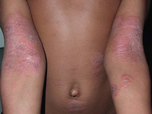
Emotional stress can exacerbate AD in some patients who may respond to frustration, anxiety, embarrassment, or other psychologically stressful events with a perceived increase in pruritus and subsequent scratching. This may be particularly important in the adolescent population, where even very mild skin disease may be considered ‘disfiguring.’ In some cases, patients may use scratching for secondary gain and in others scratching has simply become habitual. Relaxation, biofeedback, and behavioral modification techniques may be helpful in such patients. Psychosocial evaluation and counseling should be considered in families where emotional triggers appear to function as obstacles to disease management, or where quality of life is clearly affected. Therapeutic education has been shown beneficial in managing AD, and may be accomplished through extended in-office discussion, written handouts, eczema ‘school programs’ and/or web resources, which include video training modules (e.g., www.eczemacenter.org). Families may also benefit from support groups such as the US National Eczema Association (www.nationaleczema.org) and the UK National Eczema Society (www.eczema.org).

Specific investigations

First-line therapies

image Education B
image Emollients B
image Topical corticosteroids A
image Wet wraps C

Second-line therapies

image Topical immunomodulators A
image Antimicrobials C
image Antihistamines/anxiolytics C
image Barrier repair products B
image Avoidance of triggers A

A double-blinded, randomized, placebo-controlled trial of cetirizine in preventing the onset of asthma in children with atopic dermatitis: 18 months’ treatment and 18 months’ posttreatment follow-up.

Warner JO, ETAC Study Group. Early Treatment of the Atopic Child. J Allergy Clin Immunol 2001; 108: 929–37.

Infants between 1 and 2 years of age with AD in this double-blinded trial were randomized to 0.25 mg/kg body weight cetirizine administered twice daily compared with placebo. After 18 months of treatment, follow-up continued for a further 18 months. Although there was no difference in the cumulative prevalence of asthma between active and placebo treatment in the intention-to-treat population (p = 0.7), cetirizine did appear to delay (and in some cases to prevent) the development of asthma in those patients sensitized to grass pollen and, to a lesser extent, house dust mites.

Third-line therapies

image Dust mite reduction A
image Timing of solid food introduction A
image Phototherapy A
image Azathioprine A
image Cyclosporine A
image Methotrexate B
image Mycophenolate mofetil C
image Interferon-γ A
image Efaluzimab E
image Systemic corticosteroids B

Azathioprine in severe adult atopic dermatitis: a double-blind, placebo-controlled, crossover trial.

Berth-Jones J, Takwale A, Tan E, Barclay G, Agarwal S, Ahmed I, et al. Br J Dermatol 2002; 147: 324–30.

In this double-blind crossover trial, adult patients with severe AD were treated with azathioprine 2.5 mg/kg daily and placebo for 3 months each. There was a significant difference in favor of azathioprine in the improvement of the six area, six sign atopic dermatitis (SASSAD) sign score, decreasing by 26% during treatment with azathioprine, versus 3% on placebo. Pruritus, sleep disturbance, and disruption of work/daytime activity all improved significantly on active treatment, but the difference in mean improvement between azathioprine and placebo was statistically significant only for disruption of work/daytime activity. The authors suggested that a longer period of treatment might have further improved the eczema. Gastrointestinal disturbances and deranged liver enzymes were common. Of the 37 patients enrolled, 12 prematurely terminated treatment with azathioprine, and four with placebo.

Cyclosporine in the treatment of patients with atopic eczema – a systematic review and meta-analysis.

Schmitt J, Schmitt N, Meurer M. J Eur Acad Dermatol Venereol 2007; 21: 606–19.

A systematic review of the effectiveness of systemic cyclosporine in patients with severe atopic eczema was conducted. Fifteen studies comprising a total of 602 patients were included. In all studies analyzed, cyclosporine consistently decreased the severity of atopic eczema. Data from 12 of the 15 studies were pooled and showed a dose-related response with a mean decrease in disease severity of 22% (95% CI: 8–36%) under low-dose cyclosporine (3 mg/kg) and 40% (95% CI: 29–51%) at dosages ≥4 mg/kg after 2 weeks of treatment. After 6 to 8 weeks the relative effectiveness was 55% (95% CI: 48–62%). Effectiveness of cyclosporine was similar in adults and children, but tolerability may be better in children.

Double-blind, controlled, crossover study of cyclosporine in adults with severe refractory atopic dermatitis.

Sowden JM, Berth-Jones J, Ross J, Motley RJ, Marks R, Finlay AY, et al. Lancet 1991; 338: 137–40.

A multicenter, randomized, double-blind, controlled crossover clinical trial was conducted on 33 patients with severe refractory atopic dermatitis, to determine the effects of cyclosporine (5 mg/kg/day) on their health-related quality of life. After treatment with cyclosporine, patients indicated significant improvement in quality of life; however, there was either no correlation, or only a very poor correlation, between the quality of life parameters and clinical measures of extent and activity of eczema. Twenty patients receiving cyclosporine reported adverse events, compared with eight taking placebo. When cyclosporine was stopped, relapse was noted; however, the mean scores for disease activity and extent of disease in these patients were less than their baseline values.

Cyclosporine in atopic dermatitis: time to relapse and effect of intermittent therapy.

Granlund H, Erkko P, Sinisalo M, Reitamo S. Br J Dermatol 1994; 132: 106–12.

Forty-three patients with severe AD were treated with a 6-week course of cyclosporine 5 mg/kg daily, and then re-treated after a follow-up of 6 to 26 weeks (depending on the time to relapse) with an identical course of cyclosporine. A significant reduction in disease activity was observed after 2 weeks of cyclosporine treatment. After both treatment periods, approximately half of the patients relapsed after 2 weeks; after 6 weeks follow-up the relapse rates were 71% and 90%, respectively, for the two treatment periods. Notably, after the first treatment period, five patients did not relapse during the 26-week follow-up, and for the second treatment period two did not relapse. All of these seven patients were still in remission at one year.

Cyclosporine in atopic dermatitis: review of the literature and outline of a Belgian consensus.

Naeyaert JM, Lachapelle JM, Degreef H, de la Brassinne M, Heenen M, Lambert J. Dermatology 1999; 198: 145–52.

This excellent review summarizes all the major trials of cyclosporine for AD and gives practical recommendations for the clinician. These authors recommend that cyclosporine be reserved for adults with severe AD and only used in children with recalcitrant disease for short periods. A starting dosage of 2.5 mg/kg daily can be adjusted up after 2 weeks, depending on response, to a maximum dose of 5 mg/kg daily. Screening for gynecologic or prostate malignancy, and skin biopsy to exclude cutaneous T-cell lymphoma, as well as close monitoring of renal function and blood pressure, are recommended.