Arthroscopic Microfracture and Chondroplasty

Published on 11/04/2015 by admin

Filed under Orthopaedics

Last modified 22/04/2025

Print this page

rate 1 star rate 2 star rate 3 star rate 4 star rate 5 star
Your rating: none, Average: 0 (0 votes)

This article have been viewed 2025 times

CHAPTER 22 Arthroscopic Microfracture and Chondroplasty

Introduction

Articular cartilage defects are one of the pathologies that are most commonly encountered during hip arthroscopy. Limited information is available regarding the best treatment of these lesions; however, microfracture has been demonstrated to be effective for the treatment of articular cartilage defects of the knee and other weight-bearing joints, and several small series and case reports have been published that have described the use of microfracture for the treatment of articular cartilage defects in the hip. Several investigators are conducting ongoing outcome studies with regard to this treatment.

There are many conditions that can cause chondral defects that necessitate microfracture and chondroplasty, and these conditions can be acute, chronic, traumatic, or atraumatic. Defects can be full or partial thickness. Acute causes include hip dislocation, hip subluxation, and direct blows to the hip. Chronic causes include labral pathology, femoral acetabular impingement, loose bodies, dysplasia of the hip, a history of slipped capital femoral epiphysis, avascular necrosis, and degenerative joint disease.

It is critical to note that the diagnosis of these lesions is often only established intraoperatively, and patients should be aware that labral pathology is often associated with articular cartilage damage. The postoperative rehabilitation process and the patient’s prognosis can be significantly different if such a lesion is discovered, and the patient and surgeon should be prepared for this possibility. A working knowledge of chondroplasty and microfracture will be helpful for any orthopedic surgeon who attempts to treat these diseases.

Brief history and physical examination

As with other musculoskeletal conditions, a careful history and physical examination are warranted. An understanding of the acuity and cause of the injury has important prognostic indications. Acute injuries (e.g., hip dislocations, subluxation events) are often associated with significant articular cartilage lesions that are sometimes asymptomatic. A direct lateral blow to the greater trochanter (e.g., a fall) can cause a medial impaction injury to the femoral head. A more gradual onset of pain or discomfort suggests a degenerative or impingement cause of the articular cartilage injury.

The location, frequency, and radiation of pain should be determined. Furthermore, alleviating or worsening symptoms or activities should be noted. Attention should be paid to any presence of mechanical symptoms (e.g., clicking, snapping, popping), because chondral defects can be associated with labral injuries. Defects associated with impingement may result in a history of pain with daily activities that involve hip flexion (e.g., prolonged sitting).

Physical examination features will be similar to those of other intra-articular pathologies, with pain usually experienced deep in the groin rather than posteriorly or laterally. The passive and active range of motion should be assessed. Specific testing for impingement symptoms should be performed, which includes the assessment of pain with combined hip flexion, internal rotation, and adduction. A loss of internal rotation with the hip flexed suggests a potential cam- or pincer-type deformity. Limited range of motion during the logrolling of the supine patient’s leg suggests either synovitis or another, more diffuse pathology (e.g., arthritis, synovial chondromatosis). Unfortunately, there is no single clinical examination that reveals the presence or absence of a chondral defect. If a specific area of the joint is suspected of being damaged, weight-bearing activity that loads the area of the defect may elicit symptoms.

Imaging and diagnostic studies

Plain radiographs may initially provide the most information. Degenerative joint disease, dysplasia, loose bodies, avascular necrosis (AVN), cam- or pincer-type impingement, and a history of slipped capital femoral epiphysis (SCFE) may be apparent on plain films. We recommend the use of a weight-bearing anteroposterior pelvic film and a frog-leg lateral view to evaluate bony morphology around the hip joint, with attention paid to the femoral head–neck offset and crossover lesions of the acetabulum in addition to the standard evaluation of loose bodies. A joint space narrowing of more than 2 mm to 3 mm is indicative of arthritis, and we would not recommend microfracture for this patient population. A pure cross-table lateral view has also been advocated by some authors to obtain additional information about the femoral head–neck offset.

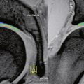
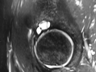
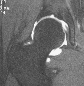
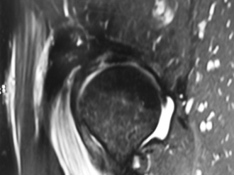
Magnetic resonance imaging may be helpful to obtain a cause of the pain; however, many authors have noted that articular cartilage defects are frequently not clearly identified by the imaging sequences and resolutions that are often used (Keeney CORR 2004). Secondary signs of articular cartilage damage may be identified, such as subchondral bony cysts, bone edema (Figure 22-1), and joint effusions. High-resolution images may demonstrate distinct defects (Figure 22-2), articular cartilage thinning, or heterogeneity of the articular cartilage signal (Figure 22-3). The use of a surface coil with high-resolution, cartilage-sensitive images in the axial, coronal, and sagittal planes may provide a higher spatial resolution that could be helpful for diagnosis. Magnetic resonance arthrography improves the ability to evaluate the articular surface, but current techniques lack reliable sensitivity. In the future, the use of delayed gadolinium-enhanced MRI of cartilage (dGEMRIC) may provide additional information about the proteoglycan content of the articular cartilage.

Surgical technique

The patient should be positioned on a fracture table or with a hip distraction system and in either a supine or lateral position, depending on surgeon preference. In most cases, the author prefers to have the patient in the supine position, with a commercially available hip distraction system placed on the leg that allows for the free movement of the limb. The hip is generally slightly internally rotated, flexed, and in neutral coronal alignment. The affected extremity is placed in 25 lb to 50 lb of longitudinal traction to distract the joint. A well-padded perineal post should be used to decrease the risk of pudendal nerve injury. The joint can then be distracted 7 mm to 15 mm under fluoroscopic guidance. The patient should be prepared and draped in the usual sterile fashion. Typically, after the surgical preparation solution has been applied and has dried, a large, clear drape with an adhesive central section (i.e., a “shower curtain” drape) is applied to the lateral aspect of the hip, and the top of the drape is allowed to cover the contralateral limb.

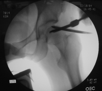
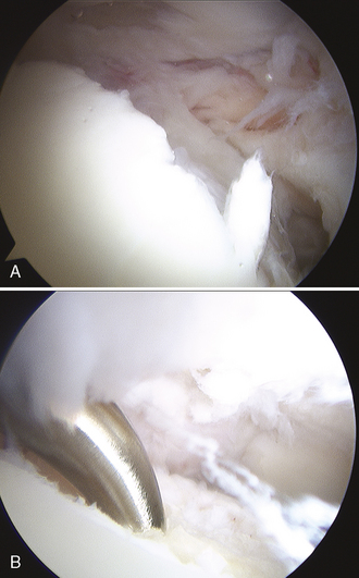
A spinal needle is then placed into the hip joint, and the stylet is removed; this breaks the vacuum seal and decreases the force needed to distract the joint. If necessary, the needle should be repositioned to avoid the labrum. This initial portal should be placed so that the tip of the needle enters the fovea of the acetabulum; this helps to ensure the good visualization of the joint. After the appropriate placement of the initial spinal needle and the guidewire, portal placement with the use of cannulated trocars can be performed to allow for access to the joint for visualization and instrumentation. A thorough diagnostic examination of the hip should occur, and this should involve the use of multiple portals. In some cases, defects on the femoral head are relatively medial (Figure 22-4), and careful instrument placement (Figure 22-5) is necessary to identify and treat defects (Figure 22-6, A and B).

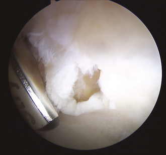
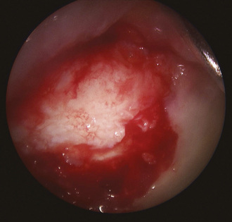
After a defect is identified, the area should be debrided thoroughly with a motorized shaver, curette, or arthroscopic knife. All unstable cartilage should be debrided down to exposed bone until smooth vertical walls of healthy cartilage remain (Figure 22-7). Delaminated articular cartilage flaps are common adjacent to cam-type labral detachment lesions and should be removed (Figure 22-8). Thorough chondroplasty helps to ensure that the fibrin clot that forms after the microfracture stays within the defect.

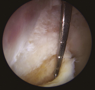
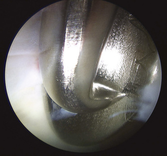
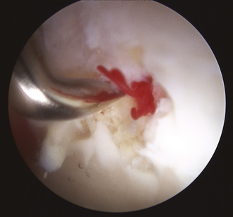
Arthroscopic awls should be placed through portals that allow for access to the defect. The insertion and positioning of these instruments are significantly aided by the use of a slotted cannula (Figure 22-9). The semicylindric shape of this cannula allows irregularly shaped tools to enter into the joint. Subsequently, the slotted cannula can be removed to allow for the freer movement of the awl. As a result of the direction of instrument insertion into the joint, higher-degree angle-tipped awls (i.e., up to 90 degrees) are often used (Figure 22-10). Gentle punctures through the subchondral bone are created and spaced 3 mm to 4 mm apart to cover the area of the defect. If they are placed closer to one another, a bone fragment may break off, and the clot may not be retained. In addition, excessive force can cause the fracture holes to spread. Special attention should be paid to the depth of the awl puncture. A puncture depth of 2 mm to 4 mm will ensure that blood and fat droplets are discharged from the holes. The verification of an adequate depth of penetration can be performed by the application of suction to the area to see if blood or fat emerges (Figure 22-11). When the surgeon is satisfied with the microfracture, the instruments and the arthroscopic fluid should be removed from the joint. The incisions should be closed in the surgeon’s preferred manner, and a sterile dressing is then applied.

Postoperative rehabilitation

Before proceeding to the operating department for any hip arthroscopy, it is vital to have a well-informed discussion with the patient regarding his or her ability to comply with weight-bearing restrictions. This is a mandatory conversation as a result of the limitations of current radiologic techniques to discover cartilage defects that may only be found incidentally during hip arthroscopy. Microfracture may not have a successful result if patients do not understand and consent to weight-bearing limitations before they proceed into the operating department. In addition, range-of-motion limitations are indicated if concomitant labral repair is performed.

Rehabilitation is similar to that of knee microfracture methods. A period of 6 to 8 weeks of toe-touch weight bearing with crutches is implemented. Passive range-of-motion exercises begin immediately postoperatively, and a continuous passive motion machine is extremely valuable for improving the quality of the repair and for significantly decreasing postoperative hip stiffness and hip flexor tendinitis. Physical therapy may also begin during this period, with a focus on range-of-motion exercises and an avoidance of extreme hip flexion, adduction, and internal rotation. Nonresistive stationary bicycling as tolerated may also begin, although patients will typically be more comfortable if the seat is raised to avoid hip flexion of more than 90 degrees. After 8 weeks, weight bearing may be advanced to full capacity, and physical therapy sessions can begin to focus on strengthening exercises. Athletes can typically return to sports 4 to 6 months postoperatively.

Results

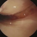
In our experience, extensive areas of delamination or articular cartilage lesions are indicative of degenerative disease and are typically not successfully treated with microfracture. In addition, we have observed that hip arthritis can progress extremely rapidly over a period of several months and that it is unlikely in these cases that the treatment of focal defects will alter the course of the disease. The most successful results are found among young patients with acute pathology without any degenerative condition. Articular cartilage lesions associated with labral pathology and femoroacetabular impingement have had less satisfactory results if the underlying impingement pathology is untreated. At times, this requires labral detachment, the debridement of a pincer lesion that typically encompasses part of the cartilage defect, and labral refixation (Figure 22-12).

image

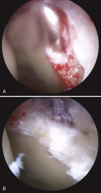
Figure 22–12 A, Anchor insertion into the debrided acetabular rim, as seen in Figure 22–7. B, The same area after repair with the arthroscopic mattress stitch.

Results of the treatment of chondral defects with microfracture are somewhat difficult to interpret, because concomitant procedures are performed in the vast majority of cases, and it is difficult to identify the specific benefit that can be attributed to the microfracture procedure itself. However, subjective patient outcome data and second-look arthroscopies have demonstrated the efficacy of microfracture for the treatment of these defects.

Multiple authors have noted that the incidence of chondral defects is high when a labral injury is present, most likely as a result of femoroacetabular impingement. McCarthy noted that, in a series of 436 hip arthroscopies, 54% of defects occurred in the anterior acetabular quadrant.

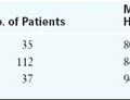
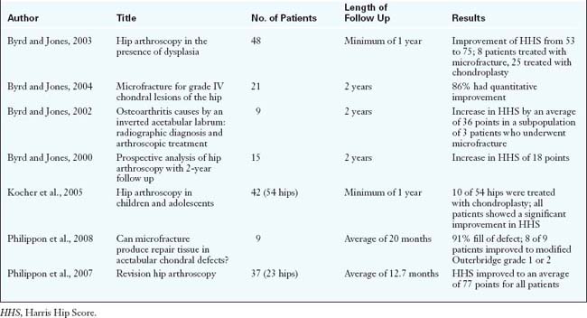
There are only a limited number of studies that have evaluated the efficacy of microfracture (Table 22-1). Byrd and Jones looked at 21 cases of microfracture (19 acetabular cases, 1 femoral head case, and 1 case that involved both of these areas) and found an improvement in the mean Harris Hip Score from 51.4 to 75.2. Magnetic resonance imaging and arthrography revealed the pathology in only 19% of cases, and the average defect size was 12.2 mm2 (range, 6 mm2 to 17.5 mm2). Labral tears were found in 17 of these cases, and degenerative ligamentum teres tears were found in 3 cases. The mean age of the patients was 35 years. At a minimum of 2 years of follow up, the authors found that 86% of patients had improved from the time of surgery. These authors also reported about 9 patients who were treated for inverted articular labrums. Three patients had chondral defects that were treated with microfracture, and these patients subsequently had the best outcomes of the group: they returned to their original baseline of activity and increased their Harris Hip Scores by an average of 36 points. In another study of arthroscopy and dysplasia, the investigators performed 8 microfracture procedures, with similar improvements seen in the Harris Hip Scores. Again, almost all of these patients had concomitant labral debridement.

McCarthy noted that, in a series of 10 elite athletes (including 4 who had chondral defects), all patients returned to compete in their sports. Similarly, Bharam noted that, in a series of 28 professional athletes, 19 had chondral defects in addition to other pathology; these patients were also able to return to full sports activity.

Kocher and colleagues reported about chondroplasty of the acetabulum and the femoral head in 10 pediatric cases. These authors noted that there was an overall increase in the Harris Hip Scores from 53.1 to 82.1. However, the series did not isolate the improvement in hip scores for the chondroplasty group, because the chondroplasties were likely performed with other procedures.

Our experience with microfracture has been good for patients with small acute defects, who have returned to full athletic function. Patients with avascular necrosis have been treated with chondroplasty and microfracture of the femoral head in areas of articular cartilage loss and the retrograde injection of demineralized bone matrix into a cleared area of necrotic bone. These patients have done well clinically without subsequent collapse.

Philippon and colleagues recently reported about a series of 9 patients who underwent second-look procedures for acetabular defects who had been treated with microfracture. All of these patients had labral pathology. The average defect size was 163 mm2 (range, 40 mm2 to 240 mm2). These patients had an average fill of 91%. The single patient in the study who did poorly had advanced osteoarthritis, which was noted as a contraindication for the procedure. One of the patients went on to total hip arthroplasty at 5.5 years postoperatively, despite 95% fill being found during a second-look arthroscopy 3 years after the procedure. Furthermore, Philippon and colleagues reported that microfracture or chondroplasty occurred in 70% of the revision cases.

Evidence exists to support these promising techniques, which have been well described for other joints. Improved data collection and analysis will help to further refine indications for this procedure. In addition, improved imaging of the articular cartilage will be helpful for the preoperative and postoperative evaluation of defects and their healing. Finally, a randomized controlled study that demonstrates the effectiveness of these techniques would also be welcome.

Annotated references and suggested readings

Bharam S., Draovitch P., Fu F.H., Philippon M.J. Return to competition in pro athletes with traumatic labral tears in the hip. Abstract. In American Orthopaedic Society for Sports Medicine. FL: Orlando; June 2002. (AOSSM)

Byrd J.W.T., Jones K.S. Hip arthroscopy in the presence of dysplasia. Arthroscopy. 2003;19:1055-1060.

Byrd and Jones looked at 48 patients with either dysplasia or borderline dysplasia. From this population, they reported the results of various procedures that were performed, including chondroplasty and microfracture. The Harris Hip Score improved from 53 to 75 in the chondral damage group..

Byrd J.W.T., Jones K.S. Microfracture for grade IV chondral lesions of the hip. Arthroscopy. 2004;20:e41.

This abstract provides 2 years of follow up for 21 patients who received microfracture for their grade IV lesions. Overall, 86% of patients improved. Interestingly, the abstract also comments on the ineffectiveness of magnetic resonance imaging and arthrography to diagnose the articular pathology that was discovered intraoperatively..

Byrd J.W.T., Jones K.S. Osteoarthritis caused by an inverted acetabular labrum: radiographic diagnosis and arthroscopic treatment. Arthroscopy. 2002;18:741-747.

This study found that 9 out of 220 arthroscopic hip patients developed osteoarthritis as a result of an inverted acetabular labrum. The authors discovered that a subset of 3 patients who were treated with microfracture did the best out of the group: their Harris Hip Scores increased the most, and they returned to normal activity..

Byrd J.W.T., Jones K.S. Prospective analysis of hip arthroscopy. Arthroscopy. 2000;16:578-587.

This article is a case series report about 38 procedures that were performed in 35 patients. Chondral injury was treated and resulted in an overall improvement in the Harris Hip Score of 18 points. Specific treatments were not listed..

Crawford K., Philippon M.J., Sekiya J.K., Rodkey W.G., Steadman J.R. Microfracture of the hip in athletes. Clin Sports Med. 2006;25:327-335.

The authors provide an excellent review of the current literature that supports microfracture. In addition, this article supplies information about the workup and treatment of microfracture..

Frisbie D.D., Oxford J.T., Southwood L., et al. Early events in cartilage repair after subchondral bone microfracture. Clin Orthop. 2003;407:215-227.

This excellent basic science article describes and demonstrates changes in subchondral bone after microfracture..

Keeney J.A., Peelle M.W., Jackson J., Rubin D., Maloney W.J., Clohisy J.C. Magnetic resonance arthrography versus arthroscopy in the evaluation of articular hip pathology. Clin Orthop Relat Res. 2004;249:163-169.

Kocher M.S., Kim Y.J., Millis M.B., et al. Hip arthroscopy in children and adolescents. J Pediatr Orthop. 2005;25:680-686.

This study looked at 54 arthroscopies in 42 pediatric patients. Included in the cumulative data are 10 cases of chondroplasty. Significant improvement in the Harris Hip Score occurred with a minimum of 1 year of follow up..

McCarthy J., Barsoum W., Puri L., Lee J.A., Murphy S., Cooke P. The role of hip arthroscopy in the elite athlete. Clin Orthop Relat Res. 2003;406:71-74.

McCarthy J.C., Noble P.C., Schuck M.R., Wright J., Lee J. The Otto E. Aufranc Award: the role of labral lesions to development of early degenerative hip disease. Clin Orthop Relat Res. 2001;393:25-37.

Mithoefer K., Williams R.J., Warren R.F., Wickiewicz T.L., Marx R.G. High-impact athletics after knee articular cartilage repair: a prospective evaluation of the microfracture technique. Am J Sports Med. 2006;34(9):1413-1418. 3rd

Philippon M.J., Schenker M.L., Briggs K.K., Kuppersmith D.A., Maxwell R.B., Stubbs A.J. Revision hip arthroscopy. Am J Sports Med. 2007;35:1918-1921.

Philippon and colleagues provide a look at 37 patients who required revision hip arthroscopy. In the study, impingement accounted for most revisions, with chondral defects being a close second. Outcomes demonstrated that patients regain some function within the first year..

Philippon M.J., Schenker M.L., Biggs K.K., Maxwell R.B. Can microfracture produce repair tissue in acetabular chondral defects? Arthroscopy. 2008;24:46-50.

The authors provide a case series regarding second looks at acetabular defects that underwent microfracture. Eight of 9 patients had 95% to 100% coverage with grade 1 or 2 appearance of cartilage at an average follow up of 20 months..

Shindle M.K., Voos J.E., Heyworth B.E., et al. Hip arthroscopy in the athletic patient: current techniques and spectrum of disease. J Bone Joint Surg Am. 2007;89:29-43.

This article provides a good review of the history and diagnostic features of hip disease. It also provides pearls to consider with regard to the radiologic workup..