Arthroscopic Iliotibial Band Lengthening and Bursectomy for Recalcitrant Trochanteric Bursitis and Coxa Saltans Externa

Published on 11/04/2015 by admin

Filed under Orthopaedics

Last modified 22/04/2025

Print this page

rate 1 star rate 2 star rate 3 star rate 4 star rate 5 star
Your rating: none, Average: 0 (0 votes)

This article have been viewed 3413 times

CHAPTER 16 Arthroscopic Iliotibial Band Lengthening and Bursectomy for Recalcitrant Trochanteric Bursitis and Coxa Saltans Externa

Introduction

Pain over the lateral aspect of the hip can be attributed to a number of conditions. The differential diagnosis includes degenerative joint disease of the hip, avascular necrosis of the femoral head, stress fracture of the femoral neck, infection, entrapment neuropathies, referred pain from lumbar disc disease, sciatica, snapping hip, and trochanteric bursitis. Trochanteric bursitis is a very common condition that is treated by both orthopedic surgeons and general practitioners and can be a source of disabling lateral hip pain. A dull, intermittent, aching pain in the region of the greater trochanter characterizes the condition. Occasionally, the pain radiates to the lateral thigh or the buttocks. The condition is thought to be a result of repetitive microtrauma and friction between the iliotibial band (ITB) and the greater trochanter, with subsequent inflammation of the interposed bursa. Degeneration of the gluteal tendons at the trochanteric attachment can also occur.

Some patients present with complaints of pain and an audible snapping sensation near the lateral aspect of the hip. External snapping hip or coxa saltans is caused by a tight ITB sliding over the greater trochanter with repetitive flexion and extension of the hip. The condition must be differentiated from an internal snapping hip, which is commonly attributed to the sliding of the iliopsoas tendon over the femoral head or the iliopectineal eminence, and from intra-articular causes of snapping hip, such as loose bodies, labral tears, and synovial chondromatosis. Asymptomatic external coxa saltans does not require treatment, but occasionally patients develop associated trochanteric bursitis with resultant pain and disability.

Most patients with trochanteric bursitis respond to nonoperative treatment that typically consists of activity modification, nonsteroidal anti-inflammatory medications, the stretching of a tight ITB, physical therapy with modalities (e.g., heat, ultrasound), and local injections of corticosteroids and anesthetics directly into the bursa. For the small group of patients whose symptoms fail to resolve with nonoperative therapy, the excision of the trochanteric bursa is recommended. Historically, this procedure has been performed with the use of an open approach; however, several authors have recently reported similar success when performing the bursectomy arthroscopically. In patients who have a tight ITB and associated external snapping hip, an ITB lengthening should be performed concurrently with the bursectomy.

Surgical technique

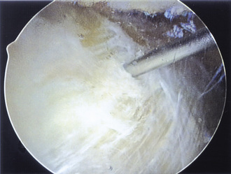
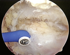
Our preferred surgical technique for recalcitrant trochanteric bursitis is an arthroscopic trochanteric bursectomy. Patients are positioned in the lateral decubitus position with the affected extremity facing up. A deflatable beanbag is used to stabilize the patient, and all bony prominences are well padded. The leg is draped freely so that the hip can be taken through a full range of motion (Figure 16-1). The greater trochanter is palpated and outlined on the skin with a marker. A spinal needle is then inserted directly onto the trochanteric prominence. The needle is then withdrawn slightly by several millimeters, and approximately 30 mL to 40 mL of normal saline is injected into the bursa, thus creating a space underneath the ITB. We prefer to leave the needle in for the purpose of localization. Next, a proximal portal is created 2 cm to 3 cm proximal to the trochanter, and a distal portal is created 2 cm to 3 cm distal to the trochanter. The skin is incised in line with the long axis of the femur. A 4-mm, 30-degree arthroscope is introduced directly into the subcutaneous tissues above the ITB. A 4.5-mm shaver is placed in the other portal and localized to the arthroscope with a triangulation technique (Figure 16-2). The fat directly above the ITB is removed to allow for full visualization of the ITB (Figure 16-3). An arthroscopic ablation probe can also be used to maintain hemostasis and concurrently to remove adherent tissues.
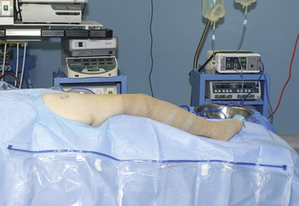
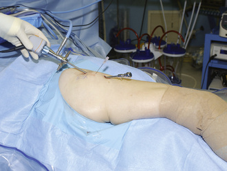
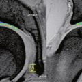
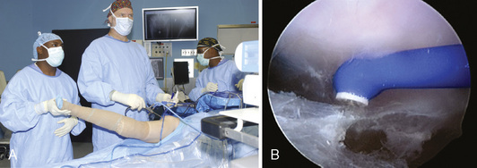
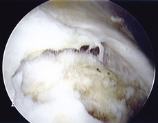
After the ITB is clearly identified, the arthroscopic ablator is used to create a longitudinal incision approximately 7 cm to 8 cm in length in line with the fibers of the ITB just slightly posterior to its midline and the trochanteric prominence; this exposes the trochanteric bursa (Figure 16-4). The surgical assistant abducts the leg to further relax the incised ITB (Figure 16-5) and to allow the surgeon to advance the arthroscope and instruments underneath the ITB. Next, the shaver and the ablator are used to thoroughly debride the bursa and its thick, fibrous adhesions. Having the assistant slowly internally and externally rotate the leg brings the posterior and anterior portions of the bursa, respectively, into view. Extreme internal rotation is avoided, because this maneuver places the posteriorly located sciatic nerve at risk. Coagulation is frequently necessary to maintain hemostasis and visualization.

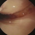
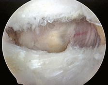
After the bursa has been completely excised, the tendinous attachments of the gluteus minimus and the gluteus medius to the greater trochanter can be seen. Occasionally, irritation and scuffing of the tendinous insertions are noted, and, infrequently, tears and avulsions of the tendons (so-called rotator cuff tears of the hip) are present (Figure 16-6). Early in our experience, we treated the tears with gentle debridement, but, more recently, we have repaired these tears with nonabsorbable sutures with the use of a technique that is similar to the marginal convergence repair technique used for rotator cuff tears in the shoulder. After bursal excision has been completed and hemostasis is ensured, the instruments are withdrawn, and excess fluid is expressed from the portals. The portals are then closed with nylon suture, and a compression dressing and ice pack are applied.

In patients with associated external coxa saltans, an ITB lengthening procedure is necessary in addition to the standard trochanteric bursectomy. After the usual slightly posterior longitudinal incision is made in the ITB, the arthroscopic ablator is used to gently resect its anterior and posterior edges. Additional tissue is removed near the center of the midline cut to create an elliptical resection of the ITB (Figure 16-7). The posterior resection is slightly greater than the anterior resection, because the snapping usually results from the thickened posterior portion of the ITB flipping over the greater trochanter. An alternative method is to perform a crisscross secondary transverse incision, thereby effectively creating a cruciate-type release. The hip is taken through a range of motion to ensure adequate decompression and resection. The bursa is then resected as previously described.

Technical Pearls

Results and outcomes

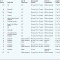
In general, both open and arthroscopic treatments of trochanteric bursitis result in a high percentage of good outcomes (Table 16-1). Physicians agree that, in the majority of patients with this condition, a conservative approach will be successful; only a select few patients will require operative intervention. For a successful result in patients with external coxa saltans, the ITB should be effectively lengthened by one of several open or arthroscopic methods, such as a Z-plasty, an ellipsoidal-shaped or diamond-shaped resection, a cruciate or cross-shaped incision, or a simple longitudinal release.

Annotated references

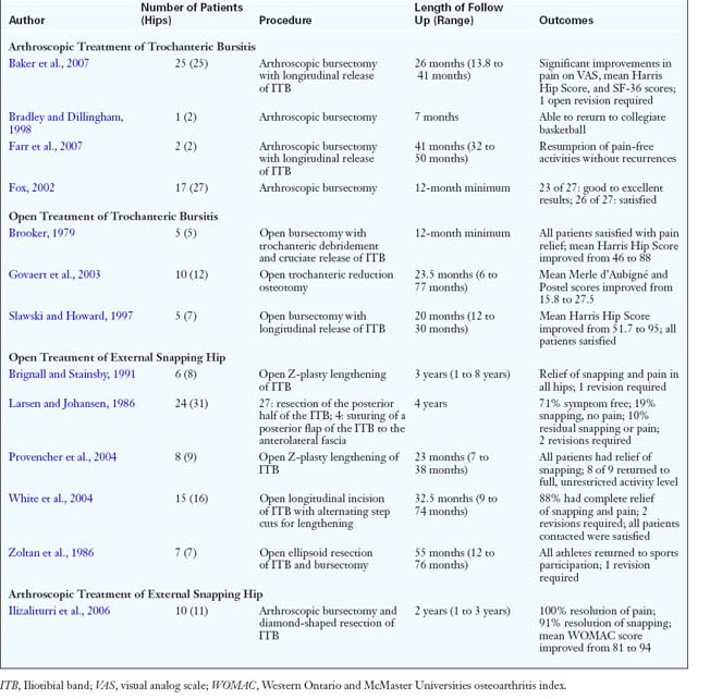
Baker C.L.Jr, Massie R.V., Hurt W.G., et al. Arthroscopic bursectomy for recalcitrant trochanteric bursitis. Arthroscopy.. 2007;23:827-832.

Twenty-five patients were reviewed at a mean of 26 months after undergoing arthroscopic bursectomy. There were significant improvements in mean Harris Hip Scores (51 to 77), mean Visual Analog Scale (VAS) scores for pain (7.2 to 3.1), mean physical function scores on the Short Form 36 (SF-36) (33.6 to 54), and mean pain scores on the SF-36 (28.7 to 51.5). One patient developed a seroma that required repeat surgery, and another patient required open revision bursectomy for relief..

Bradley D.M., Dillingham M.F. Bursoscopy of the trochanteric bursa. Arthroscopy.. 1998;14:884-887.

This article is a case report of a Division I male basketball player with bilateral trochanteric bursitis who was treated successfully with bursoscopy and bursectomy..

Brignall C.G., Stainsby G.D. The snapping hip: treatment by Z-plasty. J Bone Joint Surg Br.. 1991;73:253-254.

Six patients (8 hips) with external coxa saltans were treated with an open Z-plasty lengthening of the ITB. One hip required revision lengthening, and snapping was relieved in all patients at a mean length of follow-up of 3 years..

Brooker A.F.Jr. The surgical approach to refractory trochanteric bursitis. Johns Hopkins Med J.. 1979;145:98-100.

Five patients underwent open excision of the bursa, debridement of the greater trochanteric prominence, and ITB release. A circular defect was created for 1 patient’s release, and a cruciate incision in the ITB was performed for the remaining 4 patients. All of the patients had satisfactory pain relief at a minimum follow-up of 1 year..

Farr D., Selesnick H., Janecki C., et al. Arthroscopic bursectomy with concomitant iliotibial band release for the treatment of recalcitrant trochanteric bursitis. Arthroscopy.. 2007;23:905. e1–5. Epub 2007 Jan 25

Two patients were treated with arthroscopic resection of the trochanteric bursa in combination with a longitudinal release of the ITB with the use of an electrocautery probe. At an average of 41 months of follow-up, there were no recurrences with the resumption of the full activity level..

Fox J.L. The role of arthroscopic bursectomy in the treatment of trochanteric bursitis. Arthroscopy.. 2002;18:E34.

Twenty-seven patients underwent arthroscopic bursectomy, with good to excellent results obtained in 23 of the patients. At 5 years of follow-up, there were three recurrences. No treatment was directed at the ITB..

Gordon E.J. Trochanteric bursitis and tendinitis. Clin Orthop.. 1961;20:193-202.

The signs and symptoms of a group of 61 patients with trochanteric bursitis are presented. The author details the method and success of conservative treatment. The diagnosis and correction of associated disorders are emphasized..

Govaert L.H., van der Vis H.M., Marti R.K., et al. Trochanteric reduction osteotomy as a treatment for refractory trochanteric bursitis. J Bone Joint Surg Br.. 2003;85:199-203.

Ten patients (12 hips) with refractory trochanteric bursitis had a 5-mm to 10-mm-thick slice of the greater trochanter removed, with the remaining trochanter advanced and fixed distally and medially. The authors concluded that the procedure is safe and effective with good pain relief at a mean of 23.5 months after surgery..

Ilizaliturri V.M.Jr, Martinez-Escalante F.A., Chaidez P.A., et al. Endoscopic iliotibial band release for external snapping hip syndrome. Arthroscopy.. 2006;22:505-510.

Ten patients (11 hips) with external coxa saltans underwent endoscopic trochanteric bursectomy and resection of a diamond-shaped portion of the ITB, and they were evaluated at an average follow-up of 2 years. Pain was relieved in 100% of hips, and snapping was relieved in 91% of hips..

Kagan A.II. Rotator cuff tears of the hip. Clin Orthop Relat Res.. 1999;368:135-140.

The author reports about seven patients who were found during surgery for trochanteric bursitis to have partial tears of the gluteus medius tendon attachment to the trochanter. All of these tears were repaired with heavy nonabsorbable suture. At a mean follow-up of 45 months, all patients were free of pain..

Larsen E., Johansen J. Snapping hip. Acta Orthop Scand.. 1986;57:168-170.

In 31 patients (31 hips) with external snapping hip, 27 underwent the resection of the posterior half of the ITB and 4 underwent the suturing of a posterior flap of the iliotibial tract to the anterolateral fascia. At an average follow-up time of 4 years, 71% of patients (22 hips) were symptom free. The femoral neck angle was significantly decreased in affected patients as compared with healthy controls (128 degrees versus 134 degrees)..

Provencher M.T., Hofmeister M.P., Muldoon M.P. The surgical treatment of external coxa saltans (the snapping hip) by Z-plasty of the iliotibial band. Am J Sports Med.. 2004;32:470-476.

Nine patients with symptomatic hips with external coxa saltans were treated with an open Z-plasty lengthening of the ITB. At an average follow-up of 23 months, all patients had complete resolution of snapping symptoms, and all but 1 patient returned to a full activity level. The authors felt that this technique provided predictable, excellent results..

Slawski D.P., Howard R.F. Surgical management of refractory trochanteric bursitis. Am J Sports Med.. 1997;25:86-89.

Five patients (7 hips) underwent open trochanteric bursectomy via a longitudinal incision in the ITB. All patients reported marked improvement postoperatively with a return to all activities. Occasional discomfort was noted after athletic activity in the majority of hips..

White R.A., Hughes M.S., Burd T., et al. A new operative approach in the correction of external coxa saltans: the snapping hip. Am J Sports Med.. 2004;32:1504-1508.

Sixteen patients (17 hips) with external snapping hip underwent open ITB release with a longitudinal incision and alternating transverse step cuts. Fourteen of the 16 hips (88%) evaluated during follow-up had resolution of pain and snapping. Two patients required revision release at 3 and 6 months after the initial surgery..

Zoltan D.J., Clancy W.G.Jr, Keene J.S. A new operative approach to snapping hip and refractory trochanteric bursitis in athletes. Am J Sports Med.. 1986;14:201-204.

Seven athletes with external coxa saltans and associated greater trochanteric bursitis were treated with an open ellipsoidal excision of the ITB and the removal of the trochanteric bursa. The procedure was performed under local anesthesia to allow for the dynamic assessment of elimination of snapping. At a mean follow-up of 55 months, all patients had returned to sports activities, although one had required a more extensive revision resection for relief..