Anesthetic considerations for weight loss surgery

Published on 07/02/2015 by admin

Filed under Anesthesiology

Last modified 22/04/2025

Print this page

rate 1 star rate 2 star rate 3 star rate 4 star rate 5 star
Your rating: none, Average: 0 (0 votes)

This article have been viewed 1312 times

Anesthetic considerations for weight loss surgery

Brian P. McGlinch, MD

The frequency with which weight loss surgery (WLS) is performed has increased significantly over the past 15 years, in part due to the increasing prevalence of obesity, but also because of the understanding that diet and exercise are often ineffective in producing sustained weight loss, the overwhelming evidence that WLS results in significant and sustained weight loss, that reductions occur in obesity-related comorbid conditions, and that the procedures themselves are associated with low rates of morbidity and mortality. Patients who are potential candidates for WLS typically undergo extensive medical evaluations for underlying obesity-related comorbid conditions (e.g., obstructive sleep apnea, diabetes, reactive airway disease, hyperlipidemia, gastroesophageal reflux disease), have treatment of these conditions, and improve their physical conditioning. As a result, the patient who presents for WLS is usually medically and physiologically optimized for surgery and represents a surprisingly low risk for experiencing untoward perioperative events. At the time of this writing, WLS is overwhelmingly performed laparoscopically; this chapter will focus on this surgical approach.

Obesity

Obesity is not solely a North American phenomenon. All areas of the world and all socioeconomic classes demonstrate a rapidly increasing prevalence of obesity. Obesity is defined as a body mass index (BMI, weight in kilograms divided by the square of the height in meters) greater than 30 kg/m2. Morbid obesity is present at a BMI of 35 kg/m2 in people who have weight-related comorbid conditions (i.e., hyperlipidemia, diabetes mellitus, obstructive sleep apnea, reactive airway disease) or a BMI of more than 40 kg/m2 in the absence of weight-related comorbid conditions. Excessive caloric intake only partly contributes to obesity. Psychiatric, physiologic, and metabolic components are all likely factors that contribute to the obesity phenomenon. Emerging research identifying digestive hormones that influence eating behavior, metabolism, and weight gain (e.g., ghrelin, peptide YY) could potentially lead to nonsurgical therapies for obesity. However, at this time, only surgical interventions have been demonstrated to be effective in producing a significant sustained weight loss in the treatment of obesity.

Surgical procedures for weight loss

Current WLS involves restrictive or restrictive-malabsorption components. Restrictive procedures (adjustable gastric banding, gastric sleeve resection) create a small stomach but do not alter how food is digested. The most common restrictive procedure at this time is the laparoscopic adjustable gastric banding procedure. The U.S. Food and Drug Administration approved the adjustable gastric band for clinical use in 2001, and its application has increased annually since its introduction. In this procedure, a limited dissection of connective tissue is performed at the top of the stomach, and an inflatable band is passed that encircles the upper stomach (Figure 164-1, A). The band can be adjusted via a port attached to the body wall by adding or withdrawing saline. The surgical risk is considered to be very low. In select patients, this is being performed as an outpatient procedure.

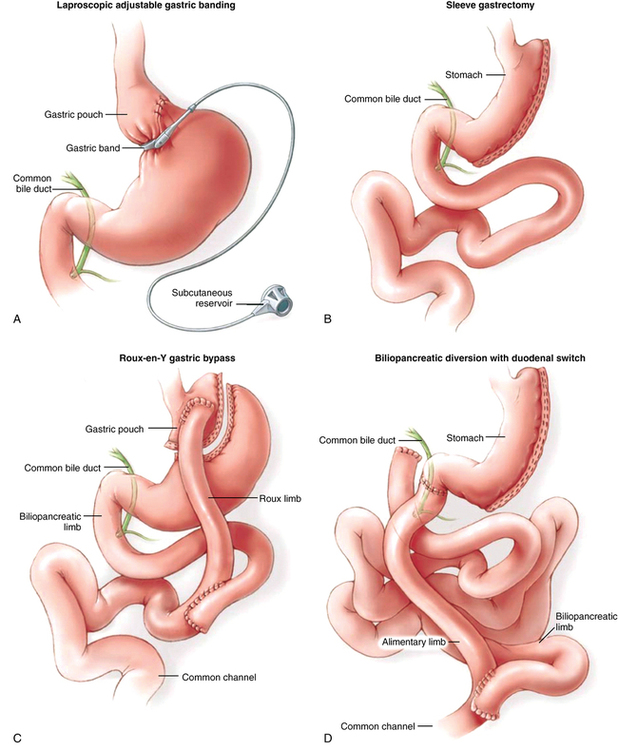
The gastric sleeve resection is typically performed laparoscopically and reduces stomach volume to approximately 100 mL by externally stapling the stomach to exclude the fundus and greater curvature to form a narrow tube along the lesser curvature of the stomach (Figure 164-1, B). Both laparoscopic adjustable gastric banding and gastric sleeve resection procedures result in significant weight loss, providing that the patients receiving these interventions comply with proper eating habits, particularly those who avoid high-caloric content (e.g., milkshakes, candy, ice cream). Blood loss is minimal. Surgical duration is relatively brief. Complications are infrequent.

The laparoscopic Roux-en-Y gastric bypass is currently the most commonly performed WLS in the United States. This procedure creates a small gastric pouch (30 mL) that empties into a limb of bowel that excludes a large portion of the small intestine (restrictive and maldigestive interventions) (Figure 164-1, C). As a result, satiety is achieved at relatively low volumes (restriction), and the surface area of small bowel that can absorb calories and nutrients is bypassed (maldigestion), resulting in reliable, sustained weight loss. Blood loss is minimal.

The biliopancreatic diversion/duodenal switch procedure is a more complex restrictive-malabsorptive procedure normally reserved for use in patients with a BMI in excess of 50 kg/m2. With this procedure, a gastric sleeve resection is performed, the stomach is separated from the duodenum, and the small bowel is divided at a point proximal (approximately 100 cm) to the terminal ileum. The stomach is reanastomosed to the distal limb of small bowel, creating an “alimentary channel” where food enters but is not digested. Biliary and pancreatic fluids drain into the duodenum (now separated from the stomach) and contact food where this “biliopancreatic limb” of proximal small bowel is anastomosed to the limb of the alimentary channel (Figure 164-1, D). Digestive enzymes contact food late in the process, and the short segment of small bowel that is available to absorb nutrients and calories is significantly restricted. Compared with the Roux-en-Y gastric bypass, the biliopancreatic diversion/duodenal switch procedure requires considerably longer time, but blood loss and fluid shifts are not significantly different.

Anesthetic management of patients undergoing weight loss surgery

Fortunately, patients undergoing WLS have typically undergone a thorough preoperative evaluation based upon symptoms. Evaluations that are otherwise not indicated by symptoms and clinical findings are unnecessary. Patients without symptoms of obstructive sleep apnea do not need a polysomnographic study. Patients without cardiac symptoms do not need to have a stress test or echocardiogram unless other clinical signs (i.e., heart murmurs, pulmonary edema) warrant evaluation independent of the anticipated operation. In most circumstances, diseases typically associated with morbid obesity (e.g., obstructive sleep apnea, hyperlipidemia, diabetes, hypertension, restrictive airway disease) have been evaluated and treated, resulting in optimization of underlying conditions in advance of the operation. The anesthesia provider remains responsible for ensuring that the necessary clinical studies have been completed, that the patient is devoid of acute illness, and that other confounding issues are absent (e.g., the patient did not recently take anticoagulants) prior to inducing anesthesia.

The obese patient should not lie flat on an operating table. Obesity decreases lung compliance, reduces expiratory reserve volume, and decreases functional residual capacity well beyond that seen in nonobese patients. Dependent areas of lung collapse and close, contributing to a ventilation-perfusion mismatch. In the presence of obesity-induced increased O2 demand and CO2 production, these physiologic changes in pulmonary function occurring in the supine position often lead to rapid O2 desaturations and hypercarbia during airway management and subsequent mechanical ventilation during anesthesia. Placing ramping cushions or blankets behind the patient’s upper torso and neck, as well as providing 25-degree reverse Trendelenburg positioning, provides greater lung volumes, a reduced tendency for atelectasis formation and intrapulmonary shunting, and a 23% increase in O2 saturations, compared with the supine position. Applying 10 cm H2O of positive end-expiratory pressure (PEEP) during induction of anesthesia prolongs the duration of O2 saturations above 92%, as compared with patients of similar size not receiving PEEP. Reestablishing these interventions (i.e., torso elevation, reverse Trendelenburg position, and application of PEEP) at the end of the surgical procedure should be considered, given the benefits demonstrated at anesthetic induction.

Compared with the nonobese patient, the obese patient will require an increased minute ventilation to maintain normocarbia. Establishing a pneumoperitoneum reduces ventilation compliance more in obese patients than it does in nonobese patients. However, oxygenation is affected by neither the presence of the pneumoperitoneum nor the Trendelenburg position commonly used in WLS. The application of PEEP is useful in improving oxygenation during these operations. However, improved oxygenation in response to PEEP is present only as long as the patient remains intubated. Regardless of the improvement in oxygenation provided by various amounts of PEEP intraoperatively (as determined by intraoperative blood gas analysis), postextubation blood gases are not significantly different from baseline nor from values from patients who do not receive PEEP intraoperatively, suggesting that there is no “best” amount of PEEP during WLS. The clinician should base ventilator-management strategies on individual patient information and the results of laboratory studies and clinical examination.

Insufflation of the peritoneum represents an important period for anesthetic management. Intraperitoneal pressure potentially obstructs venous return from the lower body and also encroaches on intrathoracic space. In most laparoscopic WLS, insufflation of the peritoneum, accompanied by maximal reverse Trendelenburg positioning (to facilitate surgical visualization of the stomach), results in significantly decreased venous return and cardiac preload. Profound hypotension and reflex bradycardia (from the abrupt reduction in cardiac preload) may occur during this period and warrant prompt recognition and treatment; the patient should be rapidly returned to the supine position, the peritoneum desufflated, and vasopressor, vagolytic, or both agents administered, as indicated. These physiologic responses to pneumoperitoneum can be attenuated by fluid loading the patient prior to insufflation. Once the pneumoperitoneum is established, tachycardia and increased mean arterial pressure are common.

Complications

Laparoscopic WLS procedures represent some of the most technically difficult procedures performed by general surgeons. The surgeon’s experience performing laparoscopic procedures is likely the single most important factor that correlates with the frequency of complications. The facility in which WLS is performed also plays a significant role. Because these two aspects of WLS are, by far, the greatest contributors to complication rates, emphasis will be placed on these aspects of WLS.

Summary

Laparoscopic WLS is the only therapy yielding sustained, significant weight loss and eliminating or attenuating weight-related comorbid conditions. Due to the thorough preoperative evaluations that WLS candidates undergo, anesthesia providers infrequently require additional studies or evaluations to ensure that these patients are safe for surgery. Inducing anesthesia with the patient ramped on blankets and in a modest reverse Trendelenburg position allows for improved oxygenation and ventilation. Surgeon and facility experience with WLS play significant roles in the morbidity and mortality rates associated with these procedures. Future weight-loss strategies may involve hormone therapies, but, most likely, laparoscopic WLS will remain the predominant obesity therapy offered throughout the world.