Anesthesia in the patient with extreme obesity

Published on 13/02/2015 by admin

Filed under Anesthesiology

Last modified 22/04/2025

Print this page

rate 1 star rate 2 star rate 3 star rate 4 star rate 5 star
Your rating: none, Average: 1 (1 votes)

This article have been viewed 1403 times

Anesthesia in the patient with extreme obesity

James P. Conterato, MD

General

Obesity has rapidly become an endemic disorder of Western society. Its presentation is now a daily phenomenon for the practicing anesthesia provider. Obesity is a condition of excess body fat, its derivation coming from the Latin word obesus, meaning fattened by eating. Body fat is a relative state, but in healthy Western societies, body fat averages for women are 20% to 30%, for men 18% to 25%, and for ultrafit marathoners approximately 7%. Obesity is a disorder that has genetic, socioeconomic, and endocrine causes. In developing countries, it correlates with a higher socioeconomic status, whereas in Western society, it more frequently occurs in lower socioeconomic classes. The original definitions of obesity are derived from the concept of ideal body weight calculations, developed by insurance companies to describe those population groups with the lowest mortality rates for age, and calculated as follows:

< ?xml:namespace prefix = "mml" />Ideal body weight (kg) = height (cm) – x

image

where x equals 105 for women and 100 for men. Body mass index (BMI) is a more relevant measure of height-weight relationships and is the currently accepted standard used to stratify obesity, although evidence has accumulated that the distribution of fat (central and intra-abdominal vs. peripheral) may best correlate with morbidity and mortality rates, recognizing that central intra-abdominal fat is a more metabolically active, proinflammatory substrate. BMI is calculated as body weight (in kilograms) divided by height (in meters) squared. The World Health Organization and the Centers for Disease Control and Prevention have defined levels of obesity (Table 163-1). Obesity can be further subdivided into morbid obesity (defined as a BMI >35 and <54.9) and super or extreme morbid obesity (BMI >55).

Table 163-1

Classification of Weight by Body Mass Index (BMI)

Category BMI (kg/m2) Obesity Class
Underweight <18.5  
Normal weight 18.5-24.9  
Overweight 25.0-29.9  
Obesity 30.0-34.9 Obesity class I
  35.0-39.9 Obesity class II
Extreme obesity ≥40 Obesity class III

Adapted from Obesity: Preventing and Managing the Global Epidemic. Geneva, Switzerland, World Health Organization; 1997. Report No. 894.

The inflection point for risk is a BMI greater than 30, at which point an exponential increase occurs in multiple risks for morbidity and mortality. Fat-distribution analyses suggest that the waist-to-hip ratio may correlate best with risk and dramatically increases at values of greater than 1 for women and greater than 0.8 for men.

The literature is replete with documentation that the morbidly obese are at increased risk for developing a wide variety of comorbid conditions, including a threefold to fourfold increased incidence of the age-related risks for diabetes, hypertension, cerebrovascular disease, and ischemic heart disease. Obesity predisposes an individual to develop the now well-recognized metabolic syndrome, consisting of hypertension, insulin resistance with adult-onset diabetes (type 2), and hyperlipidemia. It represents a proinflammatory and prothrombotic state associated with vascular endothelial dysfunction, which dramatically increases one’s risk of developing atherosclerotic disease. Additionally, meta-analyses demonstrate increased incidences of essentially all solid-tissue cancers except esophageal cancer, and of deep venous thrombosis and thromboembolism, osteoarthritis, gout, chronic back pain, asthma, infertility, impotence, and gallbladder disease in obese people. Other potentially key considerations are the cardiorespiratory complications of obstructive sleep apnea, apnea-hypoventilation syndrome (pickwickian syndrome), and cardiomyopathy of obesity.

The anesthesia provider faces potential issues in caring for obese patients that encompass these comorbid conditions and impact airway management, cardiorespiratory physiology, pharmacokinetics and pharmacodynamics of drugs, and positioning concerns, to name a few.

Physiology and pathophysiology of morbid obesity

Cardiovascular effects

Each kilogram of fat develops approximately 3000 m of blood vessels and requires a progressive increase in cardiac output that averages 100 mL/min. As a result, circulating blood volumes in the systemic and pulmonary circuits increase, resulting in increased cardiac preload and afterload. Ventricular dilatation and increased stroke volume develop, which eventually elicit ventricular hypertrophy, all of which increase cardiac work. The systemic hypertension and cardiomegaly seen are a result of increases in circulating blood volumes and cardiac output, as are the development of hyperinsulinemia-induced sympathetic activity and tone and progressive peripheral insulin resistance eliciting increased pressor effects of renin and angiotensin II. The development of pulmonary hypertension is a result of increased circulating blood volume, increased sympathetic tone, and chronic arterial hypoxemia (see following discussion under “Airway and Pulmonary Effects”).

The constellation of events that lead to hypertrophy-induced diastolic dysfunction can eventually result in the failure of hypertrophy compensating for ventricular dilatation, resulting in eventual systolic dysfunction—what is described as obesity-induced cardiomyopathy. Patients lose the ability to compensate for additional work stress on the cardiovascular system, relying more on increases in heart rate than stroke volume. Studies have documented an impaired response to exercise in the morbidly obese, with imageO2 max rarely reaching above 25 mL of O2 per kilogram per minute, indicating that levels of work that are submaximal in normal patients can only be met by an increased anaerobic debt in morbidly obese individuals.

Airway and pulmonary effects

Obesity imposes a restrictive ventilatory defect on the patient because of weight gain involving the chest wall and because of excessive diaphragmatic loading from abdominal fat, impeding the normal excursion of the diaphragm and significantly increasing the work of breathing. Both of these phenomena contribute to a decrease in pulmonary compliance. However, the major cause of the decrement in pulmonary compliance appears to be due to the increase in pulmonary blood volume. This restriction results in a decrease in total lung capacity and functional residual capacity (FRC), mainly from the loss of expiratory reserve volume. FRC decreases exponentially with increasing BMI, often reaching a point at which closing capacity is greater than FRC during tidal breathing, resulting in increased ventilation/perfusion mismatching in the awake state and increased shunt fraction (images/imaget) under general anesthesia. At rest, most morbidly obese patients experience only a modest decrease in PaO2 and maintain PaCO2 via hyperventilation (low tidal volume, increased respiratory rate). On average, a 1-mm Hg drop in PaO2 and 1-mm Hg increase in PAO2 − PaO2 occur with each additional 10-kg weight gain. These patients tolerate apnea poorly and experience up to a 50% drop in FRC on induction of general anesthesia (vs. 20% in nonobese patients). Reported images/imaget for this group of patients undergoing general anesthesia can reach 10% to 25%, versus the normal 2% to 5%. The expanded pulmonary blood volume and loss of FRC also increase pulmonary vascular resistance, which then also contributes to the increased work of breathing. This loss of FRC can also accelerate the rate of uptake of inhalation anesthetic agents (shorter time constant in the lungs).

Airway anatomy becomes distorted and airway, management becomes more challenging for the anesthesia provider as the patient’s level of obesity becomes more extreme. Patients may have enlarged face and cheeks, large tongue, impaired mouth opening and cervical range of motion, increasing soft tissue encroachment in the palate and pharynx, short neck, and large breasts, all of which may contribute to difficult mask ventilation, difficult direct laryngoscopy, or difficult intubation. Difficult mask ventilation has been reported to occur in up to one third of morbidly obese patients. Although older literature has suggested an incidence of difficult intubation as high as 30% in these patients, more recent prospective trials document that, in general, this is not the case and that the only two variables that appear predictive of difficulty intubating patients in this population are an increased neck circumference and a Mallampati score of 3 or more.

As BMI exceeds 30 kg/m2, the risk of a patient having obstructive sleep apnea increases. This syndrome is due to the normal reduction in upper airway tone seen during stage 4 and rapid eye movement sleep. The excessive deposition of soft tissue in the upper airway in the obese individual accentuates this tendency and can result in periodic obstruction of the airway during sleep. Obstructive sleep apnea consists of both apneas and hypopneas and is best delineated via formal sleep evaluations with polysomnography. For more information on this topic, see Chapter 108, Obstructive Sleep Apnea.

With progressive worsening of obstructive sleep apnea, desensitization of breathing control can evolve, resulting in degrees of chronic hypoxemia and hypercarbia. Up to 50% of individuals with a BMI greater than 50 kg/m2 have evidence of daytime hypoventilation. Eventual physiologic changes develop that consist of secondary polycythemia, pulmonary hypertension, and right ventricular failure. This apnea hypoventilation syndrome has also been referred to as the pickwickian syndrome, although the term is no longer in use.

Gastrointestinal and hepatic effects

Study results have suggested that the obese patient is at increased risk of aspiration due to higher residual gastric volumes and lower gastric pH. However, recent studies have questioned these conclusions, indicating no greater increased aspiration risk per se. Obese individuals have normal or accelerated rates of gastric clearance, and the incidence of gastric reflux does not differ significantly from that of the general population. However, difficult airway management by mask may predispose some to an increased aspiration risk.

Short of significant liver dysfunction due to obesity (nonalcoholic steatohepatitis with cirrhosis), there is no evidence of impaired hepatic function in individuals with morbid obesity. Neither is there any proven greater tendency to inhalation agent–induced hepatitis.

Anesthesia for the morbidly obese patient

A routine general health examination, specifically focusing on cardiac, pulmonary, airway, and metabolic issues (Table 163-2), should be performed preoperatively.

Table 163-2

Preoperative Assessment of the Morbidly Obese Patient

System Assessment
Cardiovascular Obtain baseline electrocardiogram
  Assess BP control
  Examine for symptoms or evidence of right or left ventricular dysfunction
  Examine for presence of CAD
  Obtain appropriate guided studies of heart function (echocardiography) or CAD (noninvasive or invasive evaluation of coronary circulation)
  Ensure appropriate preoperative drug administration for any cardiovascular comorbid conditions
Pulmonary Assess exercise tolerance
  Evaluate for symptoms or history of OSA
  Determine compliance with CPAP or BiPAP and ensure that patient brings personal CPAP device to hospital, if on therapy*
  Obtain baseline ABGs on room air
  Obtain PFTs if appropriate
Airway Perform a basic airway examination
  Look for evidence of impaired oral or cervical ROM
  Determine Mallampati score and neck circumference
  Inquire about previous difficult mask airway or intubation
Laboratory studies Obtain electrolyte, blood glucose, and serum hemoglobin concentrations
  Phlebotomize patients with a hemoglobin concentration >17 g/dL
Gastrointestinal Premedicate with metoclopramide, histamine H2 blocker, or PPI, as appropriate
Monitoring Consider the needs for adequate intravenous access and BP monitoring
  A central venous catheter may be needed to obtain reliable venous access
  BP cuffs should be of appropriate size (cuff bladder at least 75% of arm circumference)
  In some cases, the use of direct arterial monitoring may be more reliable or necessary
Miscellaneous Ensure that plans are made for the use of an appropriately sized OR bed that can accommodate the patient’s weight and size

ABGs, Arterial blood gases; BiPAP, bilevel positive airway pressure; BP, blood pressure; CAD, coronary artery disease; CPAP, continuous positive airway pressure; OR, operating room; OSA, obstructive sleep apnea; PFTs, pulmonary function tests; PPI, proton pump inhibitor; ROM, range of motion.

*Refer to the American Society of Anesthesiology’s Practice Guidelines for the Perioperative Management of Patients with Obstructive Sleep Apnea. An updated report by the American Society of Anesthesiologists Task Force on Perioperative Management of Patients with Obstructive Sleep Apnea. Anesthesiology. 2013 Dec 16. [Epub ahead of print].

Intraoperative anesthetic techniques

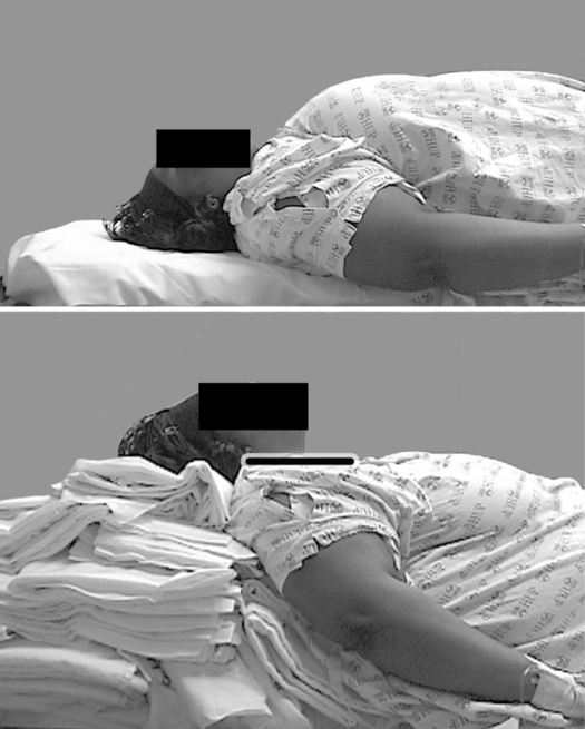
Modification of the anesthetic technique is necessary to minimize complications related to obesity. Because of physical limitations imposed by the patient’s girth, the anesthesia provider should ensure that proper “ramping” of the patient has been performed, such that the sternal notch is parallel to the angle of the mandible (Figure 163-1). This facilitates alignment of the intraoral axes for intubation. If a history of or major concerns regarding difficult intubation exist, awake fiberoptic intubation may be a reasonable option. Preoxygenation for 3 to 5 min with the patient in a mild reverse Trendelenburg position while using 10 cm H2O of continuous positive airway pressure plus 10 cm H2O of peak end-expiratory pressure with mask ventilation has been shown to significantly mitigate the initial drop in FRC on induction and to prolong time to desaturation. Appropriately sized oral and nasal airways should be available and used to maintain airway patency. An inability to use a mask should always be anticipated. The use of an intubating laryngeal mask airway can be highly effective in achieving successful airway maintenance when conventional mask techniques have failed.

Intraoperatively, alveolar recruitment using titrated peak end-expiratory pressure and some degree of head-up (beach-chair) positioning should be maintained. This is especially true when the operation requires introduction of a pneumoperitoneum. This technique has been proved to be more effective than the use of large tidal volume ventilation. Judicious use of colloids should be employed to maintain intravascular volume and prevent negative hemodynamic effects of excess peak end-expiratory pressure. Because of the mechanical limitations on the respiratory system and the increased sensitivity of morbidly obese patients to the effects of sedatives, opioids, and inhalation agents, spontaneous ventilation should not be used in these patients.

There is no evidence to support the use of one inhalation agent over another. Emergence times are essentially similar, especially when the agents are used in conjunction with processed electroencephalographic monitoring. The use of N2O should be avoided, especially in the setting of pulmonary hypertension.

The dosing of intravenously administered anesthetic agents varies with lipid and solvent solubility. Lean body mass (ideal body weight + 20%) is a good estimate for determining the dose of hydrophilic drugs, such as nondepolarizing neuromuscular blocking agents, as well as remifentanil that, although lipid soluble, behaves similarly in obese and nonobese individuals. The dose of lipid-soluble drugs and succinylcholine (increased plasma cholinesterase activity) should be based on the patient’s current total body weight.

The use of multimodal analgesic agents that mitigate the use of opioids should be considered. The use of nonsteroidal anti-inflammatory agents, COX-2 inhibitors, intravenously administered lidocaine infusions, and subanesthetic doses of ketamine all help to prevent additive respiratory depression. Additionally, a marked sparing of the use of opioidergic agents without respiratory inhibition has been shown with the use of intraoperative and postoperative dexmedetomidine infusions.

Regional anesthesia

Regional anesthesia is more difficult to perform in the morbidly obese patient. Landmarks are obscured, increasing the number of attempts and the potential for complications. Nonetheless, a properly performed regional blockade (peripheral or neuraxial) circumvents many of the negative respiratory and hemodynamic effects of general anesthesia. The use of ultrasound guidance for both peripheral and neuraxial blocks can markedly enhance success and accuracy.

Determining the proper doses of local anesthetic agents is more unpredictable for neuraxial blocks because morbidly obese patients have smaller cerebrospinal fluid and epidural volumes because of venous epidural engorgement and potential epidural lipomatosis. The correct dose may be decreased by as much as 20% from normal. To avoid the potentially disastrous effects of an unintended high intrathecal block, consider using a combined spinal-epidural technique, which allows more conservative intrathecal dosing with the option to augment levels via an epidural catheter.