Chapter 294 Adult Tapeworm Infections
Cestodes are segmented flat worms popularly referred to as tapeworms. The family is large with a wide range of sizes (8 mm to 10 m) and morbidities. Their life cycle is usually distributed between 2 hosts, although some species, such as Taenia solium, can complete development in 1 host and others, such as Diphyllobothrium latum, require 3 hosts. A consistent theme in cestode developmental biology is that the adult, sexually replicating stages inhabit the gastrointestinal tract and have low pathogenicity, whereas, the asexually reproducing intermediate stages are tissue invasive and are potentially the cause of very serious morbidity. Depending on the species, humans can be host to either stage or both (Table 294-1). The most important invasive cestodes, T. solium (Chapter 295) and Echinococcus species (Chapter 296), are presented in subsequent chapters. The differential distribution of adult versus intermediate stages also influences diagnostic approaches. Infection with the adult worm can be easily diagnosed by finding eggs or segments of adult worms in the stool, whereas the invasive stage of the parasite cannot be observed in any easily sampled fluid. Infection with an intermediate stage, therefore, must be diagnosed by serologic tests, imaging, or invasive procedures.
Taeniasis (Taenia Saginata and Taenia Solium)
Etiology
The adult beef tapeworm (Taenia saginata) and the pork tapeworm (Taenia solium) are large parasites (4-10 m) named for their intermediate hosts and are found only in the human intestine. Like the adult stage of all tapeworms, their body is a series of hundreds or thousands of flattened segments (proglottids) whose most anterior segment (scolex) anchors the parasite to the bowel wall. New segments arise from the tail of the scolex followed by progressively more mature ones. The gravid terminal segments contain 50,000-100,000 eggs, and the eggs or the intact proglottids themselves pass out of the body in the stool. These 2 tapeworms differ most significantly in that the intermediate stage of the pork tapeworm (cysticercus) can also infect humans and cause significant morbidity (Chapter 295). A 3rd species found in Asia (Taenia asiatica) cannot be distinguished from the beef tapeworm, but its intermediate stage infects the liver of pigs and may produce invasive disease in humans.
Diagnosis
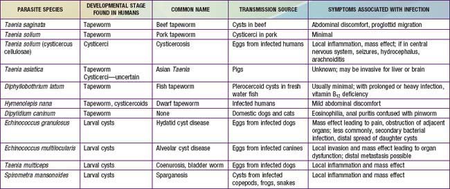
It is important to identify the infecting species of tapeworm. Carriers of adult pork tapeworms are at increased risk for transmitting eggs with the pathogenic intermediate stage (cysticercus) to themselves or others, whereas children infected with the beef tapeworm are a risk only to livestock. Because proglottids are generally passed intact, visual examination for gravid proglottids in the stool is a sensitive test; these segments may be used to identify species. Eggs, by contrast, are often absent from stool and cannot reliably distinguish between T. saginata and T. solium (Fig. 294-1). If the parasite is completely expelled, the scolex of each species is diagnostic. The scolex of T. saginata has only a set of 4 anteriorly oriented suckers, whereas T. solium is armed with a double row of hooks in addition to suckers. The proglottids of T. saginata have more than 20 branches from a central uterine structure, and those of T. solium have 10 or fewer. When in doubt, more proglottids should be obtained or the sample should be referred to a laboratory with parasitologic expertise. Only molecular methods can be used to distinguish T. saginata from T. asiatica.
Diphyllobothriasis (Diphyllobothrium Latum)
Diagnosis
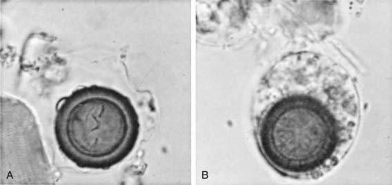
Parasitologic examination of the stool is useful because eggs are abundant in the feces and have morphology distinct from that of all other tapeworms. The eggs are ovoid and have an operculum, which is a cap structure at one end that opens to release the embryo (Fig. 294-2). The worm itself has a distinct scolex and proglottid morphology; however, these are not likely to be passed spontaneously.
Anantaphruti MT, Yamasaki H, Nakao M, et al. Sympatric occurrence of Taenia solium, T. saginata, and T. asiatica, Thailand. Emerg Infect Dis. 2007;13:1413-1416.
Craig P, Ito A. Intestinal cestodes. Curr Opin Infect Dis. 2007;20:524-532.
Samkari A, Kiska DL, Riddell SW, et al. Dipylidium caninum mimicking recurrent Enterobius vermicularis (pinworm) infection. Clin Pediatr. 2008;47:397-399.
Scholz T, Garcia HH, Kuchta R, et al. Update on the human broad tapeworm (genus Diphyllobothrium), including clinical relevance. Clin Microbiol Rev. 2009;22:146-160.


