91
Adnexal Neoplasms
• Adnexal neoplasms are tumors, more commonly benign, that show features of cutaneous adnexal structures (Fig. 91.1), e.g. hair (Table 91.1), sebaceous glands (Table 91.2), apocrine glands and eccrine glands (Table 91.3).
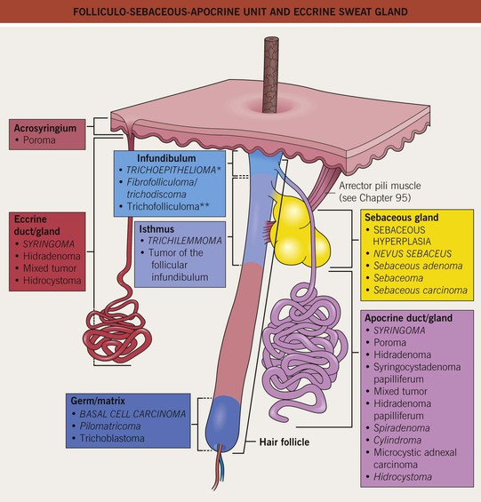
Fig. 91.1 Folliculo-sebaceous-apocrine unit and eccrine sweat gland. Adnexal tumors showing differentiation toward the hair follicle, sebaceous gland, apocrine gland, and eccrine gland are listed in correspondingly colored boxes. Important entities are in capital letters; entities associated with syndromes are in italics (see Tables 91.1–91.3). *Can show germinal differentiation; **differentiation toward entire follicle.
Table 91.1
Benign and malignant tumors with follicular differentiation.
Cysts (e.g. epidermoid and pilar) are discussed in Chapter 90. BHD encodes folliculin, a tumor suppressor; CYLD encodes a deubiquinating enzyme; APC encodes a protein that regulates cell adhesion/migration.
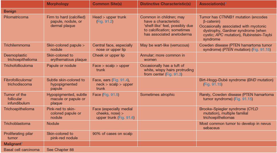
* Patients generally have multiple tumors when associated with a syndrome.
† Rare tumors include pilomatrical carcinoma and trichilemmal carcinoma.
Table 91.2
Benign and malignant tumors with sebaceous differentiation.
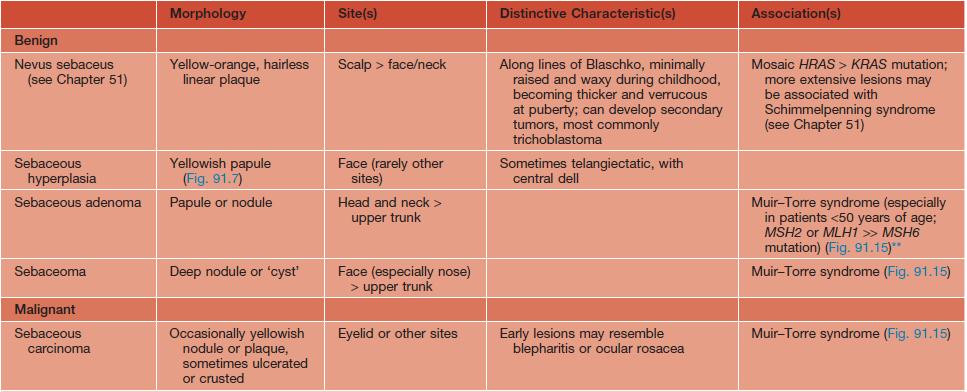
** MSH2, MLH1, and MSH6 encode mismatch repair proteins.
Table 91.3
Benign and malignant tumors with apocrine/eccrine (glandular) differentiation.
Cysts (e.g. hidrocystoma) are discussed in Chapter 90.
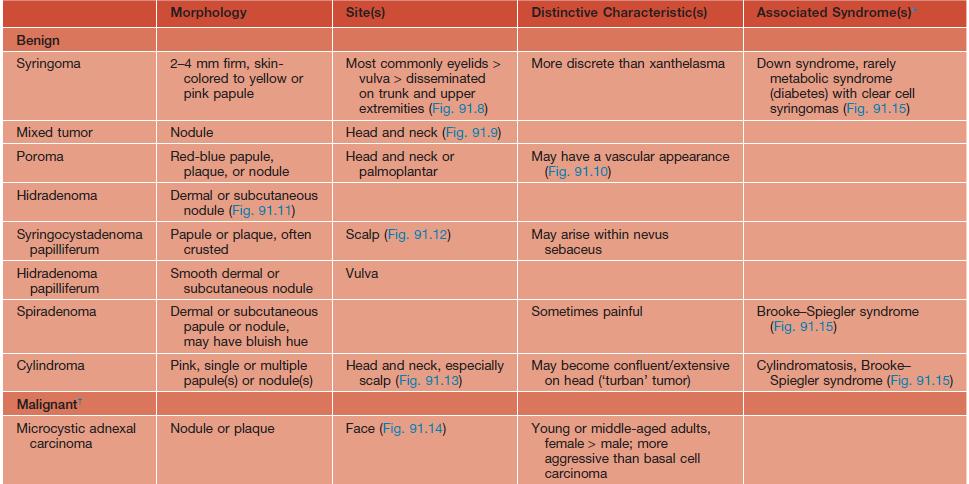
* Patients generally have multiple tumors when associated with a syndrome.
† Other rare tumors include porocarcinoma and adenoid cystic carcinoma.
Fig. 91.3 Trichofolliculoma. Wispy vellus hairs emerge from a skin-colored papule with a dilated central pore.
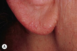
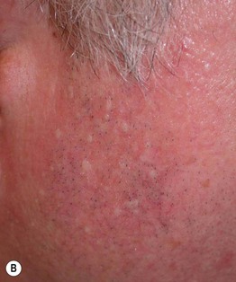
Fig. 91.4 Fibrofolliculomas in association with Birt–Hogg–Dubé syndrome. A Several skin-colored papules are present on the ear. B Multiple smooth papules of the lateral cheek that are relatively hypopigmented and lack the telangiectasias of the background skin.
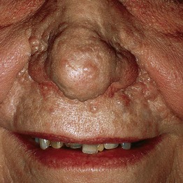
Fig. 91.6 Multiple familial trichoepitheliomas. Numerous skin-colored papules and nodules on the mid-face.
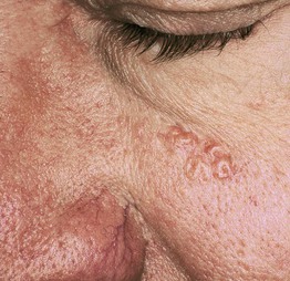
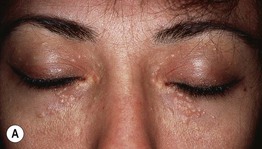
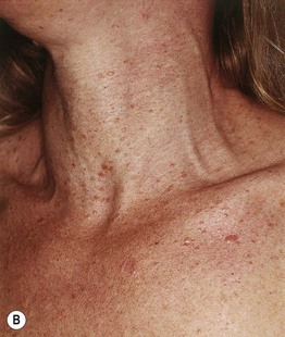
Fig. 91.8 Syringomas. A Aggregated skin-colored papules on the eyelids. B Multiple skin-colored to pink, smooth papules on the neck and upper chest. The lesions favor the ventral surface of the trunk, a distribution pattern referred to as ‘en demicuirasse’. B, Courtesy, Jean L. Bolognia, MD.
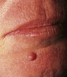

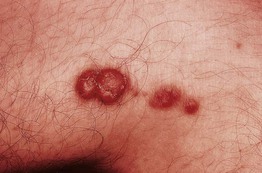
Fig. 91.12 Syringocystadenoma papilliferum. Grouped papules and nodules. The possibility of an associated nevus sebaceus needs to be considered.
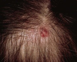
Fig. 91.13 Cylindroma. Whereas numerous tumors aggregated on the scalp are called turban tumors, solitary lesions usually present as smooth erythematous nodules.
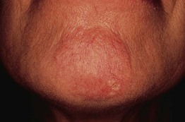
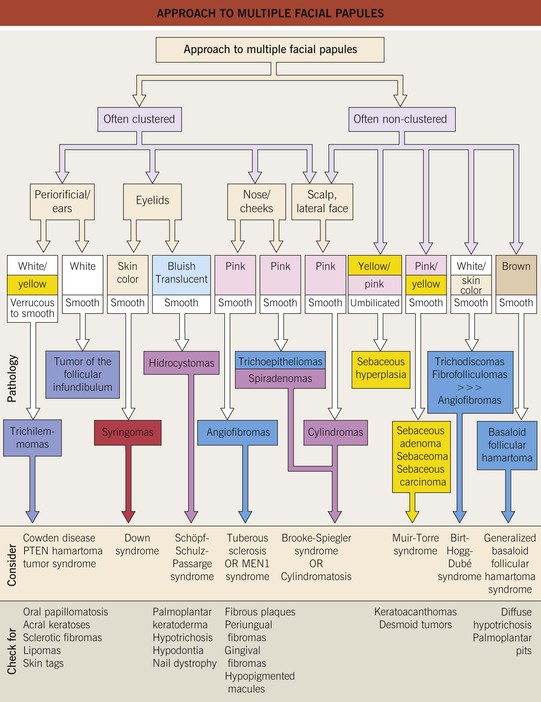
Fig. 91.15 Approach to multiple facial papules. As a general rule, multiple lesions are suggestive of a syndrome, but exceptions are syringoma and tumor of the follicular infundibulum (rarely syndromic). Other causes of multiple facial papules include BCCs, miliary osteomas, milia, verrucae, and acne. MEN, multiple endocrine neoplasia.
For further information see Ch. 111. From Dermatology, Third Edition.


