Published on 18/03/2015 by admin
Filed under Dermatology
Last modified 22/04/2025
This article have been viewed 2879 times
Jonathan E. Blume and Daniel Caplivski
Evidence Levels: A Double-blind study B Clinical trial ≥ 20 subjects C Clinical trial < 20 subjects D Series ≥ 5 subjects E Anecdotal case reports
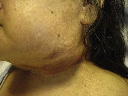
Actinomycosis is often an indolent infection that may be difficult to recognize initially. It is caused by an anaerobic Gram positive rod that is a normal human commensal. Infections caused by Actinomyces spp. are usually the result of introduction of the bacteria into a normally sterile space from the oropharynx, gastrointestinal tract or vaginal tract. The organism may be difficult to recover in the microbiology laboratory because it grows slowly and is ideally cultured under anaerobic conditions. Microscopically, it has a similar appearance to Nocardia spp.; however, it does not retain the modified acid fast stain and the colonies may have a molar tooth appearance. Infections of the cervical region are typified by the slow induration of the skin at the angle of the jaw that progresses over several weeks or months. A mass may be palpable and, due to the slow progression and absence of systemic inflammatory symptoms, the infection can often be confused with other conditions such as malignancies. Patients may notice the discharge of small yellow grains known as ‘sulfur granules.’ These are macroscopic colonies of the organism that may be cultured for confirmation, but their absence should not exclude the diagnosis from consideration.
Actinomyces spp. are universally susceptible to penicillin. The severity of the illness will dictate whether the patient requires intravenous or oral formulations of the antibiotic, but the general principle is that prolonged treatments for 6 months or more are often required for cure. In patients who are penicillin allergic, the tetracycline class of antibiotics is a useful substitute. Surgical removal of the abscess may also be required in order to ensure complete resolution. Treatment failures are rare, but may be due to co-infecting organisms from the oropharynx. In order to treat these organisms, the combination of a β-lactam antibiotic with a β-lactamase inhibitor may be sufficient. Carbapenems such as imipenem, meropenem, or ertapenem are also effective.
Wong VK, Turmezei TD, Weston VC. BMJ 2011; 343: d6099.
Lerner PI. Antimicrob Agents Chemother 1974; 5: 302–9.
Holmberg K. Microbiol Sci 1987; 4: 72–8.
An excellent review of the diagnosis of actinomycosis.
The most accurate way to diagnose actinomycosis is via culture – usually a difficult task, which requires thioglycolate or brain–heart-enriched agar at 37°C under anaerobic or microaerophilic conditions. ‘Molar-tooth’ and ‘breadcrumb’ colonies may take up to 3 weeks to grow. Unfortunately, definitive identification cannot be based on colony morphology and requires the measurement of physiological and biochemical characteristics (e.g., sensitivity to oxygen, presence of preformed enzymes).
Because cultures of Actinomyces spp. are often unsuccessful, observation of ‘sulfur granules’ on a peripheral smear or histology often helps make the diagnosis. The granules are bacterial colonies which on hematoxylin and eosin staining have a basophilic central area surrounded by a zone of eosinophilic ‘clubs’. Other typical histologic findings include extensive fibrosis, chronic granulation tissue, sinus tracts, and scattered microabscesses.
Immunofluorescent staining of Actinomyces spp. is available and can be used on clinical material, granules, and formalin-fixed tissues. The direct immunoperoxidase technique can specifically show Actinomyces spp. in formalin-fixed sections via light microscopy. These techniques, as well as gene sequencing (see below), are promising diagnostic modalities given the difficulty of culture and histologic identification.
Oostman O, Smego RA. Curr Infect Dis Resp 2005; 7: 170–4.
Culture isolation of Actinomyces spp. and microscopic visualization of Gram-positive, non-acid-fast, thin, branching filaments remain the best methods of diagnosing cervicofacial actinomycosis.
Woo PCY, Fung AMY, Lau SKP, Hon E, Yuen KY. Diagn Microbiol Infect Dis 2002; 43: 113–18.
Actinomyces odontolyticus was identified by rRNA gene sequencing. Because the 16 S ribosomal RNA gene is conserved within a species, it can be used to identify a specific species of bacteria.
Park JK, Lee HK, Ha HK, Choi HY, Choi CG. AJNR Am J Neuroradiol 2003; 24: 331–5.
Findings on CT and MRI may be helpful in distinguishing cervicofacial actinomycosis from malignant neoplasms, tuberculosis, and fungal infections.
Smith AJ, Hall V, Thakker B, Gemmell CG. J Antimicrob Chemother 2005; 56: 407–9.
The authors tested the susceptibility of 87 strains of Actinomyces to 12 different antimicrobial agents. All isolates were susceptible to penicillin and amoxicillin.
Smego RA, Foglia G. Clin Infect Dis 1998; 26: 1255–63.
The authors recommend 2 months of oral penicillin V (2–4 g/day divided every 6 hours) or a tetracycline (e.g., oral doxycycline 100 mg twice daily) for mild cervicofacial disease. For more complicated infections, parenteral penicillin G (10–20 million U/day divided every 6 hours) for 4 to 6 weeks, followed by 6 to 12 months of oral penicillin V (2–4 g/day divided every 6 hours) is suggested. A tetracycline, erythromycin, clindamycin, or cephalosporins are advocated for patients allergic to penicillin.
Peabody JW, Seabury JH. Am J Med 1960; 28: 99–115.
The authors review the treatment of actinomycosis and state that penicillin is the drug of choice.
Martin MV. Br Dent J 1984; 156: 252–4.
Ten patients with cervicofacial actinomycosis were cured in less than 6 weeks with a combination of amoxicillin (500 mg four times daily) and surgery.
Cevera JJ, Butehorn HF, Shapiro J, Setzen G. Laryngoscope 2003; 113: 2108–11.
A 39-year-old woman who developed actinomycosis of the thyroid gland after tooth extraction was cured with thyroidectomy and 6 months of ceftriaxone (1 g intravenously every 12 hours).
Skoutelis A, Petrochilos J, Bassaris H. Clin Infect Dis 1994; 19: 161–2.
A 38-year-old patient with pulmonary actinomycosis was successfully treated with a 3-week course of daily ceftriaxone (2 g intravenously), followed by 3 months of daily oral ampicillin (no dose listed but typically given 500 mg orally every 6 hours).
Badgett JT, Adams G. Pediatr Infect Dis J 1987; 6: 221–2.
de Vries J, Bentley KC. Int J Clin Pharmacol 1974; 9: 46–8.
A 60-year-old man with cervicofacial actinomycosis that was resistant to penicillin and tetracycline responded fully to a 1-month course of clindamycin (150 mg four times a day).
Fass RJ, Scholand JF, Hodges GR, Saslaw S. Ann Intern Med 1973; 78: 853–9.
Four patients with cervicofacial actinomycosis and one with thoracic actinomycosis were successfully treated with a combination of intravenous (1.8–2.7 g/day) and oral (0.9–1.2 g/day) clindamycin.
Mert A, Bilir M, Bahar H, Torun M, Tabak F, Ozturk R, et al. Int J Infect Dis 2001; 5: 112–14.
A 35-year-old man with primary actinomycosis of the hand was cured with 1 month of intravenous ampicillin (12 g/day), followed by 11 months of oral doxycycline (200 mg/day).
de Souza E, Katz DA, Dworzack DL, Longo G. J Urol 1985; 133: 290–1.
A case of acute prostatitis due to Actinomyces spp. was cured with long-term erythromycin (500 mg intravenously every 6 hours followed by 500 mg orally every 6 hours), chosen because of its excellent penetration into prostatic secretions.
Bradley P. Br J Oral Surg 1971; 9: 54–6.
A 58-year-old man with actinomycosis of the temporomandibular joint was cured with a 12-week course of erythromycin (500 mg six times a day).
Yew WW, Wong PC, Wong CF, Chau CH. Clin Infect Dis 1994; 19: 983–4.
Yew WW, Wong PC, Lee J, Fung SL, Wong CF, Chan CY. Monaldi Arch Chest Dis 1994; 54: 126–9.
Seven of eight patients with pulmonary actinomycosis were successfully treated with a 4-week course of parenteral imipenem–cilastatin (500 mg intravenously every 8 hours).
Takeda H, Mitsuhashi Y, Kondo S. J Dermatol 1998; 25: 37–40.
A patient with primary cutaneous disseminated actinomycosis was cured with a 3-month course of intravenous minocycline (2 mg/kg/day).
Martin MV. Br J Oral Maxillofac Surg 1985; 23: 428–34.
Six patients with cervicofacial actinomycosis were cured with eight to 16 weeks of oral minocycline (250 mg four times a day). There were no recurrences after 1 year.
Langloh JT, Lauerman WC. J Pediatr Orthop 1987; 7: 222–3.
Surgical drainage followed by a 6-week course of oral tetracycline (500 mg orally every 6 hours) cured a case of actinomycosis of the quadriceps.
Goldstein EJ, Citron DM, Merriam CV, Warren YA, Tyrrell KL, Fernandez HT. Antimicrob Agents Chemother 2006; 50: 3507–13.
Tigecycline (50 mg intravenously every 12 hours, with a 100 mg loading dose) had good in vitro activity against isolates of Actinomyces spp..
Macfarlane DJ, Tucker LG, Kemp RJ. J Infect 1993; 27: 177–80.
Ferreira D de F, Amado J, Neves S, Taveira N, Carvalho A, Nogueira R. J Bras Pneumol 2008; 34: 245–8.
Morrone N, De Castro Pereira CA, Saito M, Dourado AM, Pereira Da Silva Mendes ES. G Ital Chemioter 1982; 29: 121–4.
King JW, White MC. Arch Intern Med 1981; 141: 1234–5.
Shauly Y, Nachum Z, Gdal-On M, Melamed S, Miller B. Graefes Arch Clin Exp Ophthalmol 1993; 231: 429–31.
A 52-year-old patient with treatment-resistant lacrimal canaliculitis due to A. israelii was cured with hyperbaric oxygen.
Manheim SD, Voleti C, Ludwig A, Jacobson JH. J Am Med Assoc 1969; 210: 552–3.
After failing to respond to surgery and intravenous penicillin, a 63-year-old patient with perirectal actinomycosis was cured with hyperbaric oxygen.
Treatment of Skin Disease Comprehensive Therapeutic Strategies 4e
WhatsApp us

 Penicillin
 Amoxicillin
 Ceftriaxone
 Clindamycin
 Doxycycline
 Erythromycin
 Imipenem
 Minocycline
 Tetracycline
 Ciprofloxacin
 Levofloxacin
 Rifampin
 Hyperbaric oxygen