Published on 19/03/2015 by admin
Filed under Dermatology
Last modified 22/04/2025
This article have been viewed 1742 times
Sherrif F. Ibrahim and Marc D. Brown
Evidence Levels: A Double-blind study B Clinical trial ≥ 20 subjects C Clinical trial < 20 subjects D Series ≥ 5 subjects E Anecdotal case reports
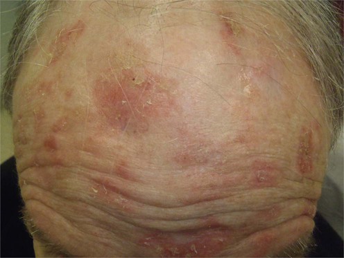
Actinic keratoses (AK) are ill-defined pink to skin-colored, scaly papules found on chronically sun-exposed areas in light-skinned individuals. They most frequently appear on the face, ears, balding scalp, extensor forearms, and dorsal hands. AKs are a strong predictor for the development of squamous cell carcinoma (SCC) and, to a lesser extent, basal cell carcinoma. Australians have the highest reported prevalence, which approaches 60%, and in the US AKs are the second most common reason for visits to the dermatologist.
Actinic keratoses are common dysplastic intra-epidermal lesions that are considered to be precursors to SCC. Reports have varied as to the rates of progression to invasive SCC, from 0.025% to over 25% per year, and AKs are commonly located adjacent to SCC histologically. For these reasons, most practitioners advocate the treatment of AKs, as considerable morbidity and potential mortality can be associated with invasive disease. However, there have been no randomized controlled studies demonstrating a reduction in the frequency of SCC with treatment of AKs.
The diagnosis of AK is primarily clinical, and because of their superficial nature, a variety of effective management approaches exist. Biopsy of suspected AKs is typically not warranted; however, in patients with a history of multiple skin cancers, immunosuppressed patients, and lesions in high-risk areas such as the lip or ear, clinicians should have a low threshold for biopsy to rule out invasive SCC. Indications for biopsy include tenderness, rapid growth or thickening of lesions, bleeding, hyperkeratosis, and failure to respond to treatment.
Prevention of AKs through sun avoidance and diligent use of broad-spectrum sunscreens and blocking agents is an important aspect of management. This has been shown to prevent the development of new AKs and reduce the incidence of non-melanoma skin cancers.
With cumulative sun exposure and advancing age, rates of AK development increase, necessitating either ablative or topical treatment. Cryotherapy with liquid nitrogen is by far the most commonly employed therapeutic modality because it can be performed quickly and effectively in the office setting. However, given the common appearance of AKs on a background of diffuse actinic damage, individual lesions may be poorly defined and involve large, contiguous areas requiring field treatment with topical agents such as 5-fluorouracil (5-FU) or imiquimod. The latter has recently been shown to have high rates of treatment success with durable results and has become an accepted first-line therapy with a newer 3.75% formulation recently introduced. A novel agent, ingenol mebutate, is derived from the Euphorbia peplus plant and has been approved as an additional topical agent for the field treatment of AKs. The advantages of topical approaches are that they are patient-administered, non-invasive, carry little risk of scarring or pigmentary change, and can be used for anatomically difficult or cosmetically sensitive areas. However, these agents require adequate patient compliance and are often accompanied by prolonged erythema lasting several weeks. Photodynamic therapy (PDT) with aminolevulinic acid (ALA) or methyl aminolevulinate (MAL) has continued to become more widespread, given its proven therapeutic results and excellent cosmetic outcome. PDT offers a physician-administered approach to field treatment with shorter periods of inflammation and erythema than several topical agents, and thus many studies indicate higher patient satisfaction. Variations in the light dose, light source, sensitizing agent and its application time, and frequency of treatments may improve efficacy. Head-to-head trials of different treatment approaches are difficult to perform, as variations in treatment protocols make direct comparisons challenging. Recently, there has been a growing trend towards combination therapy, such as topical agents either before or after cryotherapy, or sequential use of multiple topical modalities with varying mechanisms of action. Other approaches such as laser resurfacing, chemical peels, and dermabrasion may be considered in certain situations when lesions have failed the above treatments, or if severe photodamage is present. Finally, for recalcitrant or hyperkeratotic lesions, curettage or excision may be appropriate.
In selected cases:
Biopsy
Ehrig T, Cockerell C, Piacquadio D, Dromgoole S. Dermatol Surg 2006; 32: 1261–5.
A total of 271 clinically diagnosed AKs were biopsied. Clinical diagnosis was in agreement with histology 91% of the time, with about one in 25 lesions revealing invasive SCC.
Venna SS, Lee D, Stadecker MJ, Rogers GS. Arch Dermatol 2005; 141: 507–9.
Seventeen of 23 lesions (74%) with classic features of AK in patients with a history of previous skin cancer were confirmed histologically. Five lesions (22%) were shown to be SCC.
Actinic keratoses are typically diagnosed clinically. However, there should be a low threshold to biopsy tender, hyperkeratotic, large, or recalcitrant lesions to exclude malignancy.
Darlington S, Williams G, Neale R, Frost C, Green A. Arch Dermatol 2003; 139: 451–5
In this Australian study, 1621 adults were randomized to either daily use of sunscreen or application at their usual discretionary rate. There was a 24% reduction in AKs in the daily use group.
Thompson SC, Jolley D, Marks R. N Engl J Med 1993; 14: 1147–51.
In a 6-month randomized, placebo-controlled trial of 588 patients in Australia, SPF 17 sunscreen applied daily was found to both reduce the development of new AKs and increase the remission of existing AKs compared to a vehicle cream.
Thai K, Fergin P, Freeman M, Vinciullo C, Francis D, Spelman L, et al. Int J Dermatol 2004; 43: 687–92.
In this prospective multicenter study, 90 patients with 421 AKs on the face and scalp were treated with cryotherapy with a single freeze–thaw cycle using different freeze times. The patients were reviewed 3 months later. Overall, the complete response (CR) rate was 67.2%, varying from 39% for freeze times less than 5 seconds to 83% for times longer than 20 seconds. The authors also found that hypopigmentation was present in 29% of CR lesions. Patients rated cosmetic outcomes as good to excellent for 94% of CR lesions.
Jorizzo J, Weiss J, Furst K, VandePol C, Levy SF. Arch Dermatol 2004; 140: 813–16.
This study demonstrates that there is a role for the combination of therapeutic modalities in the treatment of AKs. In this prospective, double-blind, randomized controlled trial, 144 patients, each with at least five AKs on the face, were randomized to receive 1 week of treatment with 0.5% 5-FU cream daily for 7 days or placebo cream. Patients were then treated with single freeze–thaw cycle cryotherapy using liquid nitrogen, with a thaw time of 10 seconds. These patients were then followed up at 4 weeks and 6 months. The authors found that, at 4 weeks, 16.7% of patients in the 5-FU group were completely clear of lesions, compared to 0% in the vehicle group (p<0.001). At 6 months post-treatment, 30% of patients in the 5-FU group were clear of lesions, compared to 7.7% of patients in the vehicle group (p<0.001).
Lebwohl M, Dinehart S, Whiting D, Lee PK, Tawfik N, Jorizzo J, et al. J Am Acad Dermatol 2004; 50: 714–21.
In this report of two phase III double-blind, vehicle-controlled studies, 436 subjects at 24 centers in the US were randomized to either 5% imiquimod or vehicle applied once daily, 2 days per week, for 16 weeks. The complete clearance rate for the treated arm was 45%, as opposed to 3.2% for the vehicle group. The median percent reduction in AK lesions was 83% for the treated group and 0% for the vehicle group, indicating that a reduced frequency of imiquimod application is quite effective.
Hanke CW, Beer KR, Stockfleth E, Wu J, Rosen T, Levy S. J Am Acad Dermatol 2010; 62: 573–81.
Swanson N, Abramovits A, Berman B, Kulp J, Rigel DS, Levy S. J Am Acad Dermatol 2010; 62: 582–90.
These two back-to-back reports demonstrate the efficacy and tolerability of lower concentration formulations of imiquimod as well as alternate dosing schedules. Interestingly, the results were essentially identical for two 3-week cycles as they were for two 2-week cycles, with the 3.75% strength imiquimod proving superior to the 2.5% in both studies and slightly less efficacious than previous studies that have investigated 5% imiquimod used twice weekly for 16 weeks.
Krawtchenko N, Roewert-Huber J, Ulrich M, Mann I, Sterry W, Stockfleth E. Br J Dermatol 2007; 157 (Suppl 2): 34–40.
This study compared the baseline and 1-year follow-up rates of clinical clearance, histological clearance, and cosmetic outcomes of imiquimod, 5-FU, and cryosurgery for the treatment of AKs. Patients were randomized to one of the three treatment groups. Clinical clearance was achieved in 68% of those treated with cryosurgery, 96% with 5-FU, and 85% with imiquimod. Histological clearance rate was 32% for cryosurgery, 67% for 5-FU, and 73% for imiquimod. The 1-year sustained clearance rate was 28% for cryosurgery, 54% for 5-FU, and 73% for imiquimod. The patients treated with imiquimod were also judged to have superior cosmetic outcomes.
Braathen LR, Szeimies R, Basset-Seguin N, Bissonnette R, Foley P, Pariser D, et al. J Am Acad Dermatol 2007; 56: 125–43.
This is a comprehensive evidence-based review and treatment recommendations for the use of PDT to treat non-melanoma skin cancers, including AKs. The authors review each of the main clinical trials using PDT to treat AKs, and conclude that PDT with MAL and ALA is highly effective, offering excellent cosmetic results, and should be considered as first-line therapy.
Morton C, Campbell S, Gupta G, Keohane S, Lear J, Zaki I, et al. Br J Dermatol 2006; 155: 1029–36.
In this 24-week study subjects received one treatment session of PDT to one side of their face and two cycles of freeze–thaw cryosurgery to the other. Of the 1501 lesions treated, PDT resulted in a higher rate of cure (87% vs 76% reduction from baseline). Both subjects and investigators preferred PDT and also felt it had a better cosmetic outcome. Both treatment regimens were deemed safe and well tolerated.
Kaufmann R, Spelman L, Weightman W, Reifenberger J, Szeimies RM, Verhaeghe E, et al. Br J Dermatol 2008; 158: 994–9.
This was another intra-individual trial that treated one side of the body (non-face/scalp) with a single course of PDT and the other with cryotherapy. For the 1343 lesions treated, both treatment modalities had high efficacy rates at 24 weeks, although cryosurgery performed better (78% for PDT and 88% for cryosurgery). Investigator and patient assessment of cosmetic outcome was much higher for PDT than for cryosurgery (79% vs 56% of lesions having ‘excellent cosmetic outcome’ based on investigator evaluation, 50% vs 22% based on patient evaluation).
Piacquadio DJ, Chen DM, Farber HF, Fowler JF Jr, Glazer SD, Goodman JJ, et al. Arch Dermatol 2004; 140: 41–6.
In this randomized, placebo-controlled study 243 patients were randomized to receive vehicle or ALA followed by PDT within 14–18 hours. Clinical response rate was based on complete clearing of 75% of lesions measured at weeks 8 and 12. Of the PDT-treated group, 77% had a complete response by week 8 and 89% by week 12. This compared to 18% and 13% for the placebo group. Most patients experienced erythema and edema at the treated sites, which improved within 4 weeks of therapy. Stinging, burning, or pain occurred during the treatments, but resolved within 24 hours.
Hadley J, Tristani-Firouzi P, Hull C, Florell S, Cotter M, Hadley M. Dermatol Surg 2012; 38: 722–7.
In this split-faced trial, 61 patients received 5% imiquimod twice weekly to half the face and two sessions of ALA-PDT to the other side of the face. No significant difference was noted with respect to partial or total clearance rate; however, ALA-PDT was superior when mean lesion reduction rate was evaluated.
Kang S, Goldfarb MT, Weiss JS, Metz RD, Hamilton TA, Voorhees JJ, Griffiths CE. J Am Acad Dermatol 2003; 49: 83–90.
In this prospective randomized, vehicle-controlled study 90 patients received either 0.1% adapalene gel, 0.3% adapalene gel or vehicle gel, initially daily for 4 weeks, then twice daily for up to 9 months. Overall, 62% (p<0.01) of those who received 0.1% adapalene gel and 66% (p<0.01) of those who received 0.3% adapalene gel showed at least a moderate improvement of their AKs, compared to 34% of patients receiving a vehicle cream. Adapalene gel recipients reported a higher level of mild erythema, peeling, and dryness compared to control groups.
Pirard D, Vereecken P, Melot C, Heenen M. Arch Dermatol Res 2005; 297: 185–9.
This meta-analysis pooled 364 patients from three studies that compared diclofenac to hyaluronan vehicle gel. Overall, patients treated with diclofenac had a significantly higher rate of complete clearance of lesions. They concluded that complete response rates were 39% with a mean treatment duration of 75 ± 21 days. Mild-to-moderate skin irritation was the major side effect of the treatment group.
Martin GM, Stockfleth E. J Drugs Dermatol 2012; 11: 600–8.
This comprehensive review draws from 17 publications beyond the initial phase III clinical trials that lead to the approval of diclofenac sodium for the treatment of AKs. It discusses the data with regards to treatment efficacy, tolerability, and performance compared to other topical field agents, as well as its use in combination treatment strategies.
Lebwohl M, Swanson N, Anderson LL, Melgaard A, Xu Z, Berman B. N Engl J Med 2012; 366: 1010–19.
This multicenter, randomized, double-blind report investigated the use of 0.015% ingenol mebutate used once daily for 3 consecutive days for AKs on the face and scalp, as well as 0.05% ingenol mebutate (IM) used once daily for 2 consecutive days for AKs on the trunk and extremities. All analyses were conducted at day 57. The rate of complete clearance was 42% for the face/scalp group and 34% for the trunk/extremities group. Local skin reactions peaked between day 3 and day 8 and were mild to moderate in nature. Hypertrophic or hyperkeratotic lesions were excluded from the study.
Anderson L, Schmieder GJ, Werschler WP, Tschen EH, Ling MR, Stough DB, Katsamas J. J Am Acad Dermatol 2009; 60: 934–43.
A total of 222 patients from 22 US centers with non-facial AKs were randomized to receive vehicle for 3 days, 0.025% ingenol mebutate (IM) gel for 3 days, vehicle for 1 day followed by 0.05% IM gel for 2 days, or 0.05% IM gel for 3 days. Partial clearance rate (>75% reduction in baseline AKs) as well as complete clearance rate was statistically significantly higher in all three treatment groups over baseline and these results were dose-dependent. Partial and complete clearance rates were 28%/20% for the 0.025% × 3 days group, 34%/24% for the 0.05% × 2 days group, and 43%/31% for the 0.05% × 3 days cohort. The most common local skin responses seen in all groups included erythema, scaling, crusting, swelling, erosion/ulceration, vesiculation, and pigmentary changes and were largely resolved by day 15. Patient satisfaction was high.
Iyer S, Friedli A, Bowes L, Kricorian G, Fitzpatrick RE. Lasers Surg Med 2004; 34: 114–19.
In this retrospective study of 24 patients with over 30 AKs on the face treated with full-face ultrapulse CO2 laser or erbium Er : YAG laser resurfacing, the authors found that 21 patients remained lesion free for at least 1 year.
Ostertag JU, Quaedvlieg PJ, Neumann MH, Krekels GA. Dermatol Surg 2006; 32: 261–7.
A retrospective case–control study of 25 patients who underwent laser resurfacing for widespread AKs on the scalp and/or face. Forty-four percent had no recurrence in an average follow-up period of 39 months (range 7–70 months).
Katz TM, Goldberg LH, Marquez D, Kimyai-Asadi A, Polder KD, et al. J Am Acad Dermatol 2011; 65: 349–56.
In this study, 14 patients underwent five treatments of non-ablative fractional resurfacing with 32–40% surface area coverage at 2- to 4-week intervals. Clinical images and biopsies were evaluated. Although clinical images had a reduced number of AKs, virtually all histologic specimens were positive for features of AK and/or squamous cell carcinoma. It was concluded that fractional non-ablative resurfacing was not adequate therapy for AKs.
Ostertag JU, Quaedvlieg PJ, van der Geer S, Nelemans P, Christianen ME, Neumann MH, Krekels GA. Lasers Surg Med 2006; 38: 731–9.
A prospective randomized trial of 55 patients comparing 5-FU with Er : YAG laser resurfacing. At 3, 6, and 12 months there were significantly fewer recurrences in the laser group than in the 5-FU group. Side effects were more common in the group treated with laser resurfacing, and included erythema and hypopigmentation.
Witheiler DD, Lawrence N, Cox SE, Cruz C, Cockerell CJ, Freemen RG. Dermatol Surg 1997; 23: 191–6.
In this prospective study, 15 patients with severe facial AKs were treated on one side of the face with a single application of Jessner’s solution – a medium-depth chemical peel – and the other side with twice-daily applications of topical 5-FU 5% for 3 weeks. The authors found that both treatments resulted in a similar reduction of AKs at 12 months, with an increase in the number of AKs in both groups from 12 to 32 months. The authors concluded that both treatments were similarly efficacious in the treatment of AK, and that re-treatment for recurrences might be necessary after 12 months.
Coleman WP, Yardborough JM, Mandy SH. Dermatol Surg 1996; 22: 17–21.
In this retrospective study, 23 patients who had undergone dermabrasion for facial AKs were followed for 2 to 5 years. The authors found that the benefits of dermabrasion diminished with time: 1 year post dermabrasion, 22 patients remained clear of AKs. Of 13 patients who were followed up for 5 years, seven remained clear. The authors concluded that dermabrasion provided long-term clearance of AKs in some patients.
Recalcitrant treatment-resistant AKs may be treated with physically destructive approaches that include curettage and surgical excision. Both techniques enable tissue to be obtained for the purposes of histological analysis. Surgical excision is particularly useful in lesions suspected of being SCC because this technique enables the clinician to treat the lesion and establish the diagnosis. Widespread, extensive AKs may benefit from field treatments of a destructive nature. These include ablative laser resurfacing, dermabrasion, and chemical peels.
Treatment of Skin Disease Comprehensive Therapeutic Strategies 4e
WhatsApp us

 Sunscreens
 Cryosurgery
 Topical 5-FU
 Imiquimod
 Photodynamic therapy
 Topical adapalene
 Topical diclofenac
 Topical ingenol mebutate
 Curettage
 Excision
 Laser resurfacing
 Chemical peels
 Dermabrasion