[level-membership-for-dermatology-category]
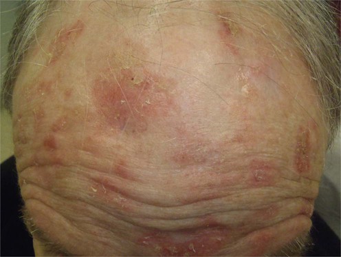
Actinic keratoses
Specific investigations

First-line therapies
 Sunscreens
 Cryosurgery
 Topical 5-FU
 Imiquimod
 Photodynamic therapy
Second-line therapies
 Topical adapalene
 Topical diclofenac
 Topical ingenol mebutate
Third-line therapies
 Curettage
 Excision
 Laser resurfacing
 Chemical peels
 Dermabrasion
[/level-membership-for-dermatology-category][not-level-membership-for-dermatology-category]
Actinic keratoses
Specific investigations
First-line therapies
Buy Membership for Dermatology Category to continue reading. Learn more here
[/not-level-membership-for-dermatology-category]
