Published on 18/03/2015 by admin
Filed under Dermatology
Last modified 22/04/2025
This article have been viewed 2416 times
Joanna E. Gach
Evidence Levels: A Double-blind study B Clinical trial ≥ 20 subjects C Clinical trial < 20 subjects D Series ≥ 5 subjects E Anecdotal case reports
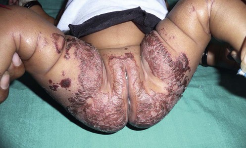
(Courtesy of Shyam B. Verma.)
Acrodermatitis enteropathica (AE) is a rare, autosomal recessive inherited disorder of zinc deficiency. It is caused by mutations in the SLC39A gene located on 8q24.3, which encodes for Zip4 intestinal zinc transporter expressed in enterocytes that absorb dietary zinc from the small intestine. The estimated prevalence is 1 in 500 000 children worldwide, without apparent predilection for race or gender. AE manifests itself shortly after birth in a bottle-fed infant, and sometime soon after weaning in a breastfed infant. Zinc within breast milk is more bioavailable to infants rather than bovine milk due to binding to a low molecular weight ligand secreted by the pancreas. Characteristic clinical signs are lesions in acral and periorificial sites; the first signs are eczematous, pink scaly plaques that can become vesicular, bullous, pustular or desquamative. They can resemble the severe diaper dermatitis of infancy. Angular cheilitis and paronychia can also be seen early. If left untreated, AE usually causes diarrhea, irritability, and alopecia, and skin lesions become secondarily infected with bacteria and Candida albicans. Impaired physical and mental development is seen in advanced disease. Appropriate supplementation of zinc in the infant’s diet results in a rapid improvement.
The diagnosis of AE is applied only to inherited zinc deficiency; non-inherited zinc deficiency is called acquired zinc deficiency. Long-term therapy and management of zinc deficiency vary depending on the severity of the disorder.
Chronic dermatitis in periorificial and acral areas should suggest the possibility of zinc deficiency, but establishing the diagnosis of AE may be difficult. The first step is laboratory determination of blood plasma or serum zinc levels. Blood sample needs to be drawn into a trace element-free bottle with a stainless steel needle. Avoid contact with rubber stoppers as they contain zinc, avoid hemolysis, use zinc-free anticoagulants, separate plasma or serum from cells within 45 minutes. Zinc levels will decrease in states of hypoalbuminemia because zinc binds albumin in the circulation. If the diagnosis of zinc deficiency has been confirmed, management becomes relatively simple: oral zinc supplementation produces dramatic resolution of the problem. High dose supplementation will allow for increased paracellular zinc absorption despite the absence of a functional Zip4 zinc transporter.
The patient or their family must understand the need for lifelong management of the disorder in terms of zinc supplementation and medical supervision. Discuss foods rich in zinc, such as shellfish, nuts and green leafy vegetables. Bioavailability of zinc can be reduced by phytates, which naturally occur in plant fibers and also with regular iron supplementation. With age and a more varied diet, the dose of daily zinc supplementation may be reduced. Zinc therapy should be monitored periodically with morning fasting specimen, full blood count, serum copper level, and stool examination for occult blood. Zinc supplementation has a theoretical risk of reducing copper absorption leading to refractory microcytic anemia that will not respond to iron therapy until the serum copper level is normalized.
Morning fasting blood plasma or serum zinc levels
Urine zinc excretion
Serum albumin and alkaline phosphatase
Serum copper levels
Genetic studies
The normal plasma zinc level is 70–110 µg/dL and in serum 80–120 µg/dL. Urinary zinc can be measured but is not definitive for diagnosis.
Wang K, Pugh EW, Griffen S, Doheny KF, Mostafa WZ, al-Aboosi MM, et al. Am J Hum Genet 2001; 68: 1055–70.
Küry S, Dréno B, Bézieau S, Giraudet S, Kharfi M, Kamoun R, et al. Nature Genet 2002; 31: 239–40.
Jensen SL, McCuaig C, Zembowicz A, Hurt MA. J Cutan Pathol 2008; 35: 1–13.
In most cases, oral supplementation with two to three times the recommended daily allowance of zinc salts in doses of 30–55 mg of elemental Zn2+ will be sufficient to restore normal zinc status within days to a few weeks, depending on the degree of depletion. The dose of elemental zinc must be determined by the patient’s blood or plasma zinc levels, and by body weight. In AE, zinc replacement should begin at 2–3 mg/kg/day of elemental zinc. Serum or plasma zinc should be checked every 3 to 6 months, adjusting the zinc dosage accordingly. In patients with acquired or dietary zinc deficiency treatment should begin at 0.5–1 mg/kg/day. Available forms of zinc supplementation include zinc sulfate, zinc acetate, zinc gluconate, and zinc propionate. Dosage must be based on the amount of elemental zinc present in the preparation, which varies between compounds. For example, a standard 220 mg capsule of a commercial zinc preparation contains approximately 55 mg Zn2+, which is an adequate daily dose for most deficient individuals. A commonly used preparation is Zincate, which is ZnSO4·7H2O.
A significant side effect of zinc supplementation is gastric irritation with nausea, vomiting, and gastric hemorrhage. Large accidental overdoses of zinc may cause fatal multisystem organ failure.
Barnes PM, Moynahan EJ. Proc Roy Soc Med 1973; 66: 327–9.
In 1973, Barnes and Moynahan were treating children with chronic, unresponsive AE-type acral dermatoses and having poor results. They then tried different medications, one of which was oral zinc. To their surprise, the patients receiving the zinc supplements cleared rapidly and completely.
Neldner KH. In: Freedberg IM, Eisen AZ, Wolff K, et al., eds. Fitzpatrick’s dermatology in general medicine, 6th edn. New York: McGraw-Hill, 2003; 412–18.
Overview of zinc deficiency, including a list of chronic disorders with nutrient deficiencies that can result in signs of marginal zinc deficiency.
Maverakis E, Fung MA, Lynch PJ, Draznin M, Michael DJ, Ruben B, Fazel N. J Am Acad Dermatol 2007; 56: 116–24.
An excellent review of AE and other zinc deficiency disorders.
We acknowledge Dr Kenneth H. Neldner for his contribution to this chapter.
Treatment of Skin Disease Comprehensive Therapeutic Strategies 4e
WhatsApp us


 Oral zinc supplementation