Published on 18/03/2015 by admin
Filed under Dermatology
Last modified 22/04/2025
This article have been viewed 2415 times
William Perkins
Evidence Levels: A Double-blind study B Clinical trial ≥ 20 subjects C Clinical trial < 20 subjects D Series ≥ 5 subjects E Anecdotal case reports
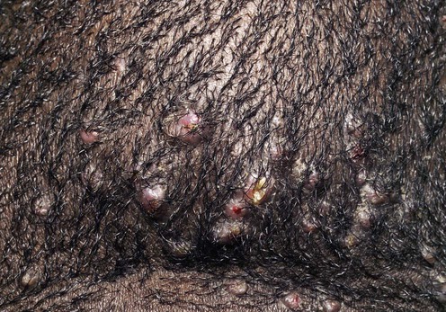
Acne keloidalis nuchae (AKN) is an idiopathic chronic inflammatory process affecting the nape of the neck and the occipital scalp; it occurs predominantly in black males. Initial features consist of papules and pustules on the occiput and posterior neck, which subsequently coalesce into plaques of dense scar tissue with central scarring alopecia. Although the etiology is unknown, the histology of early cases shows evidence of acute and chronic folliculitis, with ruptured follicles, perifolliculitis, and a foreign body granulomatous response. Later cases may show similar features, but additionally there may be hypertrophic scar formation. Close shaving of the hair has been postulated as a cause for AKN, but even during the 1960s and 1970s, with the fashion for longer hair, AKN was still seen. Physical trauma due to collars rubbing and picking by patients have all been suggested as precipitants, but none of these has been investigated in any systematic way. Whether folliculitis leading to ruptured follicles and the subsequent foreign body reaction, or the development of ingrowing hairs is the primary event, the term ‘acne keloidalis’ is a misnomer. Keloids at other sites or a family history of keloids are not features of AKN, and excision of the area does not result in keloid formation. Pseudofolliculitis barbae was associated with AKN in five of six cases in one series, but clinical or histologic evidence of superficial hair penetration is lacking. Lesions resembling AKN have been reported in those receiving long-term cyclosporine, and sarcoid papules may occasionally mimic the condition.
A clear diagnosis is a prerequisite for the management of AKN. The presence of inflammatory papules, pustules, and hypertrophic scar formation on the occipital scalp and posterior neck in a black male is pathognomonic, but cases have been described in Caucasians, and occasionally in females. Biopsy of the area is not usually required, but concerns about keloidal scarring should not inhibit obtaining histologic confirmation. Folliculitis secondary to bacterial infections, particularly staphylococcal, needs to be excluded. In staphylococcal folliculitis the pustules and papules tend to be more widely distributed across the scalp, especially over the crown. Culture will yield heavy growths of staphylococci, and the condition usually responds well to treatment with oral antibiotics, but may recur and require long-term treatment.
In view of the suggested associations with close-cropped hair and picking, it may be worthwhile enquiring about these factors. If present, these practices should be avoided.
Treatment depends on the stage of presentation. Unfortunately, the evidence base for many of the management recommendations is weak. Many patients will prefer no treatment or conservative treatment in the early stages of the disease. This is demonstrated by the fact that only 30% of patients identified in one survey had tried any treatment at all. Early disease with papules and pustules scattered across the posterior neck and occipital scalp may well be best managed by topical antiseptics, antibiotics, or potent topical corticosteroids.
With the development of hypertrophic scar formation, topical or intralesional corticosteroids may well be of benefit. Once scarring, alopecia, hypertrophic scars, and symptoms related to itch, pain, and discharging sinuses are present, treatment directed at the removal of the follicles from the affected area in their entirety is recommended.
Excisional surgery is the only treatment reported in any significant case series. The factors influencing the use of excision will be the severity of symptoms the patient is experiencing and the confidence the patient and surgeon have in the process of surgery. Scattered papules and pustules across the occipital scalp without confluent areas of hypertrophic scar formation and with only limited symptoms may lead patients to seek a more conservative treatment option.
Prioritization of the following treatments is not meant to be a strict hierarchy; for a well-developed case of AKN, the author’s treatment of choice is excision. When this is not acceptable, some of the following non-surgical approaches may be appropriate. Advice to reduce the picking (a consistently reported association) and close cropping of the hair is the first measure one should employ. This may be aided by the anti-inflammatory effect of potent topical corticosteroids. In mild early cases treatment with a topical antibiotic such as 1% clindamycin or erythromycin may be helpful. Oral anti-staphylococcal antibiotics, such as flucloxacillin or erythromycin, may be helpful, but this is not a recommendation supported by trial evidence. A very good response to flucloxacillin or erythromycin in the early stages when no scarring is present may suggest staphylococcal folliculitis rather than AKN. Long-term oral tetracycline antibiotics may be of help in some cases of early disease. Limited hypertrophic scars may respond to intralesional triamcinolone. Isotretinoin has been used with success.
Pustule swab
Deep biopsy
Investigations are not particularly helpful in AKN, and even histology tends to be a by-product of excisional treatment. If the diagnosis is in doubt, a deep biopsy to below the level of the scar tissue and follicular bulbs will confirm the diagnosis. Folliculitis secondary to Staphylococcus aureus is worth excluding, largely based on clinical features such as the distribution and the lack of hypertrophic scar formation, but positive cultures from pustules may direct topical and systemic antibiotic therapy. Pustular lesions of AKN may well grow S. aureus, but the response to treatment in the context of a simple bacterial folliculitis will be much greater than that seen in AKN.
Chu T. Practitioner 1989; 233: 307–9.
In a limited open study, 1% topical clindamycin was anecdotally found to be effective for pseudofolliculitis and acne keloidalis.
Callender VD, Young CM, Haverstock CL, Carroll CL, Feldman SR. Cutis 2005; 75: 317–21.
Sperling LC, Homoky C, Pratt L, Sau P. Arch Dermatol 2000; 136: 479–84.
Medical treatment for early papular lesions includes intralesional injections of corticosteroids, topical steroids, and topical or oral antibiotics (usually tetracycline).
Stieler W, Senff H, Janner M. Hautarzt 1988; 39: 739–42.
Oral therapy with 13-cis-retinoic acid (isotretinoin) in a 23-year-old white man resulted in a remarkable improvement within a few weeks.
If antibiotic treatment fails, oral isotretinoin may be helpful in selected cases. Once hypertrophic scarring has developed, treatment with oral or topical antibiotics is less successful and measures to control the formation of hypertrophic scar need to be employed. Potent topical corticosteroid creams may help, but intralesional injections of corticosteroids such as triamcinolone can reduce the bulk of the scar tissue. Despite these injections the process tends to continue and the treatment will need to be repeated at intervals.
Gloster HM. Arch Dermatol 2000; 136: 1376–9.
Of 25 young African-Caribbean men with extensive AKN who underwent surgical excision of AKN, 20 underwent excision with layered closure in one stage. Four patients underwent two-stage excisions with layered closure. One patient underwent excision with second-intention healing. All rated the cosmetic result of surgery as good to excellent. No patient experienced complete recurrence of acne keloidalis; 15 patients developed tiny pustules and papules within the surgical scar; five developed hypertrophic scars, all of which were successfully treated with high-potency topical and intralesional corticosteroids. Extremely large lesions should be excised in several stages.
Bajaj V, Langtry JA. Clin Exp Dermatol 2008; 33: 53–5.
Anesthesia can be achieved simply with either 0.5% or 1% lidocaine plus epinephrine (adrenaline) 1 : 200 000 or even 0.1% with epinephrine 1 : 1 000 000. The advantage of the latter is that it gives excellent longer-term control of bleeding, which can be a problem with large scalp excisions. The use of electrosurgical excision can thus be avoided, as this is associated with increased levels of postoperative pain and an increased risk of wound dehiscence.
Treatment of Skin Disease Comprehensive Therapeutic Strategies 4e
WhatsApp us


 Dissuade from picking and close hair cutting
 Topical clindamycin
 Oral anti-staphylococcal antibiotics – erythromycin or flucloxacillin
 Oral tetracycline
 Topical corticosteroids
 13-cis-retinoic acid
 Intralesional triamcinolone
 Excision to deep fat or fascia below follicles. Fusiform excision along posterior hairline where possible. Heal by second intention or primary closure as a staged procedure