CHAPTER 47. Trauma Care
Judy Stevenson
OBJECTIVES
At the conclusion of this chapter, the reader will be able to:
1. Identify the impact that the mechanism of injury has on the actual injury.
2. Describe the continuum of trauma care from prehospital to post anesthesia care unit (PACU).
3. List elements of primary and secondary assessments in the PACU as they relate to trauma care.
4. Describe total body systems management of the trauma patient.
5. Identify potential complications as they relate to trauma care and appropriate interventions to treat and/or prevent them.
6. Describe the value of a collaborative approach to care and communication.
7. Identify the types of shock and how they impact the trauma patient.
I. OVERVIEW
A. Trauma care is complex.
1. Many pathophysiological responses
2. May be single surgical intervention
3. May need repetitive surgical interventions
4. Consider multiple disciplines.
B. PACU nurse
1. Focus on vigilant continuous assessment.
2. Anticipate problems.
3. Identify subtle changes.
a. Remember medications can alter expected responses.
4. Intervene appropriately and promptly.
5. Prevent complications.
6. Offers challenge in caring
7. Requires knowledge of current research and treatment
8. Expect the unexpected since trauma happens to:
a. Children
b. Pregnant women
c. The elderly
d. The sick
e. The wealthy
f. The impoverished
g. Families
II. PREHOSPITAL
A. Goal of emergency medical services (EMS)
1. Improve field stabilization
2. Resuscitation
3. Transportation to the appropriate level trauma center
B. The Golden Hour
1. Introduced by R. Adams Crowley, MD
2. Emphasizes the importance of time in resuscitation
3. Goal is to achieve maximal survival.
4. Represents the window of opportunity to institute lifesaving and limb-saving measures
C. Prehospital phase
1. Vital information
a. Condition at the scene
(1) Age of victim
(2) Sex of victim
(3) Mechanism of injury
(4) Obvious injuries
(5) Questionable injuries
(6) Potential injuries
(7) Vital signs
(8) Intervention at the scene
(9) Intravenous (IV) fluids
(10) Response to interventions
(11) Stabilization
(12) Presence of drugs or alcohol
(13) Pertinent past medical history
(a) AMPLE
(i) Allergies
(ii) Medications
(iii) Past medical history
(iv) Last meal
(v) Events
(14) Transport time
(15) Any other pertinent information
b. Mechanism of injury (factors that can influence outcome)
(1) Motor vehicle
(a) Restraint devices
(b) Air bag deployment
(c) Patient ejection
(d) Car rolling
(e) Windshield star or shatter
(f) Speed of vehicle
(g) Where impact occurred on vehicle
(h) Other fatalities at the scene
(2) Motorcycle
(a) Speed of cycle
(b) Object with impact
(c) Front, rear, or side impact
(d) Ejection from cycle (front, rear, or side)
(e) Helmet usage
(f) Protective covering (e.g., leather jacket, gloves)
(g) Burns in addition to other injuries
(h) Other fatalities at the scene
(3) Strike with blunt object (e.g., fist, ball bat, ball)
(a) Object that struck
(b) Place struck
(c) Speed at which struck
(d) Presence of protective covering
(e) One strike or multiple strikes
(f) Injury after initial injury (e.g., fall to the ground)
(4) Strike with penetrating object (e.g., gunshot, knife, screwdriver)
(a) Object penetrated
(b) Depth of object
(c) Diameter of object
(d) Twisting or stationary
(e) Direction of penetration
(f) Location of penetration
(g) One wound or multiple wounds
(h) Entrance or exit wound
(i) Object stabilized and secure or removed
(j) Injury after initial injury (e.g., fall to the ground)
(5) Fall
(a) Height from fall
(b) Body position upon landing (e.g., feet, buttock)
(c) Incident before landing (e.g., hit head, slipped)
(d) Protective equipment (e.g., hard hat)
(6) Crush injury
(a) Weight of object
(b) Area compressed
(c) Other injuries
(d) Protective equipment
c. Victim assistance
(1) Bystanders before EMS
(a) Movement of victim before treatment
(2) EMS providers
(a) Dressings
(b) Stabilization of possible fractures
(c) Cervical collar
(d) Spine board
(e) Safety straps
(f) Airway confirmation
(g) Lifesaving interventions
(h) Vascular access
III. MECHANISM OF INJURY
A. Basic understanding
1. Related to the type of injuring forces and subsequent tissue response
2. Helps to determine the extent of potential injuries
B. Factors that influence injury
1. Amount of force: energy is unloaded onto the body.
2. Mass of the object
3. Mass of the body
4. Velocity at which the object is moving
5. Deceleration forces
a. Stop or decrease velocity of moving object
b. Examples: falls, person striking dashboard
6. Acceleration forces
a. Stationary person struck by object
b. Example: pedestrian struck by car
7. Multiple forces
a. Both deceleration and acceleration forces together
b. Example: pedestrian struck by car pushing into another vehicle
c. Three impacts involved in auto crash
(1) Automobile to object
(2) Body into automobile
(3) Organs within body
8. Blunt injury
a. Direct impact
b. Acceleration or deceleration
c. Continuous pressure, shearing, or rotary forces
d. May be less obvious and therefore more serious
e. Can leave little outward evidence of internal damage
f. Underlying tearing by rotary and shearing forces
g. Disrupts blood vessels and nerves
h. Can cause widespread epithelial and endothelial damage
i. Stimulates cells to release their constituents activating the complement
j. Coagulation cascade can begin.
k. Masks more serious complications
9. Penetrating injury
a. Definition: that which cuts or pierces
b. Multiple objects can be impaled (e.g., knife, firearms, handlebars).
c. Causes penetrating and crushing of underlying tissue
d. Produces capillary injury and destruction of tissue
e. Bullets (important factors affecting injury)
(1) Size and type of gun
(2) Velocity
(3) Range
(4) Mass
(5) Trajectory
(6) Entrance and exit wound
f. Stab wounds
(1) Length of object
(2) Force applied
(3) Angle of entry
(4) Twisting or stationary
(5) Penetrating object left in place or removed
g. Firearms
(1) More than bullets
(2) Include explosives such as bottle rockets, missiles, bombs
(3) Occurs many times more than penetrating injury occurs
h. Wounds cause disruption of tissue and cellular function.
i. Introduces debris and foreign bodies into wounds
j. May occur as local ischemia or may extend to fulminate hemorrhage
10. Compression injury
a. Blunt trauma significant to produce capillary injury and destruction
b. Contusion of tissue occurs.
c. Extravasation of blood causes discoloration, pain, and swelling.
d. Massive hematoma increases myofascial pressure.
e. Significant myofascial pressure can result in compartment syndrome.
(1) Increased pressure inside an osteofascial compartment
(2) Impedes circulation and causes cellular ischemia
(3) Results in alteration in neurovascular function
(4) Damaged muscular vessels dilate in response to histamine.
(5) Dilated vessels leak fluid into tissue; loss of capillary integrity
(6) Microvascular perfusion is impeded, and edema increases.
(7) Tissue pressure occurs.
(8) Most commonly occurs in lower leg or forearm
(9) Compartment pressure can be measured.
(a) Normal is <10 mm Hg.
(b) Greater than 30 mm Hg is significant.
(10) Fasciotomy is treatment to prevent muscle or neurovascular damage.
11. Chemical
a. Can be topical, ingested, or inhaled
b. Caustic agent
(1) Alkaline
(2) Acids
(3) Petroleum-based products
c. Damage often limited to localized area
d. Factors to include
(1) Route
(2) Amount
(3) Concentration of substance
(4) Type of substance
(5) Time lapse after exposure
(6) Prehospital treatment
12. Electrical
a. Always think of safety of rescuer.
b. Internal burn not obviously seen
c. Presents in unusual ways
(1) Burned hair on affected extremity
(2) Chest pain
(3) Thermal burn
(4) Enter and exit wounds (often hands or feet)
d. Factors that influence
(1) Voltage
(2) Time of exposure
(3) Area affected
(4) Systemic symptoms
13. Radiant
a. Events generating heat and/or flames
b. Topical or inhalation
c. Can occur in combination with other injuries
d. Burn can occur to skin and underlying structures.
e. Vasoactive chemicals released from mast cells
f. Intravascular volume lost because of tissue disruption and protein leakage
g. Hyperemia increases blood flow and increases fluid loss.
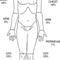
h. Seriousness of injury dependent on:
(1) Surface area
(2) Degree of burn: percent calculated on the amount of second- and third-degree burns only
(3) Presence of systemic problems
(4) Prehospital treatment
i. Inhalation reaction from radiant event
(1) Damage to respiratory vasculature can occur.
(2) Low inhaled oxygen and increased inhaled carbon dioxide can cause hypoxia.
(3) Carboxyhemoglobin levels
(4) Smoke inhalation causes:
(a) Edema of small airways
(b) Atelectasis
14. Predicable injuries
a. Can be based on specific mechanism of injury
b. All injuries cannot be predicted based on mechanism of injury.
C. Scoring systems
1. Numerous scoring mechanisms can be used.
a. Anatomic
(1) Abbreviated Injury Score (AIS)
(2) Injury Severity Score (ISS)
(3) New Injury Severity Score (NISS)
b. Physiological
(1) Glasgow Coma Scale (GCS)
(2) Revised Trauma Score (RTS)
(3) Trauma and Injury Severity Score (TRISS)
c. Injuries assigned to body parts
(1) General
(2) Head and neck
(3) Chest
(4) Abdomen
(5) Extremities and pelvis
2. Assists in determining severity of injuries
3. Assists in determining likelihood of outcome
4. Accuracy limitations can occur despite score used.
IV. STABILIZATION PHASE
A. Initial assessment, resuscitation, and stabilization
1. Initiated in emergency department (ED) or trauma center
2. Extend into operating room (OR)
3. Continue into PACU
4. Will further continue in the critical area or the surgical floor
5. Can extend even beyond discharge
a. Significance of discharge instructions cannot be overstressed.
B. Hypovolemia in trauma patient
1. Most common cause of shock
a. Result of acute blood loss
b. Result of fluid redistribution
2. Fluid resuscitation necessary
a. Prompt fluid replacement
(1) Assists in tissue perfusion
(2) Assists in delivery of oxygen to the tissues
(3) Often requires use of rapid-volume fluid infuser
(a) Can deliver IV fluids at rate of 500 to 700 mL/min
(4) Beneficial to give warm IV fluids
(a) Prevents hypothermia
b. Fluid selection
(1) Crystalloids
(a) Electrolyte solution
(i) Lactated Ringer’s (LR) closely resembles electrolyte composition of blood serum.
(ii) LR may decrease bleeding when compared with normal saline.
(b) Diffuses through capillary endothelium
(c) Distributed throughout extracellular compartment
(i) Only one fourth stays in vascular space.
(d) Common selections are LR and normal saline.
(e) Recommended first line for replacement
(f) Administration should be three to four times blood loss.
(g) Use LR in caution with suspected liver injury.
(h) Cheaper than colloids
(2) Colloids
(a) Contain protein or starch molecules
(b) Molecules remain in intravascular space.
(c) Increase osmotic pressure gradient within vascular compartment
(d) Administration is volume per volume.
(e) Half-life longer than crystalloids
(f) Common selections are:
(i) Plasma protein fraction
(ii) Dextran
(iii) Albumin
(iv) Hetastarch
(3) Hypertonic solutions
(a) Controversial in treatment
(b) Resuscitative in shock
(i) Volume expander (from extracellular source)
(c) Common selections are hypertonic or isotonic saline.
(d) Pulls fluids from extracellular space to support blood pressure (BP)
(e) Can be helpful in head injury patients
(i) Decreases brain edema
(ii) Decreases intracranial pressure
(iii) Increases cerebral pulse pressure
(f) May protect gut and inhibit acute lung injury
(g) Cautions
(i) Renal insufficiency
(ii) Hypernatremia
(4) Blood products
(a) Only blood can replace blood.
(b) Restores capacity to carry oxygen
(c) Given after fluid administration
(d) Packed red blood cells most common
(e) Universal donor is O negative.
(f) O positive can be given to nonchildbearing females and males.
(g) Type-specific blood preferred when waiting is an option
(h) Platelets may be indicated if coagulopathy suspected.
(i) Massive transfusion
(i) More than 10 to 50 units in first 12 to 24 hours
(5) Hemoglobin-based oxygen carriers/blood substitutes
(a) Modified hemoglobin molecule able to carry oxygen to tissue
(b) Longer shelf life
(c) Absence of ABO antigens
(d) No incompatible reactions
(e) Currently only available through research protocols
c. Delayed fluid administration (permissive hypotension)
(1) Can be useful in hemorrhagic patients
(2) Fluids delayed until the start of surgery
(3) Early fluid administration may delay transport.
(4) Restoration of volume can have adverse complications.
(a) Hemodilutes/disrupts body’s hemostatic mechanisms and clot formation
(5) Exacerbation of blood loss can occur from increased BP.
(a) Radial pulse is guideline.
(6) Controversial among trauma surgeons
d. Volume replacement guidelines
(1) Hemorrhage
(a) 3:1 Rule
(i) Administer 3 mL electrolyte solution to 1 mL blood loss.
(2) Burns
(a) Parkland burn formula employs LR alone for first 24 hours.
(i) Adults: LR 2 to 4 mL × Body Weight (kg) × Percent of Burn
(ii) Pediatrics: LR 3 to 4 mL × Body Weight (kg) × Percent of Burn
(iii) Add maintenance fluids with 5% dextrose in water (D 5W) to prevent hypoglycemia and to maintain adequate urine output of 1 mL/kg/hr.
(3) Combination patients (burns and hemorrhaging)
(a) Receive volume calculated for burns.
(b) Also receive volume estimates for hemorrhage loss.
e. Monitoring effective resuscitation
(1) BP goal: Systolic >90 mm Hg
(2) Hourly urine output
(a) Adults: 0.5 mL/kg per hour (30-50 mL/hr)
(b) Peds: 1 mL/kg per hour
(3) Lactate levels
(4) Base excess
3. Other causes of shock in trauma patient
a. Cardiogenic shock can result from:
(1) Cardiac tamponade
(2) Tension pneumothorax
b. Neurogenic shock
(1) Related to spinal cord injury
(2) Spinal anesthesia
c. Septic shock
(1) Usually late
(2) Caused by infectious process
V. DIAGNOSTIC STUDIES AND PROTOCOLS
A. Diagnostic tests
1. Vital role in establishing injury
2. Necessary for accurate diagnosis
3. Assists in planning effective treatment
4. X-rays
a. Lateral cervical spine
b. Upright chest anteroposterior (CXR)
(1) Repeat if initial CXR done flat (on backboard).
c. Anteroposterior pelvis
d. Any extremity with questionable injury
e. Thoracic and lumbar spine
f. Any other identified injured area
g. Soft tissue films can be helpful if impaled object suspected.
5. Computed tomography (CT) scan
a. Head (without contrast)
b. Chest
c. Abdomen
d. Pelvis
6. Ultrasound (FAST exam)
a. FAST
(1) Focused
(2) Assessment
(3) Sonography
(4) Trauma
b. Rapid, accurate, and inexpensive
c. Blunt trauma
d. Reveals presence of hemoperitoneum
e. To be positive, 200 to 500 mL fluid must be present.
f. Four areas to evaluate
(1) Hepatorenal fossa
(2) Splenorenal fossa
(3) Pericardial sac
(4) Pelvis
g. Cannot diagnose hollow visceral and retroperitoneal injuries or injuries not associated with hemoperitoneum
7. Twelve-lead electrocardiogram
a. Useful with chest injury
b. May be needed if after chest pain, additional trauma occured
8. Diagnostic peritoneal lavage
a. Controversial
b. Used only with suspected abdominal injury and severe hypotension
c. Abdominal CT scan more useful with specific injury information
d. Accuracy rate for presence of blood: 98%
e. Not useful for retroperitoneal blood
f. Before performing lavage, must place nasogastric (NG)/orogastric tube and Foley catheter to decompress bladder and stomach so that inadvertent puncture avoided
9. Arteriogram: perform if vascular injury suspected
B. Laboratory studies
1. Vital role in establishing current status
2. Common laboratory studies
a. Arterial blood gases
b. Electrolytes
c. Glucose
d. Lactate level
e. Renal function studies
f. Liver function studies
g. Coagulation studies
h. Complete blood cell count
i. Type and crossmatch
j. Urinalysis
k. Pregnancy test (childbearing females)
l. Alcohol and drug testing are controversial.
3. Assist in planning effective treatment.
VI. COLLABORATIVE APPROACH
A. Essentials
1. Begins with prehospital personnel
a. Witnesses at scene
b. First responder
c. Paramedic
d. Police
e. Fire department
f. Air ambulance personnel if activated
2. ED or trauma center
a. Emergency physician
b. Registered trained trauma nurse
c. Emergency technician and paramedic
d. Respiratory therapist
e. X-ray personnel
f. Trauma surgeon
g. Anesthesiologist
h. OR personnel
i. Chaplain
3. OR
a. OR nurse
b. Scrub technicians
c. Anesthesia provider
4. After the OR
a. PACU
b. Critical care
c. Surgical floor
d. Discharge nurse
B. Communication
1. Vital communication initiated prehospital
2. Continues throughout hospital stay
3. Comprehensive in approach
a. Physician to physician
b. ED nurse to OR nurse
c. OR nurse to PACU nurse
d. PACU nurse to floor nurse
e. Physician to family
f. Nurse to family
4. Systematic reports
a. Situation
b. Background
(1) Mechanism of injury
(2) Past medical history
c. Assessment
(1) Airway, breathing, circulation
(2) Vital signs
(3) Include diagnostic findings
(4) Treatments
(5) Suspected injuries
(6) Abnormal assessment
(7) Vital nursing information
(8) Fluids and/or blood products
d. Recommendations
(1) Pending orders
VII. POSTANESTHESIA CARE
A. Anesthesia report
1. Valuable information
a. Presenting status
(1) Name
(2) Age
(3) Surgeon
(4) Anesthesiologist
(5) Level of consciousness preoperatively
b. Significant facts pertaining to mechanism of injury
c. Prehospital phase
d. ED course
(1) Cervical spine (C spine) clearance documentation
(a) MUST be done by ED physician, trauma surgeon, or neurosurgeon
e. Operative procedure
(1) Single procedure
(2) More than one procedure
(3) More than one surgeon
f. Intubation
(1) Routine intubation
(2) Difficult intubation
(3) Rapid sequence intubation
(4) Full stomach
(5) Airway stability
(6) Intubation time and tolerance
g. Anesthetic agents
(1) Rapid sequence intubation
(2) Inhalation agents
(3) IV agents
(4) Balanced anesthesia
(5) Narcotic usage (with time of last dose)
(6) Muscle relaxants (with time of last dose)
(7) Reversal agents (with time of last dose)
(8) Antibiotics
h. Estimated blood loss
(1) Prehospital
(2) ED
(3) OR
i. Fluid resuscitation
(1) Prehospital
(2) ED
(3) OR
(4) Crystalloids
(5) Blood products
(6) Chest drains
(7) Cell saver usage
(8) Ortho refuser (orthopedic refuser system)
(9) Other drains
j. Cardiopulmonary status
(1) Vital signs
(2) Pulse oximetry
(3) Vasopressors
(4) Antidysrhythmics
(5) Arterial line
(6) Thermodilation catheter
(7) Urine output (to ensure end-organ perfusion)
k. Other identified injuries
(1) Surgical interventions
(2) Nonsurgical interventions
l. Treatment abnormalities
(1) Hypothermia
(2) Abnormal laboratory values
(3) Abnormal x-ray results
(4) Volume replacement
(5) Tetanus status
m. Treatment plans
B. Nursing assessment, primary survey
1. Airway
a. Patency
b. Proper head position
c. Ensure cervical spine protection until cleared.
(1) Do not remove cervical collar if not cleared.
d. Suctioning as needed for removal of secretions
e. Airway management as indicated
(1) Nasopharyngeal
(2) Oropharyngeal
(3) Oral or nasal endotracheal tube (ETT)
f. Continual reassessment
2. Breathing
a. Consider mechanism of injury.
(1) Blunt
(2) Penetrating
(3) Acceleration
(4) Deceleration
(5) Acceleration and deceleration
b. Location of injury
(1) Chest injury may indicate pulmonary injury.
(2) Rib fractures
(3) Pulmonary contusion
(4) Pneumothorax
(5) Tension pneumothorax
(6) Neurological event affecting respiratory status
c. Spontaneous respirations
(1) Absence of respiratory effort
(a) NOT a result of thoracic trauma
(b) Result of head trauma or drugs
(c) Respiratory rate and pattern is the most sensitive vital sign in the patient with a neurologic deficit.
d. Chest wall movement
(1) Chest wall can move without air going in or out.
e. Respiratory accessory muscle use
f. Work of breathing
g. Palpation
(1) Subcutaneous emphysema
(2) Trachea position
h. Auscultation
(1) Bilateral breath sounds
(2) Adventitious breath sounds
i. Pulse oximetry continuous
j. End-tidal carbon dioxide
k. Arterial blood gases
l. Carboxyhemoglobin level (if indicated)
m. Oxygen delivery
(1) 100% mask for spontaneous respiratory efforts
(2) Bag valve mask with assisted respiration or mechanical ventilation
3. Circulation
a. Pulses
(1) Carotid: ≥60 beats/min
(2) Radial: ≥80 beats/min
(3) Femoral: ≥70 beats/min
(4) Popliteal and dorsalis pedis pulses
(5) Bilateral
(6) Quality
(7) Rate
(8) Upper extremities to lower extremities
(9) Pulseless electrical activity (PEA) can occur in trauma patient.
(a) Tension pneumothorax
(b) Cardiac tamponade
(c) Hypovolemia
(d) Hypothermia
(e) Hypoxia
(f) Hypoglycemia
(g) Head injury
(h) Acidosis
(i) Hypokalemia
(j) Hyperkalemia
(k) Thrombosis
(i) Pulmonary
(ii) Coronary
(10) Start cardiopulmonary resuscitation if heart rate (HR) <60 beats/min in a child.
b. Cardiac monitor
(1) Rate
(2) Rhythm
(3) Dysrhythmia presence
(4) Continual cardiac monitor observance
c. BP
(1) Hypertension
(2) Hypotension
(3) Normal BP range for patient
(4) Vasopressors
(5) Monitor every 10 minutes if stable.
(6) Monitor every minute if unstable.
(7) Noninvasive cuff
(8) Arterial line
(9) Capillary refill may be more beneficial in pediatric patient.
d. Vascular access
(1) Number of sites
(2) Location of sites
(3) Type of fluids presently hanging
(4) Intake
e. Dressings
(1) Surgical dressings for drainage
(2) Drainage from nonsurgical wounds
(3) Surgical drain placement and volume of drainage
(4) Bloody drainage versus nonbloody drainage
(5) Total output
f. Urine output
(1) Ensures end-organ perfusion
(2) Essential for input and output (I&O) balance
C. Nursing assessment, secondary survey
1. General information
a. Done only after primary assessment
b. High degree of suspicion concerning mechanism of injury
c. Note any injuries not previously addressed.
(1) Swelling and bruising can take time to develop.
d. Life-threatening injuries may limit time for secondary survey in ED.
(1) Immediate surgery may be needed for life-threatening injuries.
2. Head-to-toe assessment
a. Neurological evaluation
(1) Level of consciousness (AVPU)
(a) Awake
(b) Verbal
(c) Pain response
(d) Unresponsive
(2) Appropriate verbal response
(3) Pupil reactivity and symmetry
(4) Following commands
(5) Movement of all four extremities
(6) Compare results with preoperative assessment.
(7) May need head CT if taken to OR immediately on arrival
b. Examination of head and face
(1) Abrasions
(2) Lacerations
(3) Puncture wounds
(4) Ecchymosis: raccoon eyes (periorbital bruising)
(5) Edema: facial edema can result in potential airway compromise.
(6) Gross vision exam
(7) Check for presence of contact lens.
c. Ears
(1) Ecchymosis: Battle’s sign (bruising behind ear)
(2) Drainage from nose
(a) Bloody
(b) Clear fluid (potential for cerebrospinal fluid [CSF] leak)
(i) Check for presence of glucose.
(3) Never pack or suction nose if CSF leak suspected.
(4) If CSF leak suspected and/or Battle’s sign, never insert NG tube; consider orogastric tube if needed.
d. Evaluate neck.
(1) Edema
(2) Ecchymosis
(3) Tracheal deviation
(4) Pulsating or distended neck veins
(5) Subcutaneous emphysema
e. Chest assessment
(1) Anterior, lateral, and axilla examination
(2) Lacerations
(3) Abrasions
(4) Contusions
(a) Seat belt bruising can be seen on the chest.
(5) Puncture wounds
(6) Edema
(7) Subcutaneous emphysema
(8) Chest wall symmetry
(9) Depth of respirations
(10) Reevaluation of breathing
(11) Auscultation of breath and heart sounds
(a) Adventitious sounds
(b) Murmurs
(c) Bruits
(d) Muffled heart sounds
(12) Chest pain that may indicate:
(a) Pulmonary contusion
(b) Rib fractures
(c) Cardiac contusion
f. Abdomen, pelvis, and genitalia evaluation
(1) Abrasions
(2) Contusions
(3) Edema
(4) Ecchymosis
(a) Seat belt bruising often seen across abdomen
(5) Tenderness
(6) Presence of bowel sounds
(7) Abdominal girth
(8) Pelvis examined for stability over crests and pubis
(9) Presence of priapism could indicate spinal cord injury.
(10) Urinary catheter
(a) Should be in place for all multiple trauma patients
(b) Examine urine.
(c) I&O documented
(d) Ensure adequate output to prevent rhabdomyolysis.
(11) Rectal exam
(a) Presence of blood
(b) Rectal tone
(i) Lack of rectal tone suggestive of spinal injury
(c) Trauma surgeon or ED physician
g. Extremity assessment
(1) Circulatory
(2) Sensory
(3) Motor function
(4) Range of motion
(5) Edema
(6) Ecchymosis
(7) Lacerations
(8) Abrasions
(9) Reexamine if intervention activated.
h. Back evaluation
(1) Log roll with cervical spine support.
(2) Inspect and palpate back, flanks, and buttocks.
(3) Lacerations
(4) Abrasions
(5) Ecchymosis
(6) Edema
(7) Pain
(8) Rectal exam
i. ED intervention assessment
(1) Open wounds need tetanus booster.
(2) Antibiotics often started preoperatively
(3) Pain medication
(4) Antianxiety medication
(5) Nausea medication
(6) Wound cleansing and dressings
D. Nursing interventions
1. Pain control (trauma considerations)
a. Note preoperative medications given in ED.
(1) Intramuscular medications can have long half-life.
b. Pain may be inclusive of more than the surgical site.
(1) Musculoskeletal injuries
(2) Sutured lacerations
(3) Extremity fractures
(4) Rib fractures
(5) Contusions and bruising
c. Pain evaluation (see Chapter 26)
(1) Subjective
(2) Verbal
(3) Nonverbal
(4) Hemodynamic changes (increased HR and BP)
(5) Splinting
(6) Nausea
(7) Crying
(8) Guarding
(9) Use standard pain scale to assess.
d. Pain management
(1) IV injection
(2) Patient-controlled analgesia
(3) Epidural catheter
(4) Major plexus block
(5) Music therapy
(6) Guided imagery
(7) Relaxation techniques
(8) Visitors
e. Pain goals
(1) Minimize cardiovascular depression.
(2) Minimize intracranial hypertension.
(3) Substance abusers may require higher pain doses (includes smokers).
(4) Pain management is vital to optimal care.
2. Nausea management (trauma considerations)
a. Can be of great concern for the trauma patient
b. Seldom are trauma patients prepared with nothing by mouth (NPO).
c. Consider trauma patient having anesthesia as having potentially full stomach
d. Vomiting can lead to aspiration.
e. Extubation may be delayed until gag reflex returns.
f. NG tube does not always function if food particles are large.
g. Nausea has higher incidence in trauma patient.
h. Nausea can be indication of increased intracranial pressure.
3. Psychological management
a. Emotional consideration increases postoperatively.
b. Life-threatening interventions have been implemented.
c. May be challenging postoperatively
(1) Initial shock may have worn off.
(2) Questions concerning others involved
(3) Fear of consequences
(4) Life-changing injuries
(5) Pre-event drug or alcohol use can complicate.
d. Emergence from anesthesia
(1) Orient to place and time.
(2) Brief explanation of the event
(3) Acknowledges fears
(a) Death
(b) Mutilation
(c) Change in body image
(d) Loss of control
(e) Loss of family members and friends
(4) Same information may need to be repeated several times.
(5) Be honest with information.
(6) Encourage appropriate coping skills.
(7) Psychological concepts of trauma
(a) Need for information
(b) Need for compassion
(c) Need for hope
4. Infection risks (related to trauma care)
a. May have limited past medical history
b. Large amount of unknown information may exist.
c. High risk for:
(1) Tetanus
(2) Wound infection
(3) Sepsis
(4) Communicable diseases
(5) Human immunodeficiency virus
(6) Hepatitis
(7) Sexually transmitted diseases
(8) Chicken pox
d. Universal precautions should be observed.
e. Hand washing is the best defense.
5. Nursing diagnosis: potential for:
a. Ineffective airway clearance
b. Ineffective gas exchange
c. Alterations in cardiac output
d. Alteration in tissue perfusion
e. Fluid volume deficit
f. Hypothermia
g. Risk of injury
h. Altered comfort
i. Altered thought process
j. Altered communication
k. Anxiety
l. Ineffective coping
m. Disturbance in self-concept
n. Posttraumatic stress
o. Surgical interventions and injuries dependent on circumstances
6. Nursing care
a. Vigilant continuous reassessment
b. Treatment priorities
c. Recognition of complex pathophysiological responses
d. Anticipation of subtle or overt signs of shock
e. Prevention of complications
(1) Acute respiratory distress syndrome
(2) Sepsis
(3) Acute renal failure
(4) Hypoxic liver
(5) Multisystem organ failure
VIII. SHOCK IN THE MULTITRAUMA PATIENT
A. Shock as a complication
1. Most common complication associated with traumatic injury
2. Different types of shock all exhibit problems with:
a. Delivery of oxygen to the cell
b. Delivery of nutrients to the cell
c. Inadequate tissue perfusion (cellular hypoxia)
d. Increased lactic acid level (caused by oxygen debt)
(1) Degree of rise correlates with severity and prognosis.
e. Functional impairment of cells
f. Functional impairment of organs
g. Functional impairment of body systems
h. Heart, brain, liver, kidneys, and lungs require increased oxygen.
i. Ischemia initiates complex events.
(1) Energy-dependent functions cease.
(2) Protein synthesis depleted
(3) Loss of intracellular potassium
(4) Production of lactic acid
(5) Death of vital tissue
3. Clinical manifestations of shock
a. Cool, clammy skin
b. Cyanosis
c. Restlessness
d. Altered level of consciousness
e. Altered skin temperature
f. Tachycardia
g. Dysrhythmias
h. Tachypnea
i. Pulmonary edema
j. Decreased urine output
k. Decreased end-organ perfusion
l. Increased platelet, leukocyte, and erythrocyte counts
m. Sludging of the blood
n. Metabolic acidosis
B. Types of shock
1. Hypovolemic
a. Most common type
b. Results from acute hemorrhagic loss
c. Volume shift in burn patients
2. Cardiogenic
a. Rare in trauma patients
b. Inadequate contractility of cardiac muscle
c. May be secondary to:
(1) Blunt cardiac injury
(2) Myocardial infarction (MI)
3. Distributive
a. Includes:
(1) Neurogenic
(2) Anaphylactic
(3) Septic
b. Abnormality in vascular system and maldistribution of blood volume
(1) Loss of vasomotor tone regulated by sympathetic nervous system
c. Neurogenic most common in trauma patient from spinal cord injury
4. Obstructive
a. Compression of great vessels or heart from:
(1) Tension pneumothorax
(2) Cardiac tamponade
C. Hypovolemic shock
1. Definition
a. Decrease in intravascular volume
b. Decrease in filling the intravascular compartment
2. Causes
a. Internal hemorrhage
b. External hemorrhage
c. Plasma volume loss
d. Third spacing of fluids
e. Decreased venous return
3. Classification of hemorrhage
a. Characteristic clinical manifestations according to approximate loss
b. American College of Surgeons’ Advanced Trauma Life Support Course
c. Class I
(1) Early phase
(2) Loss of as much as 750 mL
(3) Approximately 1% to 15% total blood volume loss
(4) Minimal physiological changes in:
(a) HR (<100 beats/min)
(b) BP (normal)
(c) Capillary refill (normal)
(d) Respiratory rate (14-20 breaths/min)
(e) Urine output (>30 mL/hr)
(5) Mild anxiety in response to sympathetic nervous system
(6) Treatment
(a) Rapid infusion of 1 or 2 L of balanced salt solution
(b) Maintain renal output of more than 0.5 mL/kg per hour.
d. Class II
(1) Moderate phase
(2) Loss of 750 to 1500 mL blood
(3) Approximately 15% to 30% total blood volume loss
(4) Multiple incremental physiological changes
(a) Increased anxiety
(b) Restlessness
(c) Catecholamine release
(d) HR >100 beats/min
(e) Minimal BP changes
(i) Rise in diastolic BP
(ii) Decreasing pulse pressure
(f) Slight capillary refill delay
(g) Cool, pale skin
(h) Slight depression in urine output
(5) Treatment
(a) Rapid infusion of 1 or 2 L of balanced salt solution
(b) Maintain renal output of more than 0.5 mL/kg per hour.
e. Class III
(1) Progressive phase
(2) Loss of 1500 to 2000 mL blood
(3) Approximately 30% to 40% total blood volume loss
(4) Obvious physiological changes
(a) Cerebral hypoperfusion—decreased level of consciousness
(b) Confusion
(c) Agitation
(d) Anxiety
(e) HR >120 beats/min
(f) Hypotension
(g) Capillary refill >4 seconds
(h) Deep, rapid respirations
(i) Metabolic acidosis
(j) Decreased urine output (approximately 5-15 mL/hr)
(5) Treatment
(a) Fluid administration
(b) Consider blood transfusion.
f. Class IV
(1) Hemorrhage
(2) More than 2000 mL blood loss
(3) Approximately 40% total blood volume loss
(4) Profound impact
(a) Lethargic
(b) Stuporous
(c) Unresponsive
(d) HR ≥140 beats/min
(e) Peripheral pulses weak and difficult to palpate
(f) Capillary refill >10 seconds
(g) Severe hypotension, BP difficult to obtain
(h) Cold, clammy, diaphoretic, or cyanotic skin
(i) Shallow, irregular respirations with respiratory rate >35 breaths/min
(j) No renal end-organ perfusion, resulting in anuria
(5) Treatment
(a) Fluid administration
(b) Blood administration
g. The Golden Hour
(1) Treatment within the first hour associated with lower mortality
(2) Filling the vascular tank allows:
(a) Adequate cardiac output
(b) Perfusion of tissues
D. Cardiogenic shock
1. Definition
a. Inadequate cardiac output
b. Circulatory failure
c. Impaired contractility
d. Shock secondary to acute myocardial dysfunction
(1) Systolic BP less than 80 mm Hg (<30% baseline)
(2) Cardiac index less than 2.1 L/min
e. Mortality rate: 80% to 100%
2. Causes in trauma
a. Secondary to blunt injury to heart muscle
b. Occasionally MI occurs preceding trauma event.
c. History of heart disease in association with trauma and/or anesthesia can increase likelihood of intraoperative MI.
d. Rapid fluid administration and ensuing cardiac failure
e. Disruption in normal conduction sequence (heart block, dysrhythmias)
3. Classifications of cardiogenic shock
a. Coronary
(1) Obstructive coronary artery disease interrupting blood flow
(2) Interruption of blood flow causing ischemic heart muscle
(3) Ischemic heart muscle results in decreased contraction.
(4) Decreased contraction results in inadequate cardiac output.
(5) Incidence rises with compromise of 40% left ventricular function.
(6) Increased left atrial pressure
(7) Increased pulmonary venous pressure
(8) Increased pulmonary capillary pressure
(9) Pulmonary edema
b. Noncoronary
(1) Absence of coronary artery disease
(2) Cardiac muscle damage
(a) Cardiomyopathy
(b) Valvular heart abnormalities
(c) Cardiac dysrhythmias
4. Clinical indicators
a. Systolic BP less than 80 mm Hg (<30% baseline)
b. Cardiac index less than 2.1 L/min
c. Urine output less than 20 mL/hr
d. Diminished cerebral perfusion evidenced by confusion
e. Cold, clammy, cyanotic skin
f. Classic signs and symptoms
(1) May not be seen in the hypovolemic trauma patient
(2) Pulmonary edema
(3) Jugular vein distention
(4) Hepatic congestion
g. Decreased cardiac, stroke, and left ventricular stroke work index
h. Increased pulmonary capillary wedge pressure
i. Increased pulmonary artery pressure
j. Increased systemic vascular resistance
k. Decreased systemic venous oxygen saturation
l. Decreased cardiac output
m. Respiratory and metabolic acidosis
5. Treatment
a. Early recognition
b. Improvement of myocardial oxygen supply
c. Improvement of tissue perfusion
d. Airway management, ventilation, and oxygenation
e. Correct acidosis
f. Pain relief
g. Pharmacological support to improve or correct rhythm
h. Increasing cardiac output by increasing intravascular volume
i. Vasoactive medications
(1) Dobutamine
(2) Dopamine
(3) Epinephrine
j. Decrease afterload (systemic vascular resistance)
k. When pharmacological support fails
(1) Intra-aortic balloon pump
(2) Ventricular assist device
E. Distributive shock
1. Definition
a. Also called vasogenic shock
b. Abnormal placement of the vascular volume
c. Heart pump and blood volume are normal.
d. Alteration exists within the vascular circulatory network.
e. Three types
(1) Neurogenic
(2) Anaphylactic
(3) Septic
2. Neurogenic shock
a. Definition
(1) Tremendous increase in vascular capacity
(2) Normal amount of blood incapable of adequately filling vasculature
(3) Loss of sympathetic vasomotor tone causes massive vasodilation.
(4) Venous pooling and decreased return to right side of the heart
(5) Frequently transitory
(6) Not common in occurrence except in spinal cord–injured patients
b. Causes
(1) Deep general or spinal anesthesia
(2) Loss of sympathetic vasomotor tone in the trauma patient
(a) Brain concussion or contusion of basal regions
(b) Spinal cord injury above level of T6
c. Clinical symptoms
(1) Decreased peripheral vascular resistance
(2) Decreased stroke volume
(3) Decreased cardiac output
(4) Hypotension
(5) Decreased tissue perfusion
(6) Differs from hypovolemic shock
(a) Bradycardia
(b) Warm, dry, flushed skin
d. Treatment
(1) Extensive volume expansion
(2) Vasopressors
(a) Ephedrine
(b) Neosynephrine
(3) Spinal anesthesia as causative event
(a) Head of bed flat
(b) Supine position
(c) Elevate legs if possible.
3. Anaphylactic shock
a. Definition
(1) Severe antigen-antibody reaction
(2) Relates to inflammatory process
(3) Activation of complement and arachidonic cascade
(a) Immunoglobulin E produced and binds to mast cells and basophils
(b) Mast cells trigger vasoactive contents.
(c) Histamine and vasoactive mediators released by mast cells
(d) Vasoactive mediators cause massive vasodilation.
(e) Vasoactive mediators cause increased capillary permeability.
(4) Rarely occurs in the trauma patient but should be of concern if no past medical history
b. Causes
(1) Antigen-antibody reaction
(2) Can occur with exposure to any allergen; in the trauma patient consider:
(a) Antibiotics
(b) Contrast medium
(c) Blood transfusions
c. Clinical symptoms
(1) Vary with severity
(2) Conjunctivitis
(3) Angioedema
(4) Hypotension
(5) Laryngeal edema
(6) Urticaria
(7) Bronchoconstriction
(8) Dysrhythmias
(9) Cardiac arrest
(10) One or any combinations of the preceding signs can occur.
(11) Repeat exposures can increase symptoms.
d. Treatment
(1) Removal of causative agent
(2) Discontinue blood transfusion.
(3) Oxygen
(4) Epinephrine
(a) Bronchodilator
(b) Helps restore vascular tone
(c) Increases arterial BP
(5) Aminophylline, if wheezing
(6) Diphenhydramine (Benadryl), antihistamine
(7) Steroids: decrease inflammatory process
(8) Gastric acid blocker
(a) Famotidine (Pepcid)
(b) Cimetidine (Tagamet)
(c) Nizatidine (Axid)
(d) Ranitidine (Zantac)
4. Septic shock
a. Definition
(1) Acute systemic response to invading blood-borne microorganisms
(2) Clinical syndrome on a continuum
(3) Begins with sepsis and ends with multisystem organ failure
(4) Complex cellular disease
(5) Loss of autoregulation and tissue dysfunction despite increased cardiac output
(6) Activation of kinins, complement, arachidonic, and coagulation cascades
(7) Hemodynamic instability
(a) Initial phase
(i) High cardiac output
(ii) Low systemic vascular resistance
(b) Later phase
(i) Low cardiac output
(ii) Extremely high systemic vascular resistance
(8) Myocardial depression related to severity of sepsis
(9) Release of vasoactive chemical mediators and endotoxins
(10) Decreased ventricular preload
(11) Increased capillary permeability
(a) Augments myocardial depression
(b) Produces decreased vascular volume
(12) Endotoxin stimulates complement split products.
(13) Neutrophil and platelet aggregation to the lungs
(14) Fluid collects within the pulmonary interstitium.
(15) Pulmonary compliance decreased
(16) Acute respiratory distress syndrome ensues.
(17) Profound alteration in metabolism
(18) Increased oxygen debt
(19) Rising blood lactate levels
(20) Trauma patient is predisposed because of:
(a) Contaminated wounds
(b) Poor nutritional status
(c) Preexisting disease states
(d) Altered integrity of body’s defense mechanism
(21) Principal cause of death in trauma patient surviving first 3 days
b. Causes
(1) Gram-positive microorganisms less common
(2) Gram-negative microorganisms most common
(3) Viruses
(4) Fungi
(5) Parasites
c. Clinical symptoms
(1) Warm, flushed, and dry skin
(2) Rapid respiratory rate
(3) Confusion
(4) Increased HR
(5) Hypotension
(6) High cardiac output
(7) Low systemic vascular resistance
(8) Late clinical signs: skin changes to cold and clammy.
d. Treatment
(1) Identification and elimination of infection
(2) Cultures
(3) Proper definitive antimicrobial therapy
(4) Hemodynamic monitoring
(5) Oxygenation and ventilation support
(6) Fluid administration
(7) Pharmacological support
(a) Positive inotropes
(b) Vasopressors
5. Obstructive shock
a. Definition
(1) Myocardium normal
(2) Compression to the atria
(3) Obstruction in venous return
(4) Prevents atrial filling
(5) Decrease in stroke volume
b. Causes
(1) Obstructive source
(2) Pulmonary embolism
(3) Dissecting aortic aneurysm
(4) Vena cava obstruction
(5) Cardiac tamponade
(6) Tension pneumothorax
c. Clinical symptoms dependent on causative mechanism and can include:
(1) Muffled heart sounds
(2) Jugular vein distention
(3) Tracheal deviation
(4) Diminished or absent lung sounds
(5) Hypotension
(6) Pulseless electrical activity (PEA)
d. Treatment
(1) Correct the cause.
(2) Dissecting aneurysm: surgical intervention
(3) Vena cava obstruction: surgical intervention
(4) Cardiac tamponade: cardiocentesis until surgical intervention
(5) Tension pneumothorax: needle decompression, chest tube
BIBLIOGRAPHY
1. Advanced cardiac life support provider manual. ed 5 ( 2006)American Heart Association, Dallas.
2. Advanced trauma life support course. ed 7 ( 2004)Committee on Trauma of the American College of Surgeons, Chicago.
3. Alam, H.B.; Rhee, P., New developments in fluid resuscitation, Surg Clin North Am 87 (1) ( 2007) 55–72.
4. Alspach, J.; Epgang, T., Core curriculum for critical care nursing. ed 6 ( 2006)Saunders, Philadelphia.
5. Cocchi, M.N.; Kimlin, E.; Walsh, M.; et al., Identification and resuscitation of the trauma patient in shock, Emerg Med Clin North Am 25 (3) ( 2007) 623–642.
6. Course in advanced trauma nursing: A conceptual approach. ( 2006)Emergency Nurses Association, Park Ridge, IL.
7. Davidson, J.; Griffin, R.; Higgs, S., Introducing a clinical pathway in fluid management, J Perioper Pract 17 (6) ( 2007) 248–250; 255–256.
8. Drain, C.B.; Odom-Forren, J., Perianesthesia nursing: A critical care approach. ed 5 ( 2009)Saunders, St. Louis, MO.
9. Emergency nurse pediatric course. ed 3 ( 2007)Emergency Nurses Association, Park Ridge, IL.
10. Jacob, M.; Chappell, D., Saline or albumin for fluid resuscitation in traumatic brain injury, N Engl J Med 357 (25) ( 2007) 2634–2635.
11. Mizushima, Y.; Tohira, H.; Mizobata, Y.; et al., Fluid resuscitation of trauma patients: How fast is the optimal rate?Am J Emerg Med 23 (7) ( 2005) 833–837.
12. Moore, K.M., Controversies in fluid resuscitation, J Trauma Nurs 13 (4) ( 2006) 168–172.
13. Pascual, J.L.; Maloney-Wilensky, E.; Reilly, P.M.; et al., Resuscitation of hypotensive head-injured patients: Is hypertonic saline the answer?Am Surg 74 (3) ( 2008) 253–259.
14. Pepe, P.E.; Dutton, R.P.; Fowler, R.L., Preoperative resuscitation of the trauma patient, Curr Opin Anaesthesiol 21 (2) ( 2008) 216–221.
15. Schweer, L., Pediatric trauma resuscitation: Initial fluid management, J Infus Nurs 31 (2) ( 2008) 104–111.
16. Sperry, J.L.; Minei, J.P.; Frankel, H.L.; et al., Early use of vasopressors after injury: Caution before constriction, J Trauma 64 (1) ( 2008) 9–14.
17. Trauma nursing core course. ed 6 ( 2006)Emergency Nurses Association, Park Ridge, IL.
18. Yanagawa, Y.; Sakamoto, T.; Okada, Y., Hypovolemic shock evaluated by sonographic measurement of the inferior vena cava during resuscitation in trauma patients, J Trauma 63 (6) ( 2007) 1245–1248.