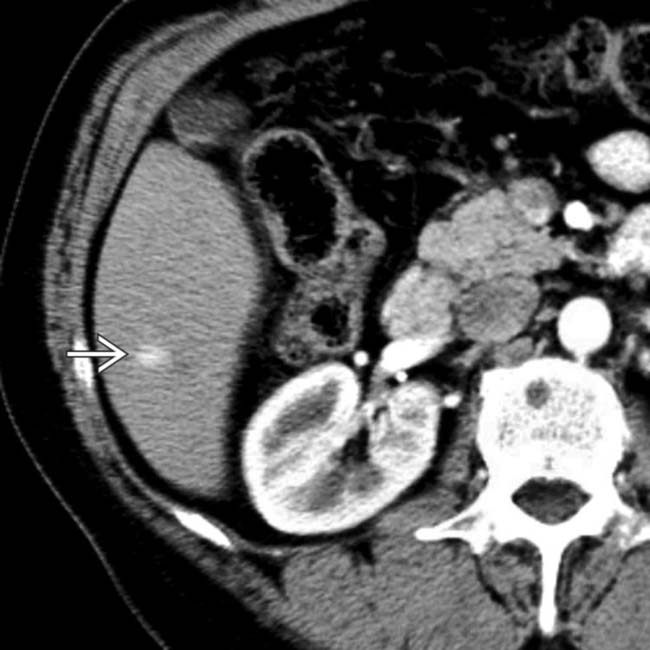
[level-membership-for-radiology-category] Wedge-shaped area of hyperattenuation with straight margins seen during arterial phase of CECT or MR


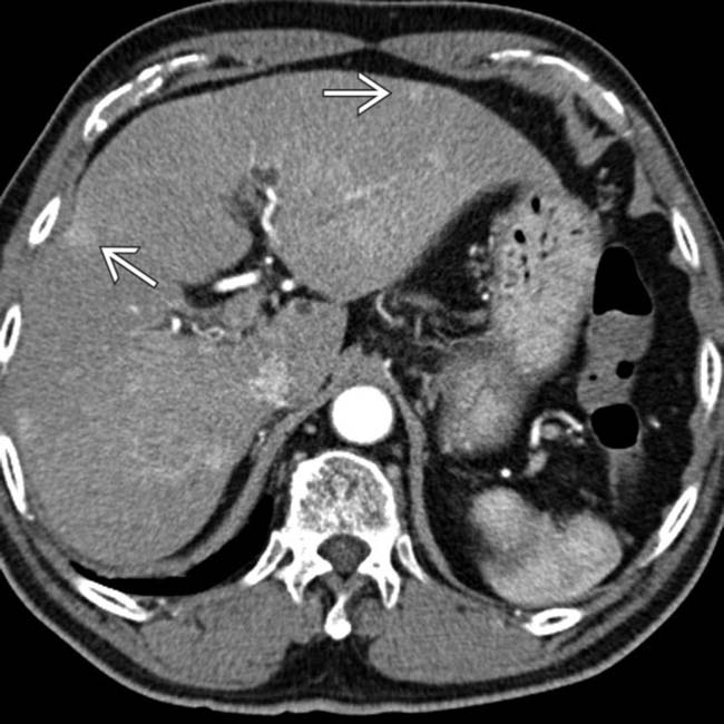
 in this 60-year-old man with cirrhosis due to chronic viral hepatitis.
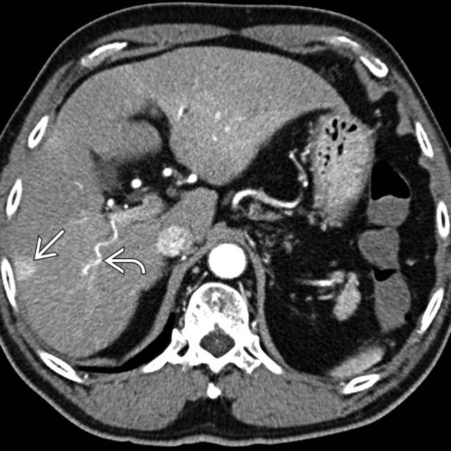
 . Also note the large, “corkscrew” hepatic arterial branch
  , a typical feature of cirrhosis. The liver has a cirrhotic morphology with wide fissures.
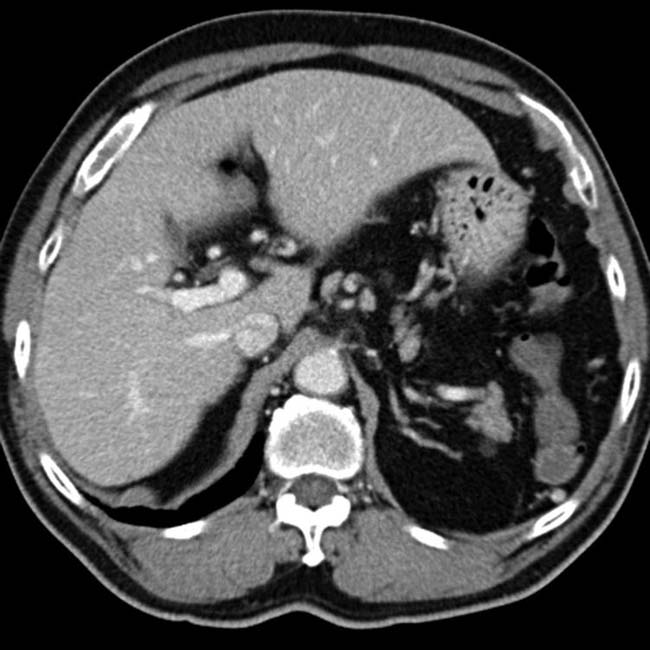


PATHOLOGY
General Features


 .
.
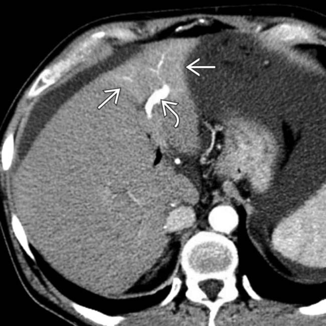
 and early enhancement of the portal vein branch that drains this segment
  , representing an arterioportal shunt, probably due to the prior liver biopsy at this site.
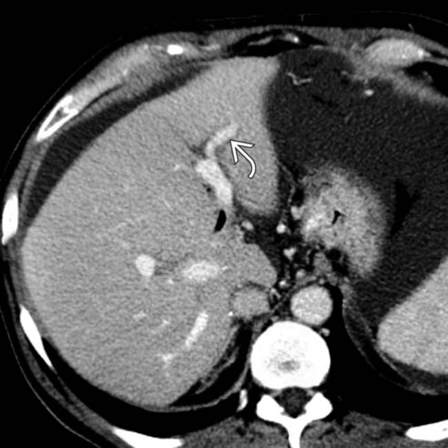
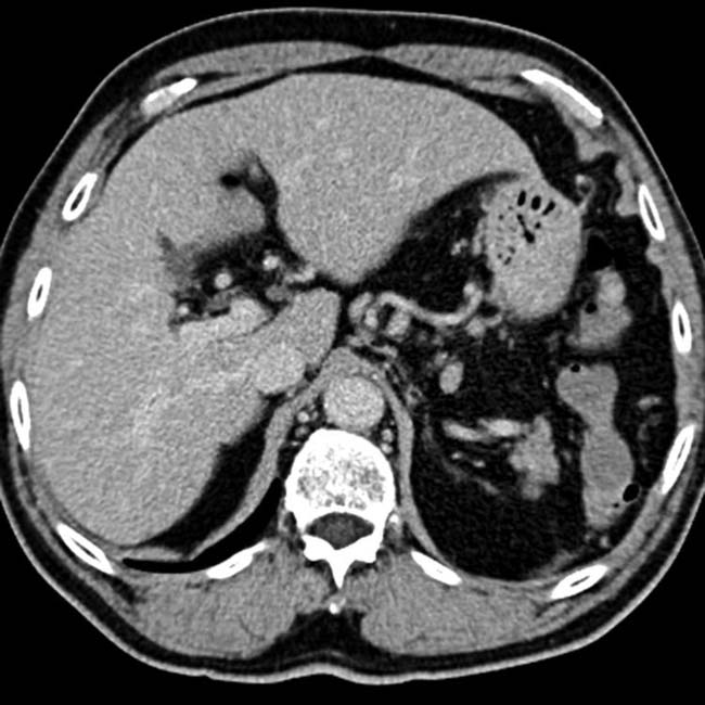
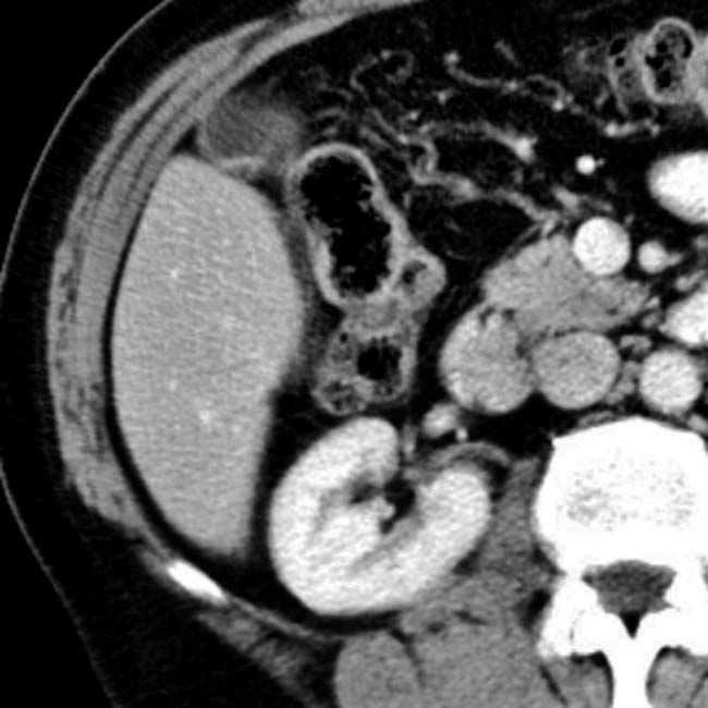
 but otherwise appears normal.
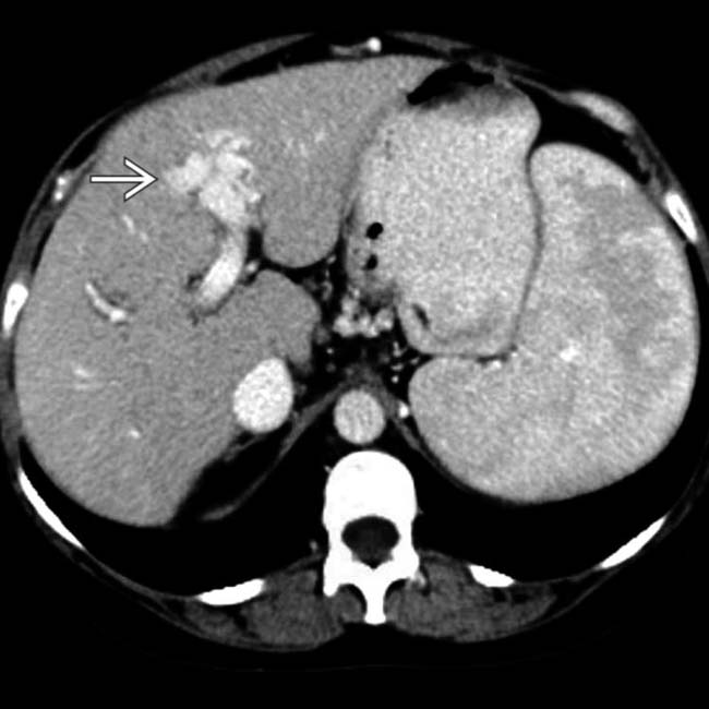
 in the left lobe, most likely the result of a prior liver biopsy at this site.
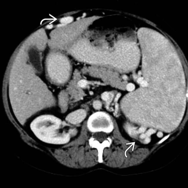
 .
.[/level-membership-for-radiology-category][not-level-membership-for-radiology-category] Wedge-shaped area of hyperattenuation with straight margins seen during arterial phase of CECT or MR
in this 60-year-old man with cirrhosis due to chronic viral hepatitis.. Also note the large, “corkscrew” hepatic arterial branch , a typical feature of cirrhosis. The liver has a cirrhotic morphology with wide fissures.IMAGING
General Features
• Best diagnostic clue
MR Findings
• Dynamic gadolinium-enhanced MR
 Arterial phase imaging (25-35 seconds after injection)
Arterial phase imaging (25-35 seconds after injection)
–
Buy Membership for Radiology Category to continue reading. Learn more here
[/not-level-membership-for-radiology-category]
