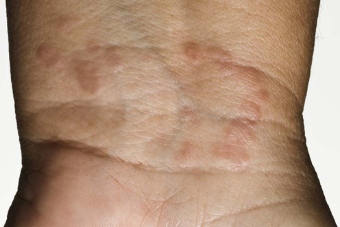
Granuloma annulare

Published on 19/03/2015 by admin

Filed under Dermatology

Last modified 19/03/2015

Print this page

rate 1 star rate 2 star rate 3 star rate 4 star rate 5 star
Your rating: none, Average: 2.2 (5 votes)

This article have been viewed 3401 times

Granuloma annulare

Hilmi F. Recica and Andrew Ilchyshyn

Evidence Levels:  A Double-blind study  B Clinical trial ≥ 20 subjects  C Clinical trial < 20 subjects  D Series ≥ 5 subjects  E Anecdotal case reports

image

Granuloma annulare (GA) is usually a benign and self limiting skin disease of unknown etiology. There are a number of variants: localized, generalized or disseminated, subcutaneous, papulo-pustular and perforating. It is characterized histologically by palisading granulomata with a central core of necrobiotic or degenerative collagen surrounded by a lympho-histiocytic infiltrate. The disease often resolves spontaneously, with no long term sequelae.

Management strategy

The localized variant of GA is often diagnosed clinically, but a skin biopsy may be required to confirm the diagnosis of the more unusual varieties such as generalized, papulo-pustular, perforating, and subcutaneous forms. Once the diagnosis is established, it is prudent to check the urine for glucose to exclude diabetes mellitus. In disseminated and generalized variants HIV, hepatitis C, and lymphomas are rare associations.

Because the majority of cases of GA are asymptomatic and self limiting, the need for active treatment should be considered carefully; in many cases, reassurance that the disease is benign and will resolve spontaneously is sufficient. Painful or unsightly lesions may justify more active treatment, though the evidence to support the efficacy of many of the treatments is poor.

Localized GA may be treated effectively with topical or intralesional steroids. Clobetasol propionate (Dermovate) lotion under occlusion with a hydrocolloid dressing, changed weekly, can be used for up to 6 weeks. Alternatively, intralesional injections of triamcinolone may be used, at intervals of 6 to 8 weeks. Cryotherapy using liquid nitrogen can be repeated at 3 or 4 weeks if required. Treatment of refractory lesions includes photodynamic therapy, UVA1, intralesional interferon; large, tumid lesions may require excision.

Generalized GA may be persistent and its unsightly appearance causes patients to seek active treatment, though the results are often disappointing. Both UVA and PUVA may be used, but relapses can occur. Isotretinoin and dapsone have been associated with improvement or clearance of generalized GA. Topical tacrolimus and pimecrolimus have been used, with greater success in generalized disease than in the localized variety. Other reported treatments for this condition include laser, fumaric acid esters, hydroxychloroquine, chlorambucil, cyclosporine, hydroxyurea, doxycycline, systemic steroids, adalimumab, infliximab, anthralin, vitamin E, allopurinol, nicotinamide, pentoxifylline, and defibrotide.

Specific investigations

Unlike necrobiosis lipoidica, in which there is a strong association with diabetes mellitus, the relationship between GA and diabetes is unclear. Nevertheless, it would seem sensible to use the diagnosis of GA as a cue to exclude undiagnosed diabetes, and at least check the urine for glucose.

Localized granuloma annulare

First-line therapies

imageIntralesional steroids B
imageCryotherapy B
imageTopical steroids E