XGC cannot be confidently distinguished from gallbladder carcinoma radiologically
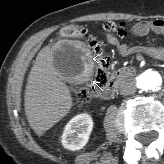
• CT: GB wall may be focally or diffusely thickened


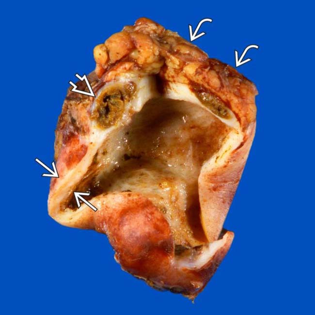
 and an intramural abscess
  . A portion of the adjacent liver
  was resected because of the high intraoperative suspicion of GB cancer. Pathology revealed xanthogranulomatous cholecystitis (XGC).
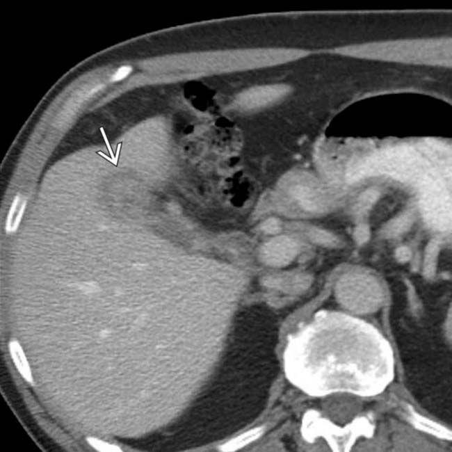
 , with an indistinct border with the liver. While the appearance was concerning for gallbladder cancer, this was found to be XGC at cholecystectomy.
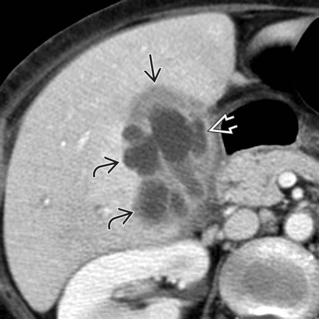
 , intramural low attenuation
  , and several pericholecystic collections
  . Low-attenuation intramural nodules in XGC are due to either abscesses or xanthogranulomas.
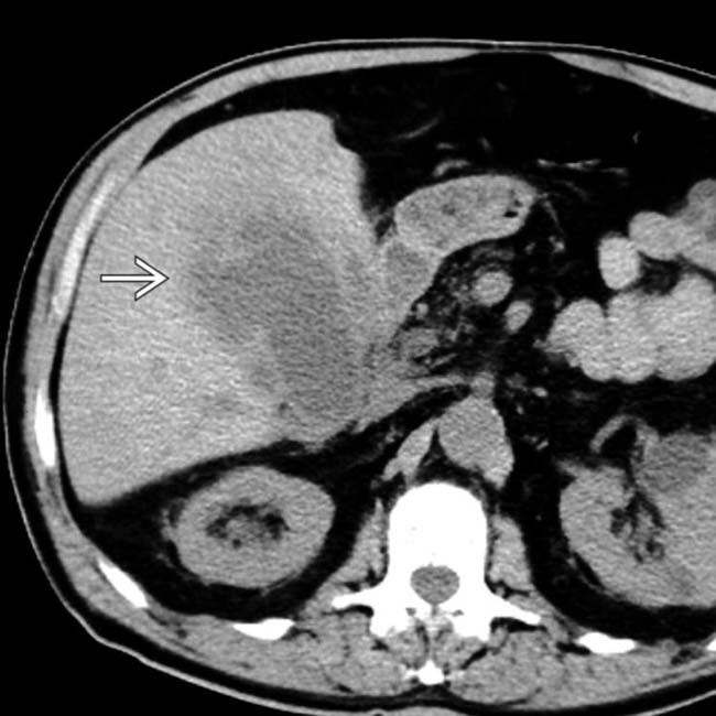
 . While this was worrisome for carcinoma, XGC was confirmed at surgery.
IMAGING
General Features


CT Findings
• Imaging findings overlap with acute cholecystitis, chronic cholecystitis, and gallbladder carcinoma
 Low-attenuation intramural nodules and bands corresponding to foamy cell infiltrate and areas of necrosis/abscess
 Low-attenuation intramural nodules and bands corresponding to foamy cell infiltrate and areas of necrosis/abscess


MR Findings


• Thickened wall often demonstrates delayed enhancement on T1WI C+ with preservation of continuous mucosal enhancement

DIFFERENTIAL DIAGNOSIS
Gallbladder Carcinoma

• Unlike XGC, frequently results in obstruction of common duct, direct invasion of liver, and bulky periportal lymphadenopathy


CLINICAL ISSUES
Presentation


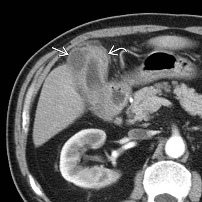
 and intramural low attenuation
  .
.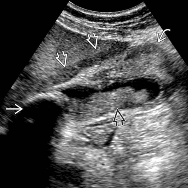
 , sludge
  , and a stone within the GB neck
  (and focal fatty sparing
  ). Preoperative differentiation between GB carcinoma and xanthogranulomatous cholecystitis (XGC) is often difficult, but the absence of ductal dilatation may suggest a chronic inflammatory process rather than neoplasia.
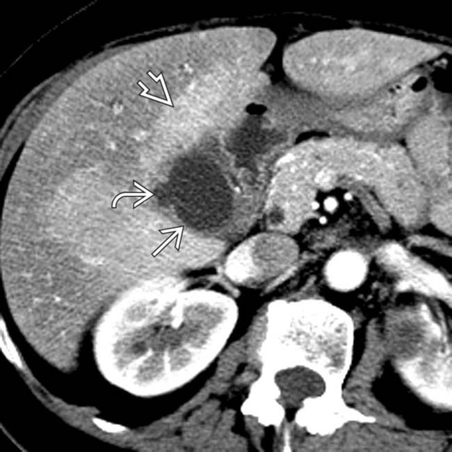
 as well as focal mural low attenuation
  . At pathology, the low attenuation corresponded to an area of necrosis filled with foamy macrophages. Note the increased hepatic perfusion, representing a THAD
  .
.
 , and sludge filling the GB lumen
  .
.
 and intramural low attenuation
  .
.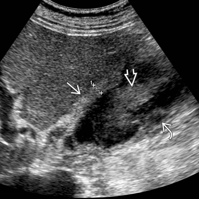
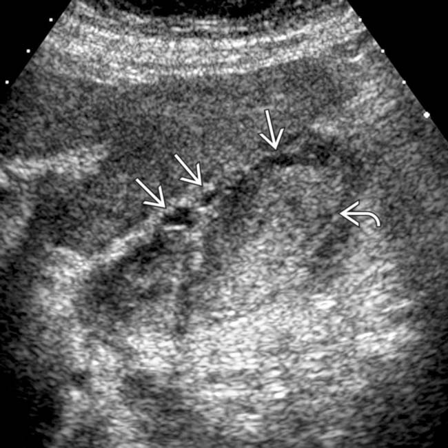
 , sludge
  , and intramural hypoechogenicity
  . Laparoscopic cholecystectomy confirmed chronic XGC. Note that the extensive adhesions and wall thickening associated with chronic inflammation often necessitate open cholecystectomy.

