Wound dressings
Gillian M. Eccleston
Chapter contents
Complications in wound healing: chronic wounds
Physical characteristics of wound dressings
Mechanical properties of dressings
Key points
Introduction
In the past, traditional fabric wound dressings were used extensively. Their primary function was considered to be to keep the wound as dry as possible by allowing evaporation of exudate. It was assumed, therefore that dressings were a passive product with only a minor role in the healing process. However, it is now realized that a wound heals faster and more successfully in a moist environment. This has led to a greater understanding of the influence that wound dressings can have on wound healing and greater attention has been given to the design of more effective dressings. Over the last two decades, a large number of new dressings has become available, based on the concept of creating an optimum environment for the treatment of wounds. However, it is emphasized that there is still no single dressing suitable for the management of all types of wounds or for the treatment of a single wound during all phases of healing.
Many of the newer dressings aim to manage chronic wounds that are difficult to treat because wound physiology is altered. Such wounds are often a problem of the elderly and bedridden. Chronic wounds, as well as compromising the quality of life of the patient, place an enormous financial burden on health services.
Some modern dressings are designed to deliver drugs or wound-healing agents directly to the affected site.
Successful design of wound dressings depends on an understanding of the healing process, the patient condition in terms of health, environment and social circumstance, and the effect that the physicochemical properties of the various dressing materials have on the wound-healing process.
Wounds and wound healing
Wounds
The protective functions of the skin are compromised by injury. A wound can be defined as a defect or a break in the skin, resulting from either mechanical or thermal damage, or as a result of the presence of an underlying medical or physiological condition. Wounds are classified according to the number of skin layers affected and the area of skin involved:
• superficial wounds involve injury to the epidermis alone
• full-thickness wounds occur when the underlying subcutaneous fat or deeper tissues are also damaged.
Wounds are described as normal (acute) if they heal rapidly with minimum scarring and chronic if they take longer than 8–12 weeks to heal.
Simple mechanical injuries such as cuts, grazes and minor burns are usually treated by the patient, whereas the more severe traumatic injuries caused by, for example, surgery, traffic accidents and fires require hospitalization. Chronic wounds require specialist nursing care. Without an understanding of all these factors, correct dressing selection is not possible; incorrect choice of dressing is potentially ineffective and wasteful in terms of nursing time.
Wound healing
Wound healing may be considered as a dynamic process in which cellular and matrix components act together to re-establish the integrity of damaged tissue and replace lost tissue. Regardless of the source or the extent of tissue damage, under normal conditions the wound-healing process occurs in a predictable fashion as four overlapping stages: inflammation, migration, proliferation and maturation (remodelling). Healing is considered to be complete when the skin surface has reformed and has regained its tensile strength.
Inflammation
Inflammation is the body’s initial response to injury and involves both cellular and vascular responses. The release of histamine and a number of other cell-mediated factors into the wound results in vasodilation, increased capillary permeation and stimulation of pain receptors. The release of a protein-rich exudate containing phagocytes and other materials from the blood capillaries onto the wound surface engulfs the debris of dead cells and bacteria (known as autolytic debridement). Fibrinogen in the exudate elicits the clotting mechanism, producing a clot or scab on the wound that causes bleeding to stop. It also gives strength and support to the injured tissue. This first stage of healing usually occurs within a few minutes to 24 hours of injury, when the wound will be red, inflamed, painful and moist.
Migration
Growth factors in the wound exudate promote the growth and migration of epithelial cells, fibroblasts and keratinocytes to the injured area to replace damaged and lost tissue. These cells regenerate from the margins, rapidly growing over the wound under the dried scab. This epithelial thickening and basal cell proliferation lasts for 2–3 days.
Proliferation
The proliferation phase involves the development of new tissue and occurs simultaneously or just after the migration phase (day 3 onwards), lasting from 5 to 20 days. Granulation tissue is formed by the infiltration of blood capillaries and lymphatic vessels into the wound, and by the supporting collagen network synthesized by fibroblasts. This process is known as granulation. The network is important for developing the tensile strength of the skin. As the proliferation continues, further epithelial cell migration across the wound takes place, providing closure and visible wound contraction. During the proliferation stage, the wound is typically beefy red in colour and moist, but not exuding.
Maturation
This final phase of wound healing (also called the ‘remodelling phase’) involves the diminution of the vasculature and enlargement of collagen fibres, which increase the tensile strength of the repair. The timescale for wound repair is from about 3 weeks to 2 years. Commonly, the tensile strength of the final scar reverts to 70–90% of that of the pre-injured tissue.
Complications in wound healing: chronic wounds
Although wound healing is a natural and predictable phenomenon and most wounds will heal uneventfully, complications can sometimes occur that lead to prolonged healing times or chronic non-healing wounds. A chronic wound fails to heal because the orderly sequence of events described above is disrupted at one or more of the wound-healing phases. A normal wound may develop into a chronic wound at any time as a result of poor primary treatment, persistent infection or disease. The most common chronic wounds include venous stasis ulcers, diabetic ulcers, ischaemic ulcers, pressure ulcers (bedsores) and ulcers due to systemic infections or malignant disease. Bacteria may gain entry to the deeper tissue of an acute or chronic wound and overcome the body’s defence mechanisms, giving rise to infection. Poor nutritional status, disease and other factors regarding the patient’s condition may reduce the ability to fight infection, as well as interfere with healing mechanisms (Table 40.1).
Table 40.1
| Nutritional status | Deficiencies in protein, vitamins (especially ascorbic acid) and minerals impair the inflammatory phase and collagen synthesis and thus prolong healing times |
| Advancing age | Elderly patients have less effective immune systems, resulting in decreased resistance to pathogens. Potential problems in healing arise from skin changes, slower metabolism and chronic health conditions such as circulatory problems and diabetes |
| Disease | Poorly controlled diabetes mellitus, renal disease, malignant disease associated with hypoproteinaemia |
| Compromised circulation | Healing delayed because inadequate nutrients, blood cells and oxygen are delivered to the wound |
| Treatment | Patients receiving drugs that compromise the immune system (e.g. steroids), chemotherapy or radiotherapy |
| Mobility | Physical inactivity can result in pressure-related skin damage |
| Obesity | Excess tension placed on wound and decreased vascularity of adipose tissue delays healing. Mobility may be reduced |
| Smoking | Decreases oxygen delivery to the wound due to vasoconstriction and coagulation of small blood vessels |
Foreign bodies introduced deep into the wound at the time of injury can cause chronic inflammatory responses that delay healing, sometimes leading to granuloma or abscess formation. Keloid and other scars that are cosmetically unacceptable may result from excess collagen production during the final phases of the wound-healing process. Underlying diseases and drugs that suppress the inflammatory process, e.g. corticosteroids, also interfere with wound healing.
Wound dressings
Dressings fall into several categories, depending on their function in the wound (occlusive, absorbent, adherent), the type of material employed to produce the dressing (polyurethane, alginate, collagen, silicone) or the physical form of the dressing (film, foam, gel). Some dressings are impregnated with medicaments such as antibacterial agents or wound debridement agents. The incorporation of pharmacological agents such as growth factors into dressings is still in its infancy, although a gel containing human platelet-derived growth factor (PDGF) is now available for treating chronic diabetic ulcers.
Dressings which make physical contact with the wound surface are referred to as primary dressings while secondary dressings cover over the primary dressing. Island dressings possess a central absorbent region that is surrounded by an adhesive portion. The properties of the common dressing types in relation to the type of wound being treated are summarized in Table 40.2. The various available types of dressings are discussed in more detail below.
Table 40.2
| Type of dressing | Key features | Uses |
| Impregnated gauze (soft paraffin or sodium chloride) | Various degrees of absorption. Inexpensive. Needs frequent changing. Dressing may stick to wound, causing pain and damage | Normal or highly exuding wounds. To apply creams or ointments to wound. Infected and necrotic wounds |
| Films | Non-absorbent. Permeable to moisture vapour, allowing some exudate to evaporate. May be transparent. Conform to contours. Adhere to wound. Impermeable to microorganisms | Later stages of wound healing where little exudate. Loss of water vapour can cause wound to dry out. Not for infected wounds and thin or fragile skin |
| Foams | Absorbent. Allow gaseous exchange. Impermeable to water and microorganisms. Can remain on wound for extended times. Thermal insulation. Some are adherent | Performance varies between dressings. Generally low to moderately exuding wounds |
| Alginates | Form hydrophilic gel on contact with wound exudate to promote moist healing. Absorbent. Physical and thermal protection. Easily washed out of the wound | Performance varies between dressings. Moderate to high exuding wounds. Haemostasis. Suitable for infected wounds. Not suitable for dry wounds |
| Gels/hydrogels | Maintain moist wound bed by balanced hydration of wound. Absorbent. Non-adherent. Require secondary dressing | Cleansing of necrotic wounds by rehydrating dead tissue and encouraging autolytic debridement |
| Hydrocolloids | Create moist environment. Absorbent. Initially impermeable to water vapour and air. Adhere to wet and dry wounds. No pain on removal. Can remain on wound for extended times. Provide insulation. Do not require secondary dressing | Performance varies between dressings. Suitable for light to moderately exuding non-infected wounds. Facilitate rehydration and autolytic debridement of sloughy or necrotic wounds |
Traditional dressings
Traditional fabric wound dressings, such as natural or synthetic bandages, cotton wool, lint and gauzes, all with varying degrees of absorbency have been used for centuries in the management of wounds. Their primary function is to keep the wound clean and dry by the evaporation of excess wound exudate. Traditional dressings are still used as primary or secondary dressings or as a part of a composite of several dressings, each having a specific function.
An example is gauze and cotton tissue (gamgee tissue) that is composed of a tubular cotton gauze wrap surrounding a layer of absorbent cotton wool. It is used to absorb exudate and is generally applied over a primary wound dressing to avoid contaminating the wound with cellulose fibres. Many commercially available dressing packs provide a selection of sterile dressings conveniently packaged together. Bandages are used to provide support, act as dressing retention materials or provide protection to clothing following the application of ointments or creams.
Gauze dressings are made from woven and non-woven fibres of cotton, rayon, polyester or a combination of these fibres. Woven products are described as fine or coarse depending on the thread count per inch. Non-woven dressings are generally more absorbent and less likely to shed fibrous material into the wound which will delay healing. Sterile gauze pads are used for packing open wounds to absorb fluid and exudate. The fibres in the dressing act to draw fluid away from the wound. The dressing needs to be changed frequently to prevent maceration of the healthy underlying tissue. Such dressings afford some bacterial protection, although this is lost if the outer surface of the dressing becomes moistened by either wound exudate or external fluids. Gauze dressings tend to adhere to wounds as fluid production diminishes, and are painful to remove. Gauze impregnated with soft paraffin is occlusive and easier to remove from the skin. Antimicrobial agents, such as silver and povidone iodine, are incorporated into some dressings to control or prevent infection, as are debriding agents, such as saline, to prevent maceration of healthy tissue.
Traditional fabric dressings provide little occlusion and allow evaporation of moisture, resulting in a dry, desiccated wound bed. They do not provide a moist environment for wound healing. Consequently, their usage for chronic wounds and burns is being replaced by the more recently developed advanced wound dressings described below.
Advanced wound dressings
Advanced wound dressings are designed to control the environment for wound healing. The primary function of some dressings is to absorb wound exudate (foams, alginates) whilst others donate fluid (hydrogels) or maintain hydration (hydrocolloids).
Vapour-permeable adhesive films
Vapour-permeable (formally known as semi-permeable) film dressings are sterile, thin films made from polyurethane, usually coated on one side with a hypoallergenic acrylic adhesive. These dressings are non-absorbent, and permeable to water vapour and gases but not to liquids or microorganisms. Their transparency allows visual observation of the wound without removal of the dressing. The films vary in thickness and size but all are elastic and highly conformable in use and so suitable for use in flexible areas such as the elbows, knees and sacral areas.
This type of dressing maintains a moist wound-healing environment, promoting the formation of granulation tissue and autolysis of necrotic tissue. However, they are not suitable for use on infected wounds or moderate-to-heavily exuding wounds where water vapour loss may occur at a slower rate than exudate formation. If the dressing is not changed frequently, the accumulation of exudate under the film may lead to skin maceration and bacterial proliferation.
Although the films developed more recently have improved water vapour permeability properties, they are still used mainly as primary or secondary dressings for partial-thickness wounds with little or no exudate, and to prevent and manage the initial stages of pressure ulcers by protecting the fragile skin. Available dressings differ in terms of film thickness and size, vapour permeability, adhesiveness, conformability and extensibility.
Foam dressings
Foam dressings are in the form of sheets of polymer foam, typically polyurethane. Some foams have additional wound contact layers (to avoid adherence when the wound is dry) and an occlusive polymeric backing layer (to prevent excess fluid loss and bacterial contamination). Absorbency is controlled by properties of the foam such as its texture, thickness and pore size. Foam dressings are superior to film dressings because, in addition to maintaining a moist environment, they are absorbent and also provide good thermal insulation. The open pore structure also gives a high moisture vapour transmission rate (HVTR).
Commercially available foam dressings range between products, with foam structures that are suitable for partial- or full-thickness wounds with minimal or moderate drainage and highly absorbent foam structures for heavily exuding wounds. They are used as primary wound dressings for absorption and insulation and as secondary dressings for wounds with packing. Sometimes the foam dressing may be prepared at the point of use and allowed to expand in volume within the cavity of the wound.
Alginate dressings
Alginate dressings are produced from calcium and sodium salts of alginic acid, which is a polysaccharide comprising mannuronic and guluronic acid units obtained from certain species of brown seaweeds. They are available as dry woven, fibrous mats and sheets for wound dressings and as thin ribbons and twisted fibrous ropes for packing into wounds. The dressings interact with wound fluid and blood to form a protective film of gel that maintains an occlusive, non-adherent moist healing environment within the contours of the wound. The interaction occurs because a partial ion exchange reaction takes place in situ between calcium ions in the dressing and sodium ions from the wound exudate, resulting in the production of a gel on the wound surface.
The gelling properties of individual dressings depend on both the relative concentrations and arrangements of the mannuronic and guluronic units as well as the amounts of calcium and sodium ions. Dressings rich in mannuronic acid tend to react readily with sodium ions, forming soft amorphous gels, whereas those rich in guluronic monomer gel less readily and form firmer gels.
Alginate dressings are generally easy to remove without pain, either by lifting the partially gelled sheet off the wound or by rinsing the gel away with water or physiological saline. They generally need a secondary dressing to prevent the alginate from drying out. Since calcium alginate is a natural haemostat, alginate-based dressings are indicated to control minor wound bleeding.
Alginate dressings are highly absorbent and are used for moderate to heavily exuding wounds. A major disadvantage of alginate dressings is that they cannot be used for dry wounds or those covered with hard necrotic tissue because, to function effectively, they need to absorb exudate from the wound. Different alginate dressings show significant differences in characteristics, such as fluid retention, adherence and dressing residues.
Hydrogel dressings
Hydrogels differ from other dressings as they have the ability to add moisture to dry wounds. Thus, they are used to facilitate autolytic debridment (explained above) in necrotic wounds, and to maintain a moist healing environment on clean granulating wounds. Hydrogel dressings are composed of three-dimensional swollen networks of hydrophilic polymers which contain a large proportion of water within their structure. They can be applied to a wound in two forms:
The two forms have different features and uses with respect to wound care.
Amorphous gel formulations.
These contain 70–95% water and are produced by dispersing natural (e.g. the alginates or carboxymethylcellulose) or synthetic (e.g. polyvinyl pyrrolidone, polyacrylamide) hydrophilic polymers in water. The rheological properties of the gels vary markedly in different commercial products according to the specific polymers used and their concentrations in the dressing. These differences influence clinical use and handling characteristics. For example, the apparent viscosity of an amorphous gel will influence its ability to fill all or part of the wound cavity, and subsequently to remain on the wound bed. Subsequent packaging, e.g. whether the dressing is enclosed in tubes, spray bottles or foil packs, will depend on how the dressings behave rheologically under shear.
Amorphous gels progressively lose structure on dilution with exudate until the polymer is dispersed. Thus, when the gel is applied directly to the wound, it is usually covered with a secondary dressing, such as a foam or gauze. Any exudate is absorbed into the gel whilst moisture evaporates through the secondary dressing. Saturated gauzes, obtained when gauze is impregnated with amorphous hydrogel, are sometimes used to fill deeper wounds.
Elastic hydrogel sheets.
These are produced by exposing dilute dispersions of polymer to an energy source such as electron-beam irradiation. The polymer forms a cross-linked structure (anywhere from 6%-30%) that physically entraps water to form a solid sheet that can be cut to fit the wound. Despite their high water content, some hydrogels can also swell further to absorb slight-to-moderate amounts of exudate. Hydrogel sheets do not require a secondary dressing because they are manufactured with a semi-permeable polymer film backing, which may or may not have adhesive borders, that controls the amount of water vapour transmitted through the dressing.
Hydrogel dressings are suitable for use at all stages of wound healing, with the exception of infected or heavily exuding wounds. They cannot absorb much exudate and an unpleasant smell may arise due to skin maceration and bacterial proliferation in infected wounds – encouraged by the large water content of the gel.
Although these dressings need to be changed frequently, they do not disturb fragile tissue and are suitable for burns and other painful wounds. Amorphous gels and impregnated gauzes are used as primary dressings, whereas hydrogel sheets may be used as primary or secondary dressings.
Hydrocolloid dressings
Hydrocolloid dressings are among the most widely used wound management products. The term ‘hydrocolloid’ has been adopted to describe the family of occlusive or semi-occlusive wound dressings obtained from colloidal gel-forming materials, such as carboxymethylcellulose, gelatin, pectin or alginate, combined with other materials including elastomers and adhesives. In their intact state, the occlusive hydrocolloid dressings are impermeable to water vapour and oxygen but, on absorption of wound exudate, a change in physical state occurs with the formation of a gel covering the wound. This seals the wound and provides a moist healing environment that allows clean wounds to granulate and necrotic wounds to debride autolytically. The gel also acts as a barrier to keep bacteria and fluids out. The dressing relieves superficial pain by covering nerve endings with gel and exudate. Angiogenesis (growth of microcapillaries) is stimulated by the dressing initially being impermeable to atmospheric oxygen. The dressing may provide insulation to prevent heat loss from the wound.
Hydrocolloid dressings are manufactured in various shapes and sizes and some are in the form of powders, wafers or pastes. They are often applied to a carrier such as a thin waterproof polyurethane film or foam sheet. The composition of the wound contact layer may differ considerably in different hydrocolloid dressings. Some are transparent, to allow the wound-healing process to be visualized. There are over 60 hydrocolloid dressings currently available that differ in their physical characteristics, such as dressing thickness (some are extremely thin), transparency, fluid-handling properties, moisture-vapour permeability, conformability, acidity/alkalinity and fluid retention These differences in physical properties govern their specific clinical applications and effectiveness.
Hydrocolloid dressings are particularly useful in paediatric wound care for management of both acute and chronic wounds as they adhere to moist and dry sites and do not cause tissue damage or pain on application or removal. Some hydrocolloid dressings are less occlusive, with higher moisture transmission rates, enhancing their ability to cope with exudate production.
Hydrocolloid dressings are used for light to moderately exuding wounds, including pressure sores, leg ulcers, minor burns and traumatic injuries. They can be left on the wound for up to 7 days provided that leakage does not occur. If leakage occurs and the bacterial barrier is broken, the wound is open to bacterial contamination and may develop an odour. Many of this type of dressing are not suitable for heavily exuding wounds or infected wounds, as they can encourage the growth of anaerobic bacteria. Some hydrocolloid dressings contain fibres that are deposited in the wound and have to be removed during dressing changes.
Bioactive dressings
Bioactive dressings deliver substances active in wound healing to the wound. They may be prepared from combinations of biopolymers that have a role in the natural wound-healing process, such as collagen, hyaluronic acid, chitosan, alginates and elastin, or may contain materials such as growth factors. Dressings that combine proteins, polymers and cells are generally described as skin substitutes, although there is no strict distinction between them and wound dressings. Technological advances in the fabrication of biomaterials and the culturing of skin cells is driving forward a new generation of engineered skin substitutes.
Tissue-engineered dressings include biodegradable films formed from, for example, collagen which acts as a scaffold onto which skin cells can be seeded for the growth of new tissues. These scaffold dressings possess mechanical properties ideally approaching those of the tissue they are to replace. When introduced into the body, they gradually degrade, leaving behind a matrix of connective tissue with the appropriate structural and mechanical properties. In the future, such scaffolds may also be used for the delivery of additional bioactive materials, such as growth factors, to a wound.
Wound management
The design and careful selection of wound dressings represent only a small part of the management of a wound. With chronic wounds, it is essential to treat the underlying cause as well as dressing the wound. Once the underlying condition is controlled and the wound has been cleaned of materials that delay healing, such as dead tissue, extraneous bacteria and debris, an appropriate dressing selection can be made according to the type of wound being treated. Wounds are generally classified by their visual appearance as necrotic, sloughy, granulating or epithelializing, each type having different dressing requirements. These are defined in Table 40.3. All wounds, regardless of type, may become infected at any stage of the healing process and subsequently develop an unpleasant odour. In this case, dressings may also be required to have antibacterial activity or be able to absorb odour.
Table 40.3
| Wound type | Appearance | Role of dressing |
| Necrotic | Often olive green or black and dry to the touch, due to presence of necrotic tissue | Remove dead tissue. Rehydrate wound bed. Maintain moist wound bed. Prevent bacterial ingress |
| Sloughy | Slough is generally yellow in colour | Remove dead tissue. Maintain moist wound bed. Absorb excess fluid. Prevent bacterial ingress |
| Granulating | Significant quantities of granulation tissue, generally red or deep pink in colour. May produce excess exudate | Maintain moist wound bed. Absorb excess fluid (if present). Provide thermal insulation. Protect from, and prevent, trauma. Prevent bacterial ingress |
| Epithelializing | A pink margin or isolated pink islands on the surface of the wound. Generally little exudate | Maintain moist wound bed. Provide thermal insulation. Protect from, and prevent, trauma. Prevent bacterial ingress |
The overall aim in the choice of dressing is to provide an environment at the surface of the wound in which healing may take place at the maximum rate consistent with the desired end result of a healed wound with an acceptable cosmetic appearance. Some of the functions that may be required of the dressing are summarized in Table 40.4. The dressing may variously be required to maintain or provide a moist environment, absorb excess exudate, promote autolytic debridement (the process of natural wound cleaning), provide thermal insulation, relieve pain, protect the wound from trauma, and combat infection and odour. In addition, it should be free from particulate contaminants, and be sterile and impermeable to microorganisms. To encourage patient compliance, dressings should be cost-effective, require infrequent changing and be available in a suitable range of forms.
Table 40.4
| Dressing function | Clinical significance to wound healing |
| Provide or maintain a moist wound surface | Prevents desiccation and cell death, enhances epidermal migration, promotes angiogenesis and connective tissue synthesis and supports autolysis by rehydration of desiccated tissue. Enhances migration of leucocytes into the wound bed and supports the accumulation of enzymes |
| Absorption. Removal of blood and excess exudate | In chronic wounds, there is excess exudate containing tissue-degrading enzymes that block the proliferation and activity of cells and break down extracellular matrix materials and growth factors, thus delaying wound healing. Excess exudate can also macerate surrounding skin |
| Debridement, i.e. wound cleansing | Necrotic tissue, foreign bodies and particles prolong the inflammatory phase and serve as a medium for bacterial growth |
| Gaseous exchange (water vapour and air) | Permeability to water vapour controls the management of exudate. Low tissue oxygen levels stimulate angiogenesis. Raised tissue oxygen stimulates epithelialization and fibroblasts |
| Protect the healing wound from bacterial invasion | Infection prolongs the inflammatory phase and delays collagen synthesis, inhibits epidermal migration and induces additional tissue damage. Infected wounds can give an unpleasant odour |
| Provision of thermal insulation | Normal tissue temperature improves the blood flow to the wound bed and enhances epidermal migration |
| Low adherence. Protect the wound from trauma | Adherent dressings may be painful and difficult to remove and cause further tissue damage |
| Cost effectiveness. Frequency of dressing change | Dressing comparisons based on treatment costs rather than unit or pack costs should be made. Although many dressings are more expensive than traditional materials, the more rapid response to treatment may save considerably on total cost |
A simple occlusive/semi-occlusive dressing that prevents the evaporation of exudate is often all that is needed to provide a moist environment at the surface of an acute clean (minimally exuding) wound. Exudate is beneficial to normal wound healing as it contains growth factors that promote the growth and migration of fibroblasts, endothelial cells and keratinocytes. However, some chronic wounds produce excess exudate that may macerate the surrounding skin and delay wound healing (see Table 40.4). Such wounds require dressings that absorb exudate and, if they require frequent changing, should be easy to remove with no tissue trauma or pain.
As mentioned above, there is no single dressing suitable for all types of wounds and often a range of different dressing types is used during the healing of a single wound. Most complex dressing systems are composites of several layers, each layer having a specific role in the wound-healing process. The primary dressing is placed in direct contact with the wound and the secondary dressing is placed over the primary dressing. The central absorbent portion of a composite is called the island dressing. Some dressings need a bandage or some form of adhesive layer to keep them in position, whilst in others there is an adhesive layer incorporated into the dressing itself. Non-adhesive dressings such as gels cause less pain and trauma on removal as they can be washed out of the wound.
Physical characteristics of wound dressings
The composition and physical properties of a wound dressing influence its ultimate performance. There are essentially three types of interrelated specifications for wound dressings:
• structural specifications, which define the composition and structure of a dressing
• performance specifications, which characterize one or more functions of a dressing and
Standard laboratory tests are generally based on specifications of the pharmacopoeias and national and international test standards. Additional non-standard tests may be performed in-house by the manufacturer or by accredited laboratories.
Structural specifications
The early monographs for dressings consisted mainly of structural specifications with limit tests for potential contaminants. Most dressing monographs were removed from pharmacopoeias when dressings were classified as medical devices.
Performance specifications
Although the success of a new dressing is ultimately determined by its clinical performance, laboratory testing can provide a rapid estimation of how a dressing will function under certain conditions. The specific property to be characterized in the laboratory will depend on the type of wound dressing, and the nature of the wound itself. It is now well established that a moist wound environment provided by wound exudate encourages more rapid wound healing, although excess exudate may cause maceration of the wound and surrounding area. Thus, a key function of advanced wound dressings is the removal of excess exudate while maintaining moisture at the wound area.
There are a number of physical tests that reflect this function by determining the absorbing properties of dressings. Such absorbency tests investigate parameters such as fluid retention, moisture-vapour transmission rate and fluid handling. Fluid affinity tests are used for dressings such as hydrogels that also donate moisture in order to promote autolytic debridement.
Fluid absorbency
The standard laboratory methods used to assess absorbency focus mainly on the dressing constituents (i.e. hydrocolloid, alginate or hydrogel). Although these methods enable comparisons of the fluid handling properties of structurally similar dressings and provide useful quality control tests, the tests are specific to individual dressing groups and the fluid handling properties of different dressing groups, e.g. alginate versus hydrocolloid, cannot be compared.
Choice of test fluid.
Although the simplest tests involve water as the test fluid, the swelling properties of some dressings, for example those prepared from alginates, are influenced by the presence of ions in the exudate. This has led to the development of a standard test solution, simply called ‘Solution A’, with an ionic composition comparable to wound exudate that has been adopted for all European standards of dressings.
Water uptake (fluid retention) test.
This is a simple gravimetric test that determines the amount of fluid absorbed and retained by the dressing (expressed as a percentage). Essentially, the increase in weight of the dressing during the absorption of fluid is measured over a given time period and used as an indication of fluid uptake and retention. Another approach uses a computer-linked video camera to determine changes in the thickness of dressings as they swell as they absorb fluid.
Moisture vapour transmission rate (MVTR).
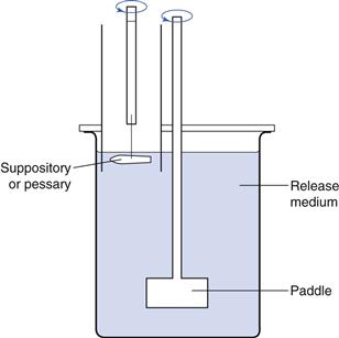
Vapour-permeable film dressings allow the loss of water vapour from wound exudate by evaporation. The MVTR may be evaluated using a Paddington cup. This consists of a cylinder (internal cross sectional area 10 cm2) with a flange at each end. A solid plate, which can be clamped into position forming a water tight seal, is fitted at one end of the cylinder and an annular ring with the same internal diameter as the cylinder fitted to the other end. The dressing is clamped between the annular ring and one of the flanges; test fluid is poured into the cup, and the plate is then clamped into position. The cylinder is weighed at the start of the experiment and then placed in an incubator to control temperature and humidity. The cylinder is removed from the incubator after a predetermined time and reweighed. The amount of fluid lost by evaporation through the back of the dressing is calculated by difference.
Fluid-handling capacity (FHC).
The absorption capacity of many dressings (e.g. alginates, hydrocolloids) is finite, limited by their size and volume. Vapour permeable films and membranes are sometimes used as a secondary dressing to allow the passage of water vapour from wound exudate located in the primary dressing. The fluid handling capacity provides information on the ability of such a composite dressing to both absorb and retain wound fluid (absorbency), whilst simultaneously losing water vapour through the outer surface of the dressing to the external environment. Both processes are important in the clinical management of wound exudate.
The FHC test determines the sum of the weight of test solution retained by the dressing and the weight of fluid lost by transmission through the dressing as moisture vapour. It is carried out in a similar manner to the MVTR test described above, except that the amount of fluid retained by the dressing is measured by removing the base of each cup and allowing any free fluid remaining in the cup that has not been absorbed by the dressing to drain away. The cup is then reweighed and the FHC of the dressing calculated by difference.
Fluid affinity
This test investigates a dressing’s ability to donate moisture to, or absorb liquid from, standard substrates. It is largely applicable to amorphous hydrogel dressings which have the ability both to donate and absorb wound exudates. The ability to donate fluid helps to facilitate the rehydration of dry necrotic tissue to promote autolytic debridement. A high fluid affinity also helps absorb excess wound exudate and liquefied tissue debris once autolytic debridement has taken place.
A typical test involves placing weighed samples of the dressing onto the surface of a series of aqueous gel plugs of gelatin (35%) or agar (2%) of known weight which are then sealed within the barrel of a syringe. Following storage at a set temperature and time (e.g. at 25 °C for 48 hours) the test material and plugs are separated and the plugs are re-weighed. The amount of donated moisture is assessed from the percentage change in weight of each plug.
Mechanical properties of dressings
Characterization of the mechanical properties of dressings is important because all dressings are required to be durable and flexible enough to accommodate the stresses caused by distortion or movement when they are applied to different areas of the body. Mechanical tests also provide useful structural parameters in quality control. Properties such as tensile strength (films), compression (foams and hydrogel sheets) and rheological properties (rehydrated gels) are widely investigated.
Tensile properties
During a tensile test, a sample of film dressing is stretched until it breaks. The applied force and displacement (amount of ‘stretch’) are measured. From this the following can be calculated.
These parameters are used to provide information about film flexibility and rigidity, and are useful in assessing the suitability of a film dressing for application to different parts of the body with various contours.
Compression tests
For dressings of greater thickness than films, for example foam and hydrogel sheets which may be able to take up large volumes of fluid but not retain the fluid even under light pressure, hardness tests can be employed. Hardness is defined as the resistance of the formulation to compression forces and is measured in units of force per unit area. The test can be used to compare the fluid handling properties of these products under varying levels of pressure.
Rheological tests
Rheological properties such as apparent viscosity, elasticity and viscoelasticity are useful for characterizing rehydrated hydrocolloid or alginate gels, and amorphous hydrogels. Optimization of rheological parameters can be an important quality control tool and help meet the requirements of such dressings, such as their ability to fill a wound and be pain free on removal.
Safety and acceptability specifications
Tests specifically designed to ensure that wound-healing products are safe and acceptable include assessments of bioadhesion, microbial performance, odour, irritancy and sensitivity. These are described below.
Bioadhesive strength
Pain and trauma due to dressing adherence are important factors for the patient when a dressing is changed. Whilst dressings that form gels do not adhere greatly, others such as gauze and non-woven fabrics can adhere strongly. In the context of dressings, adhesion is defined as the force required to detach a sample from the surface of a wound. It may be assessed in the laboratory using a Texture Analyser (a type of mechanical testing equipment) by measuring the force required to detach the dressing from the surface of model excised skin, such as porcine skin.
Bacterial barrier test
Wounds represent a potential source of cross-infection and it is important that they are isolated to prevent the ingress or egress of pathogenic microorganisms. Most dressings therefore contain a layer to prevent the transmission of organisms into or out of the wound. A test to investigate the ability of bacteria to pass through a dressing involves clamping the sterile dressing aseptically between two sterile hemispheres such that the dressing remains vertical. A fluid microbiological medium is introduced into both chambers, one of which contains a heavy inoculum of the test organism(s). The apparatus is incubated for an appropriate period, after which the chamber containing previously sterile nutrient is examined for evidence of bacterial growth.
Odour control
Certain types of wounds produce noxious odours caused by a number of volatile agents produced by microorganisms. A number of tests are available to compare the efficacy of odour adsorbing dressings, such as those containing carbon. For example, one test uses specialized apparatus to examine the ability of different dressings to prevent the passage of a volatile amine when applied to a wound model under simulated ‘in-use’ conditions.
Irritation and sensitization
As with any other preparation that is applied to the skin, the safety of the excipients must be confirmed and any possible irritation or sensitivity assessed, generally using animal models in the first instance, followed by human patch testing.
Bibliography
1. Boateng JS, Matthews KH, Stevens HNE, Eccleston GM. Wound healing dressings and drug delivery systems: A review. Journal of Pharmaceutical Sciences. 2008;97:2892–2923.
2. Cockbill S. Wounds The healing process. Hospital Pharmacist. 2002;9:255–260.
3. Debra JB, Cheri O. Wound Healing Technological Innovations and Market Overview. 2nd edn Technology Catalysts International Corporation 1998.
4. Drug Tariff (2012) Stationery Office, London (updated monthly).
5. Hess CH. Wound Care. 5th edn New York: Lippincott, Williams and Wilkins; 2005.
6. Thomas S. Surgical Dressings and Wound Management. Medetec Publications, UK 2010.