Wound construction

Published on 08/03/2015 by admin

Filed under Opthalmology

Last modified 22/04/2025

Print this page

rate 1 star rate 2 star rate 3 star rate 4 star rate 5 star
Your rating: none, Average: 0 (0 votes)

This article have been viewed 2158 times

CHAPTER 11 Wound construction

See Video image

Correct knowledge and implementation of wound architecture are paramount in creating a stable operating environment and a safe postoperative recovery.

This chapter will cover extracapsular (ECCE), small incision cataract surgery (SICS) wounds and phacoemulsification wounds. The vast majority of cataract operations in the developed world are performed via a phaco wound but occasionally it may be necessary to perform a larger wound. SICS is, at the time of writing, becoming very popular in developing countries due to its relative similarity to ECCE, low cost, and effectivity. It would be comforting to know that long-term data in terms of endothelial cell loss and glaucoma resulting from SICS will be collected and analysed but, due to the nature of the places where SICS is mainly performed around the world and the facilities available, this is unlikely.

ECCE

A backward sloping corneal wound or stepped scleral wound may be created to allow the wound to be self-sealing and allow the use of sutures which will stop the wound from opening rather than keeping it closed. This allows the tension in the sutures to be adjusted in such a way that minimal induced astigmatism is caused.

The wound will also heal at this predecided tension, which means the sutures can be left in for around 4 months, which mitigates against wound slip from early removal. The wound should be made just big enough to express the nucleus without undue force and this means an accurate preoperative assessment of the nuclear density to allow such a judgment on wound size to be made.

Scleral ECCE wound

The architecture of the wound is similar between scleral and corneal ECCE wounds but, for a scleral wound, the conjunctiva must be reflected first and sutured back in place at the end. If a very peripheral corneal wound is made then consideration should be given to moving the conjunctiva as well because if this is not done the closing sutures which start in the cornea will exit through the sclera and conjunctiva and will ‘cheese-wire’ or cut through the conjunctiva leading to inflammation, sterile conjunctival infiltrates, and loosening of the sutures.

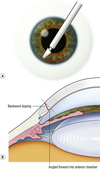
The wound is sloped backwards meaning that the blade used for the incision is pointing towards the surgeon. This makes the wound self-sealing and stable. If the wound is forward sloping it will leak and make the operation difficult and the postoperative period more risky.

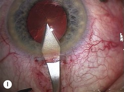
The scleral wound is made around 120°, initially backward sloping and then, when the deeper tissues are reached, a forward entry into the eye is made (Fig. 11.1). More bleeding is likely because of perforating scleral vessels and diathermy is a prerequisite. Careful wound construction and suturing can be rewarded by very little surgically induced astigmatism because of the distance from the optical centre of the cornea and the ability to leave the sutures at the desired tension.

Excessive cautery may shrink the edges of the wound and make wound apposition more difficult when closure is attempted.

Corneal ECCE wound

This should be made approximately 1 mm into clear cornea and sloped gently backwards. This ensures that the sutures when placed to close the wound will enter and exit corneal tissue and avoid the need for reflecting the conjunctiva (see Fig. 11.1).

The wound is made at least 90% depth and then a final, forward entry into the anterior chamber is made. A diamond bladed knife is best for this but a sharp steel blade is adequate, more readily available and much cheaper. Re-used steel blades blunt very quickly and should not be relied on as soon as their lack of sharpness is perceived.

The relative advantages and disadvantages of scleral and corneal ECCE wounds are summed up in Table 11.1.

Table 11.1 Comparison of corneal and scleral section

Section Advantages Disadvantages
Corneal Quicker More astigmatism
  No bleeding Endothelial damage
  ‘Mechanics easier’ (more stable) Suture care (loose etc.)
  No superior rectus suture needed Less strong wound
  Easier access for trabeculectomy later  
Scleral Less astigmatism More bleeding
  Wound alignment easier Astigmatism less easily changed (by removing sutures)
  Less endothelial damage More trabecular meshwork damage
  Suture care less (below conjunctiva) Postoperative intraocular pressure rise more likely
  Stronger wound Iris prolapse more likely
    May need superior rectus suture
    Trabeculectomy more difficult later
    Takes longer

Phaco wounds

The position, size, and shape of a phaco wound depend on a number of factors. A ‘standard’ phaco wound can be placed in the sclera, the cornea, or at the limbus. Its site may depend on surgeon preference in terms of comfort, or on optical factors such as choosing to operate to reduce astigmatism. This is often called ‘on-axis’ surgery and the wound is placed ‘on the axis of the plus cylinder’. What this means is that the wound is placed at the top of the steepest meridian of the cornea. This is the meridian with the highest number ‘K’ or keratometry reading or the smallest radius of curvature in millimeters. This will ensure that any flattening of the corneal curvature which results from the making of the cut will be beneficial in terms of reducing the steeper curve of the cornea towards that of the opposite meridian (spherizing the cornea).

The size of the wound is initially dictated by the blades provided by the company supplying the phaco probe as the keratome used to make the wound must be matched to the size of the probe and irrigating sleeve used if AC stability is to be maximized. These wounds are typically between 2.5 and 3.2 mm, but microcoaxial systems can go down to 1.8 mm. The final size of the wound will depend on the size of the implant being used and modern folding lenses will often go through an unmodified 2.75 mm wound. A technique called wound assisted delivery may be used to deliver a lens through smaller wounds, but stretching of the wound may occur in some cases.

The principle of constructing a phaco wound is the same wherever it is positioned. There should be at least one step in the wound to ensure wound stability1.

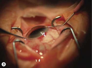
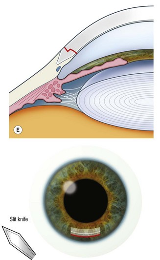
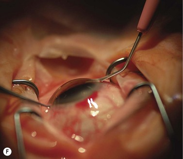
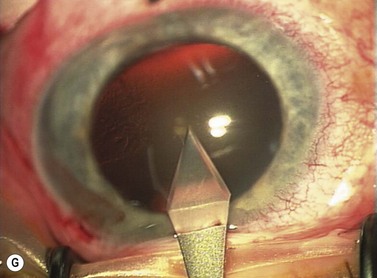
Scleral tunnel phaco wound

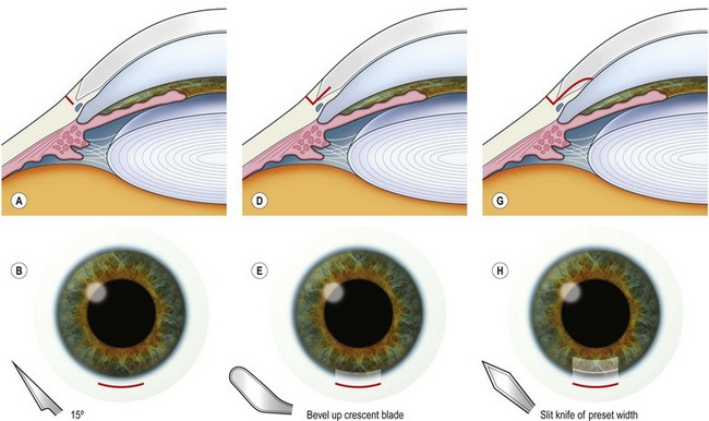
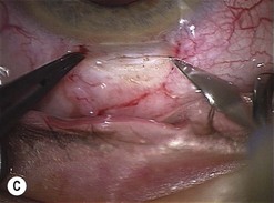
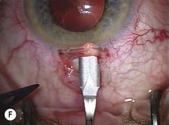
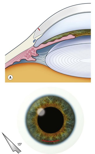
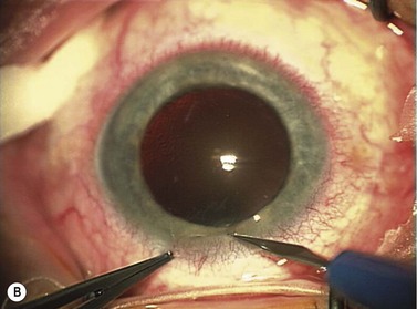
See Figure 11.2.

The conjunctiva is reflected and a frown or straight incision made approximately 3 mm long and around one-third scleral thickness. A bevel-up crescent blade is then used to dissect forwards towards the limbus until the edge of the blade is just visible in the corneal stroma. This gives a square tunnel of approximately 3 mm sides. A predefined width keratome (to match the size of the phaco probe and sleeve) is then used to enter the AC in a plane parallel to the iris. It may not be necessary to suture the wound closed at the end of the procedure, but if there is any concern about its stability this should be undertaken with an 8 or 9-0 absorbable suture. The conjunctiva should also have a suture placed to close the superficial wound.

Corneal tunnel phaco wound

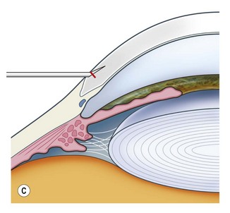
See Figure 11.3.

This wound may be constructed in the same way as the scleral tunnel albeit with a shorter length for the second stage. Some surgeons prefer to miss out the initial 3 mm groove (it has been shown by anterior segment UBM to cause slight gaping of the epithelium) and also the formal dissection forward of the corneal stroma. Instead they use the keratome to make an initial entry into the stroma, superficially and parallel to the surface of the cornea, and then, using the engaged tip of the blade, elevate the heel of the blade so its point is pointing into the AC, pushing the tip through full thickness cornea to enter it into the AC before levelling it out again to fully enter the AC parallel to the iris without damaging the lens capsule. The length of this tunnel can be altered by changing the distance traveled through the stroma before changing direction into the AC. A length of 1–2 mm is ideal and certainly adequate.

Postoperatively, the corneal stroma at the wound edges should be hydrated as this ensures greater safety from postoperative ingress of fluids into the eye and may well help to reduce the incidence of endophthalmitis2.

How to decide where to place the wound

Astigmatic considerations

For patients with less than 1.5 diopters of astigmatism and using a technique which is known to induce little astigmatism, the wound can be placed wherever it is most comfortable for the surgeon. It must be remembered that, if training is taking place, the training surgeon must be in a position to take over from the trainee if necessary, so the wound placement should be such that this is possible. For example, if a left handed trainee places the wound nasally in a right eye, a right handed trainer would find this difficult to cope with.

If more than 2 diopters of astigmatism exist then consideration should be given to placement of the wound to reduce the steep meridian astigmatism (i.e. at the head of the steep meridian).

If more than 2 diopters of difference exis between the K readings then additional maneuvers can be undertaken to reduce astigmatism. Examples of these are limbal relaxing incisions (LRIs), opposite clear corneal incisions (OCCI), arcuate keratotomies (freehand or using preset blades), which may be single or paired, or ‘T’ cuts.

Each method has a range of published studies using different nomograms which relate with or against the rule astigmatism to patient’s age and intended correction. Thus a typical formula might be to place paired arcuate cuts 8 mm apart from each other, of 3 clock hours length each and 600 microns in depth. Each nomogram is approximate and relies on experience and consistency to obtain reasonable results.

Another approach is to consider the use of toric intraocular lenses which allow routine wound placement and allow astigmatism correction with no alteration to the corneal surface. This is becoming more popular as commercial companies are now producing a greater range of lenses with which to perform these corrections.