[level-membership-for-radiology-category] Distal duodenum and jejunum are most often involved, with distal small bowel/ileum involved in severe cases
Thickened, irregular folds with sand-like micronodules
Small bowel lumen may be normal or mildly dilated
• CT
Low-density enlarged mesenteric and retroperitoneal lymph nodes that may have near fat-density
Thickened proximal small bowel folds ± submucosal edema due to hypoalbuminemia
• MR
Lymph nodes may show ↑ T1 signal due to fat
TOP DIFFERENTIAL DIAGNOSES
• Celiac disease
• Intestinal opportunistic infections
• Dysgammaglobulinemia
• Intestinal metastases and lymphoma
PATHOLOGY
• Caused by Tropheryma whipplei (probably orally acquired)
• Not all patients with Tropheryma whipplei infection develop Whipple disease, and underlying autoimmune disorders or genetic abnormalities may potentiate clinical syndrome
• Late phase: Diarrhea, malabsorption, steatorrhea, adenopathy, abdominal pain
GI symptoms generally later manifestation of disease
• Can be fatal without therapy (long-term antibiotics)
• Clinical symptoms often subside quickly after therapy
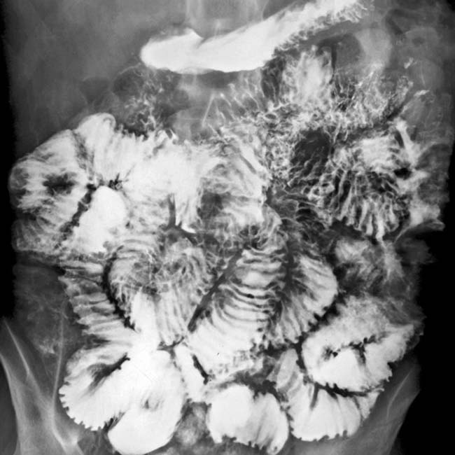
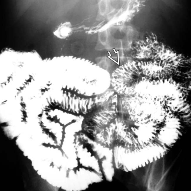
(Left) Small bowel follow-through shows nodular thickening of the jejunal folds in a patient with Whipple disease.
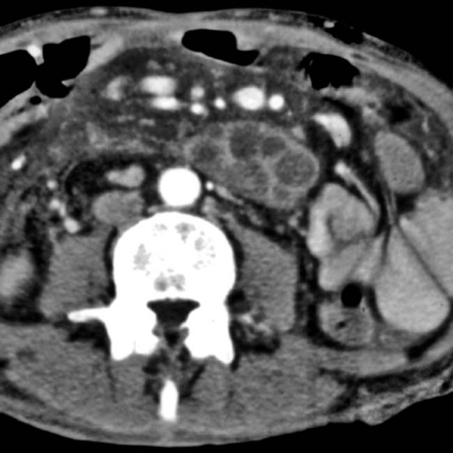
(Right) Axial CECT shows jejunal fold thickening , as well as low-attenuation mesenteric and periaortic lymphadenopathy . Endoscopic biopsy of the jejunal mucosa confirmed PAS-positive macrophages containing gram-negative, acid-fast bacilli, characteristic of Whipple disease.
(Left) Small bowel follow-through in a 40-year-old man with arthralgias and diarrhea demonstrates thickened, nodular jejunal folds.
(Right) Axial CECT in the same patient shows marked thickening of the small bowel with retroperitoneal and mesenteric lymphadenopathy , which appears lower in attenuation than in most other causes of adenopathy. Endoscopic biopsy of the jejunal mucosa revealed villi distended with macrophages full of periodic acid-Schiff-positive bacilli, diagnostic of Whipple disease.
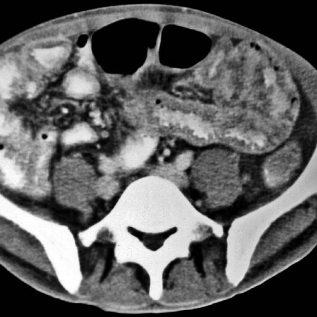
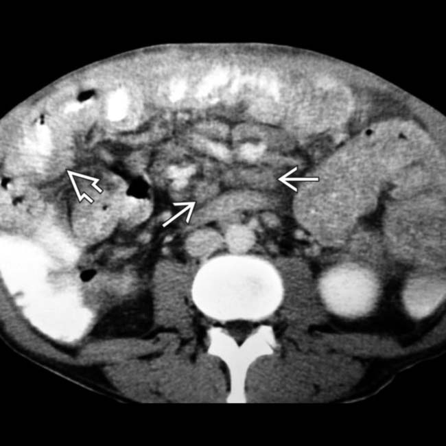
Axial CECT shows very low-density nodes in the retroperitoneum and mesentery.
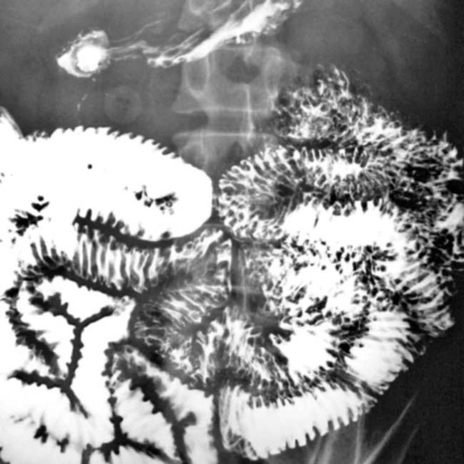
Small bowel follow-through shows micronodular fold thickening of most of the small bowel but no excess fluid.
Axial CECT shows mural thickening of long segment of jejunum.
Small bowel follow-through shows the micronodular pattern of the jejunum in a 40-year-old man with Whipple disease.
[/level-membership-for-radiology-category][not-level-membership-for-radiology-category] Distal duodenum and jejunum are most often involved, with distal small bowel/ileum involved in severe cases
Thickened, irregular folds with sand-like micronodules
Small bowel lumen may be normal or mildly dilated
• CT
Low-density enlarged mesenteric and retroperitoneal lymph nodes that may have near fat-density
Thickened proximal small bowel folds ± submucosal edema due to hypoalbuminemia
• MR
Lymph nodes may show ↑ T1 signal due to fat
TOP DIFFERENTIAL DIAGNOSES
• Celiac disease
• Intestinal opportunistic infections
• Dysgammaglobulinemia
• Intestinal metastases and lymphoma
PATHOLOGY
• Caused by Tropheryma whipplei
Buy Membership for Radiology Category to continue reading. Learn more here


 in a patient with Whipple disease.
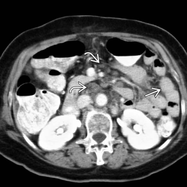
 , as well as low-attenuation mesenteric and periaortic lymphadenopathy
  . Endoscopic biopsy of the jejunal mucosa confirmed PAS-positive macrophages containing gram-negative, acid-fast bacilli, characteristic of Whipple disease.
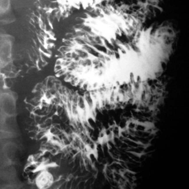
 with retroperitoneal and mesenteric lymphadenopathy
  , which appears lower in attenuation than in most other causes of adenopathy. Endoscopic biopsy of the jejunal mucosa revealed villi distended with macrophages full of periodic acid-Schiff-positive bacilli, diagnostic of Whipple disease.