von Willebrand Disease

Published on 22/03/2015 by admin

Filed under Pediatrics

Last modified 22/04/2025

Print this page

rate 1 star rate 2 star rate 3 star rate 4 star rate 5 star
Your rating: none, Average: 0 (0 votes)

This article have been viewed 2137 times

Chapter 471 von Willebrand Disease

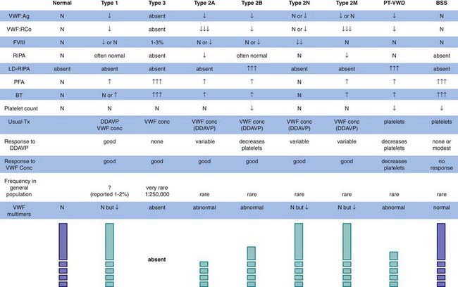
The most common hereditary bleeding disorder is von Willebrand disease (VWD), and some reports suggest that it is present in 1-2% of the general population. VWD is inherited autosomally, but most centers report more affected women than men. Because menorrhagia is a major symptom, women may be more likely to seek treatment and thus to be diagnosed. VWD is classified on the basis of whether the protein is quantitatively reduced, but not absent (type 1); qualitatively abnormal (type 2); or absent (type 3) (Fig. 471-1). Mutations in different loci that code for different functional domains of the von Willebrand factor (VWF) protein cause the different variants of VWD.

Clinical Manifestations

Patients with VWD usually have symptoms of mucocutaneous hemorrhage, including excessive bruising, epistaxis, menorrhagia, and postoperative hemorrhage, particularly after mucosal surgery, such as tonsillectomy or wisdom tooth extraction. Because a teenager’s menstrual history is usually put in the context of other family members, excessive menstrual bleeding is not always recognized as being abnormal, because others in the family may be affected with the same disorder. If a menstruating female has iron deficiency, a detailed history of bruising and other bleeding symptoms should be elicited and further hemostatic evaluation undertaken.

Because VWF is an acute-phase protein, stress will increase its level. Thus, patients may not bleed with procedures that incur major stress, such as appendectomy and childbirth, but may bleed excessively at the time of cosmetic or mucosal surgery. Bruising symptoms may diminish during pregnancy, because VWF levels may physiologically double or triple as an acute phase response. Rarely, patients with VWD may have gastrointestinal telangiectasia. This combination results in major bleeding and accounts for numerous hospital admissions for patients with severe disease. In patients with type 3, or homozygous, VWD, bleeding symptoms are much more profound. These patients are usually diagnosed early in life and may have severe epistaxis or menorrhagia that results in major blood loss and possibly shock. Patients with severe type 3 VWD may have joint hemorrhages or spontaneous central nervous system hemorrhages.

Laboratory Findings

Although patients with VWD were described historically as having a long bleeding time and a long partial thromboplastin time, these findings are frequently normal in patients with type 1 VWD. Normal results on screening tests do not preclude the diagnosis of VWD. Because there is no single assay that has demonstrated the ability to rule out VWD, if the history is suggestive of a mucocutaneous bleeding disorder, VWD testing should be undertaken, including a quantitative assay for VWF antigen, testing for VWF activity (ristocetin cofactor activity), testing for plasma factor VIII activity, determination of VWF structure (VWF multimers), and a platelet count. Although the platelet count is usually normal in most patients, those with type 2B disease or platelet-type disease (pseudo-VWD) may have lifelong thrombocytopenia. Figure 471-1 lists the variants of VWD and summarizes their laboratory findings. Levels of VWF vary with blood type (type O < A < B < AB), which can confound the clinical diagnosis of hereditary VWD, but most clinicians feel bleeding is related to the plasma level of VWF. In addition, there is controversy regarding the clinical definition of “true” VWD. Molecular genetics may clarify the diagnosis of type 1 VWD, but other genetic modifiers may exist outside the gene for VWF and significantly influence the diagnosis. The milder the patient’s phenotype, the greater the difficulty in diagnosis.

Genetics

Chromosome 12 contains the gene for VWF. In each of the type 2 variants listed in Figure 471-1, specific areas of the molecule are affected. The phenotype can guide the genetic diagnosis of the specific mutation. Investigations are underway to clarify whether all cases of type 1 VWD are related to mutations in the VWF gene on chromosome 12 or if there are genetic modifiers, such as blood type, that cause phenotypic VWD. Clinical genetic testing for VWD variants is only available in a few referral laboratories.

Von Willebrand Disease Variants

Type 1 VWD is the most common form and accounts for 85% of cases. NHLBI VWD guidelines restrict the diagnosis of VWD to those with VWF levels of <30 IU/dL. Those with levels >30 IU/dL but below the normal range are referred to as having “possible VWD” or as having “low VWF.” Bleeding symptoms include epistaxis, bruising, and menorrhagia. If bleeding is excessive, desmopressin (DDAVP) administration at a dose of 0.3 µg/kg IV will increase the level of VWF and factor VIII by 3- to 5-fold. Intranasal DDAVP (Stimate) is particularly helpful for the outpatient treatment of bleeding episodes. The dose is 150 µg (1 puff) for children weighing <50 kg and 300 µg (2 puffs) for those weighing >50 kg. A new subtype of type 1 VWD has been referred to as type 1C because the plasma VWF is low from accelerated clearance, hence the 1C designation. Such patients can be diagnosed by doing VWF:Ag levels 1 and 4 hours after desmopressin infusion to demonstrate the shortened half-life of VWF (2-4 hr) in this variant. Alternatively, determining VWF propeptide level, VWFpp, at baseline demonstrates an elevated VWFpp/VWF:Ag ratio of >2 that is caused by the accelerated clearance of VWF (and not VWFpp). In these patients, desmopressin releases adequate VWF, but levels are not maintained for the normal half-life and may require VWF concentrate infusion, with this VWF T1/2 being normal.

Type 2A VWD is caused either by the abnormal proteolysis of VWF by ADAMTS13 or by abnormal synthesis and reduced secretion. In either, only the smallest VWF multimers are present, resulting in a reduction in VWF antigen with a much greater reduction in VWF activity. Although desmopressin is safe in these patients, it is not always effective, because normal multimers are not maintained in plasma. Significant bleeding should be treated with VWF replacement therapy.

Type 2B VWD may be caused by 1 of several mutations resulting in “hyperactive” VWF. The abnormal VWF binds spontaneously to platelets, with resulting rapid clearance of VWF and platelets. The higher molecular weight multimers of VWF are preferentially cleared from the circulation, and moderate to severe thrombocytopenia is common. The laboratory diagnosis is based on the finding that the hyperactive 2B VWF binds to platelets and agglutinates them at low concentrations of ristocetin, a concentration that would not agglutinate normal platelets. If desmopressin is given to these patients, the abnormal hyperactive 2B VWF will be released and more profound thrombocytopenia might occur. Patients with 2B VWD usually respond to the infusion of VWF.

Type 2M VWD is caused by mutations that result in reduction of the platelet-binding function of VWF. Thus, levels of VWF activity are significantly lower than the levels of VWF antigen. Binding of this protein to factor VIII is normal; thus, factor VIII levels are similar to those of VWF antigen. Desmopressin will increase VWF and factor VIII levels, but the released type 2M VWF may not have sufficient activity to cause cessation of bleeding. Thus, VWF replacement therapy may need to be used if desmopressin is not clinically effective.

Type 2N VWD is caused by the reduction of factor VIII binding by VWF. This disorder has also been termed autosomal hemophilia. With this variant, platelet interaction with VWF is normal, but type 2N VWF binds weakly (or not at all) to factor VIII, resulting in rapid clearance of factor VIII. Thus, the factor VIII level is reduced much more than VWF levels. Commonly, patients who have symptomatic bleeding are compound heterozygotes who have inherited a gene for type 1 VWD from 1 parent and a gene for type 2N VWD from the other. Rarely, type 2N mutations are inherited from both parents and VWF levels are normal. In the patient who is compound heterozygous for types 1 and 2N, 1 allele makes no protein and the other allele makes a functionally abnormal protein, and as a result, all of the VWF is dysfunctional. Although desmopressin will release type 2N VWF, sustained factor VIII levels occasionally may be inadequate for normal hemostasis. A trial of desmopressin is indicated to assess the response and half-life of VWF and factor VIII after infusion. VWF replacement therapy is usually effective and because that VWF is normal, endogenous factor VIII can bind to the normal VWF with a longer maintenance of plasma factor VIII levels. Recombinant VWF is currently undergoing clinical trials.

Platelet-type (pseudo-) VWD is actually an abnormality of the GPIb receptor on platelets. This form can be considered the converse abnormality of type 2B, in that the GPIb receptor on platelets is hyperfunctional and binds plasma VWF spontaneously, resulting in thrombocytopenia and a loss of high molecular weight VWF multimers, which are indistinguishable from those seen in type 2B VWD. However, specific testing shows that this is a platelet abnormality rather than a plasma abnormality. Treatment is with transfusion of normal platelets, but if VWF level and function is particularly low, infusion with normal VWF may also be required initially for major hemorrhage.

Type 3 VWD is the homozygous or compound heterozygous inheritance of VWF deficiency. Patients exhibit undetectable plasma levels of VWF and low, but measurable, levels of factor VIII. These patients will have major hemorrhage but only rarely have joint hemorrhages. This severe, very rare form occurs in approximately 1 : 500,000 individuals. Intracranial hemorrhage, major epistaxis, and menorrhagia in women are the major features. Bleeding episodes require treatment with VWF-containing concentrates. VWF is both a plasma and a platelet protein. Because treatment with VWF-containing concentrate only corrects the plasma VWF level, patients with severe bleeding may need to be transfused with platelet concentrates to correct the deficiency of platelet VWF. Desmopressin is not effective in type 3 VWD.

Diagnosis and Differential Diagnosis

The diagnosis of VWD is dependent on the finding of a low level of at least 1 of the laboratory measures of VWF noted in Figure 471-1. The differential diagnosis of mucocutaneous bleeding includes abnormalities of platelet number, platelet function, or the vessel wall (Chapter 478). In caring for children, it is important to remember that the most common cause of such findings is trauma, especially nonaccidental trauma—child abuse.

Treatment

Treatment of VWD is directed toward increasing the plasma level of VWF and factor VIII. Because the gene for factor VIII is normal in patients with VWD, elevating the plasma concentration of VWF permits normal recovery and survival of endogenously produced factor VIII. The most common form of VWD is type 1. In these patients, the synthetic drug desmopressin induces the release of VWF from the patient’s endothelial cells. In some patients with type 2 or 1C variants, desmopressin may be similarly effective, but in other circumstances, the released VWF is dysfunctional. Patients with VWD may not respond adequately to desmopressin because they release an abnormal VWF molecule (most type 2 variants); because they have type 3 disease, in which there is no VWF to be released; or because they have accelerated clearance of released VWF (type 1C VWD). A small subset of children and adults, especially infants, do not release VWF in response to desmopressin. In these cases, replacement therapy must be used. Current replacement therapy uses plasma-derived VWF containing concentrates that also contain factor VIII. VWF distributes only to the intravascular space, because it is so large. During plasma fractionation, VWF multimers are altered to a variable extent. Therefore, 1 U/kg will increase the plasma level by 1.5%. The plasma half-life of both factor VIII and VWF is 12 hr, but the alteration of VWF during fractionation results in half-lives of 8-10 hr when concentrates are infused. Purified or recombinant VWF concentrates (containing no factor VIII) may become available in the near future. These will be useful in prophylaxis or in presurgical management. However, when used for acute bleeding, these VWF concentrates may need to be supplemented by an infusion of recombinant factor VIII for the 1st infusion, as they contain little or no factor VIII. Both VWF and factor VIII are required for normal hemostasis. If only VWF is replaced, endogenous correction of the factor VIII level takes 12-24 hr. Dental extractions and sometimes nosebleeds can be managed with both desmopressin and an antifibrinolytic agent, such as ε-aminocaproic acid (Amicar).

Bibliography

Favaloro EJ. Toward a new paradigm for the identification and functional characterization of von Willebrand disease. Semin Thromb Hemost. 2009;35:60-75.

Federici AB, Mannucci PM, Castaman G, et al. Clinical and molecular predictors of thrombocytopenia and risk of bleeding in patients with von Willebrand disease type 2B: a cohort study of 67 patients. Blood. 2009;113:526-534.

Haberichter SL, Balistreri M, Christopherson P, et al. Assay of the von Willebrand factor (VWF) propeptide to identify patients with type 1 von Willebrand disease with decreased VWF survival. Blood. 2006;108:3344-3351.

Mannucci PM. Treatment of von Willebrand’s disease. N Engl J Med. 2004;351:683-694.

Montgomery RR, Cox Gill J, Di Paola J. Hemophilia and von Willebrand disease. In: Orkin SH, Nathan DG, Ginsburg D, et al, editors. Nathan and Oski’s hematology of infancy and childhood. ed 7. Philadelphia: Saunders Elsevier; 2009:1487-1524.

Nichols WL, Hultin MB, James AH, et al. von Willebrand disease (VWD): evidence-based diagnosis and management guidelines, the National Heart, Lung, and Blood Institute (NHLBI) Expert Panel report (USA). Haemophilia. 2008;14:171-232.

Sadler JE. von Willebrand factor: two sides of a coin. J Thromb Haemost. 2005;3:1702-1709.

Sadler JE, Budde U, Eikenboom JC, et al. Update on the pathophysiology and classification of von Willebrand disease: a report of the Subcommittee on von Willebrand Factor. J Thromb Haemost. 2006;4:2103-2114.

Shankar M, Lee CA, Sabin CA, et al. von Willebrand disease in women with menorrhagia: a systematic review. BJOG. 2004;111:734-740.

Werner EJ. von Willebrand disease in children and adolescents. Pediatr Clin North Am. 1996;43:683-707.