Chapter 47 Vitamin C (Ascorbic Acid)
Deficiency
Clinical Features
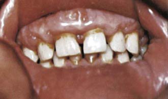
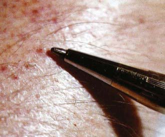
The early manifestations are irritability, loss of appetite, low-grade fever, and tenderness in the legs. These signs and symptoms are followed by leg swelling—most marked at the knees and the ankles—and pseudoparalysis. The infant might lie in the “pithed frog” position, with the hips and knees semiflexed and the feet rotated outward. Subperiosteal hemorrhages in the lower limb bones sometimes acutely increase the swelling and pain, and the condition might mimic acute osteomyelitis or arthritis. A “rosary” at the costochondral junctions and depression of the sternum are other typical features (Fig. 47-1). The angulation of scorbutic beads is usually sharper than the angulation of a rachitic rosary. Gum changes are seen in older children after teeth have erupted and are manifested as bluish purple, spongy swellings of the mucous membrane, especially over the upper incisors (Fig. 47-2). Anemia, a common finding in infants and young children with scurvy, is related to impaired iron absorption and coexistent hematopoietic nutrient deficiencies including iron, vitamin B12, and folate. Hemorrhagic manifestations of scurvy include petechiae, purpura, and ecchymoses at pressure points; epistaxis; gum bleeding; and the characteristic perifollicular hemorrhages (Fig. 47-3). Other manifestations are poor wound and fracture healing, hyperkeratosis of hair follicles, arthralgia, and muscle weakness.
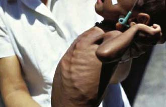
Figure 47-1 Scorbutic rosary.
(Courtesy of Dr J.D. MacLean, McGill Centre for Tropical Diseases, Montreal.)
Laboratory Findings and Diagnosis
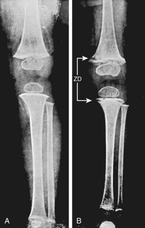
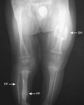
The diagnosis of vitamin C deficiency is usually based on the characteristic clinical picture, the radiographic appearance of the long bones, and a history of poor vitamin C intake. The typical radiographic changes occur at the distal ends of the long bones and are particularly common at the knees. The shafts of the long bones have a ground-glass appearance due to trabecular atrophy. The cortex is thin and dense, giving the appearance of pencil outlining of the diaphysis and epiphysis. The white line of Fränkel, an irregular but thickened white line at the metaphysis, represents the zone of well-calcified cartilage. The epiphyseal centers of ossification also have a ground-glass appearance and are surrounded by a sclerotic ring (Fig. 47-4). The more specific but late radiologic feature of scurvy is a zone of rarefaction under the white line at the metaphysis. This zone of rarefaction (Trumerfeld zone), a linear break in the bone that is proximal and parallel to the white line, represents area of debris of broken-down bone trabeculae and connective tissue. A Pelkan spur is a lateral prolongation of the white line and may be present at cortical ends. Epiphyseal separation can occur along the line of destruction, with either linear displacement or compression of the epiphysis against the shaft. Subperiosteal hemorrhages are not visible using plain radiographs during the active phase of scurvy. However, during healing the elevated periosteum becomes calcified and radiopaque, giving a dumbbell or club shape to the affected bone (Fig. 47-5). MRI can demonstrate acute as well as healing subperiosteal hematomas along with periostitis, metaphyseal changes, and heterogeneous bone marrow signal intensity.
Choh CTP, Rai S, Abdelhamid M, et al. Unrecognised scurvy. BMJ. 2010;340:150-151.
Choi SW, Park SW, Kwon YS, et al. MR imaging in a child with scurvy: a case report. Korean J Radiol. 2007;8:443-447.
Davison GW, Ashton T, George L, et al. Molecular detection of exercise-induced free radicals following ascorbate prophylaxis in type 1 diabetes mellitus: a randomised controlled trial. Diabetologia. 2008;51:2049-2059.
Fain O. Musculoskeletal manifestations of scurvy. Joint Bone Spine. 2005;72:124-128.
Gropper SS, Smith JL, Groff JL, editors. Advanced nutrition and human metabolism, ed 4, Belmont, CA: Thomson Wadsworth, 2005.
Hemilä H, Chalker E, Treacy B, Douglas B: Vitamin C for preventing and treating the common cold, Cochrane Database Syst Rev (3):CD000980, 2007.
Hemilä H, Louhiala P: Vitamin C for preventing and treating pneumonia, Cochrane Database Syst Rev (1):CD005532, 2007.
Heymann WR. Scurvy in children. J Am Acad Dermatol. 2007;57:358-359.
Nobili V, Manco M, Devito R, et al. Lifestyle intervention and antioxidant therapy in children with nonalcoholic fatty liver disease: a randomized, controlled trial. Hepatology. 2008;48:119-128.
Olmedo JM, Yiannias JA, Windgassen EB, Gornet MK. Scurvy: a disease almost forgotten. Int J Dermatol. 2006;45:909-913.
Rumbold A, Crowther CA: Vitamin C supplementation in pregnancy, Cochrane Database Syst Rev (18):CD004072, 2005.


