Virus-Associated Lymphoma

Published on 04/03/2015 by admin

Filed under Hematology, Oncology and Palliative Medicine

Last modified 22/04/2025

Print this page

rate 1 star rate 2 star rate 3 star rate 4 star rate 5 star
Your rating: none, Average: 0 (0 votes)

This article have been viewed 1658 times

Chapter 36 Virus-Associated Lymphoma

Table 36-1 Epstein-Barr Virus-Associated Lymphoma

image

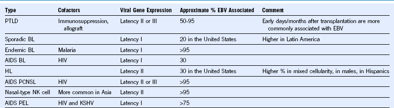
AIDS, Acquired immunodeficiency syndrome; BL, Burkitt lymphoma; EBV, Epstein-Barr virus; HIV, human immunodeficiency virus; HL, Hodgkin lymphoma; KSHV, Kaposi sarcoma—associated herpesvirus; NK, natural killer; PCNSL, primary central nervous system lymphoma; PEL, primary effusion lymphoma; PTLD, posttransplantation lymphoproliferative disorder.

image

Figure 36-1 EXAMPLES OF EPSTEIN-BARR VIRUS (EBV)—RELATED LYMPHOMAS.

Posttransplantation lymphoproliferative disorder (PTLD), polymorphic type, EBV+ occurring in the gastrointestinal tract of a 15-month-old girl following an orthotopic liver transplant (A and B). There was a mildly atypical lymphocytic infiltrate in the duodenal mucosa composed of small lymphocytes, plasma cells, and occasional large cells (A). The infiltrate was EBV+ as demonstrated by in situ hybridization for Epstein-Barr—encoded RNA (EBER) (B). Hodgkin lymphoma, nodular sclerosing type, EBV+ (C and D). The hematoxylin-eosin section shows a portion of a lymph node from a cervical lymph node biopsy of an 8-year-old girl. There are bands of sclerosis forming a cellular nodule, and within the nodule there is a mixed inflammatory infiltrate and scattered large cells with contracted cytoplasm consistent with lacunar cells (C). The immunophenotype of these cells was that of classic Hodgkin lymphoma, and the cells were EBER+ (D). EBV is seen more frequently in mixed cellularity Hodgkin lymphoma but can be seen in 10% to 40% of cases of nodular sclerosing type. It is even more frequent in cases associated with human immunodeficiency virus (HIV) and in resource-poor regions. Burkitt lymphoma, sporadic type, EBV+ (E and F). A section of the cervical lymph node biopsy of a 9-year-old girl with a rapidly enlarging neck mass is shown. The section illustrates the classic morphologic features of Burkitt lymphoma with a “starry sky” appearance, sheets of intermediate-sized cells with multiple small nucleoli and high mitotic rate. The cells were virtually all EBER+ (F). EBV can be seen in about 20% to 30% of cases of sporadic Burkitt lymphoma and is essentially always positive in endemic cases.

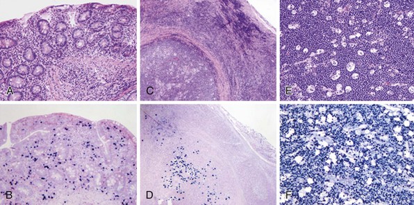
Epstein-Barr Virus-Associated Positive Posttransplant Lymphoproliferative Disorder
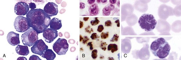
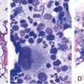
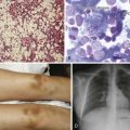
A 55-year-old renal transplant patient presents with acute renal failure 5 months after transplant. She is found on imaging to have an obstructing mass in the transplanted kidney. She undergoes kidney biopsy, and Epstein-Barr virus (EBV)—positive posttransplant lymphoproliferative disorder (PTLD) involving the transplanted organ is diagnosed. Treatment options include rituximab; decreasing immunosuppression (acknowledging the associated risk for organ rejection); changing immunosuppressive agents—switching a calcineurin inhibitor for a mammalian target of rapamycin (mTOR) inhibitor; combination chemotherapy; or, in the case of renal transplant, removal of the transplanted organ and withdrawal of immunosuppression.

Table 36-2 HIV-Associated Lymphoma

image

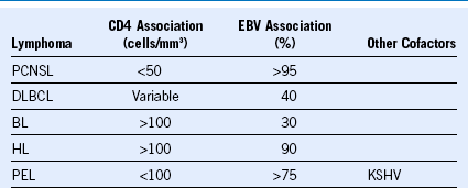
BL, Burkitt lymphoma; DLBCL, diffuse large B-cell lymphoma; EBV, Epstein-Barr virus; HIV, human immunodeficiency virus; HL, Hodgkin lymphoma; KSHV, Kaposi sarcoma—associated herpesvirus; PCNSL, primary central nervous system lymphoma; PEL, primary effusion lymphoma.