Villous Adenoma

Published on 13/07/2015 by admin

Filed under Radiology

Last modified 22/04/2025

Print this page

rate 1 star rate 2 star rate 3 star rate 4 star rate 5 star
Your rating: none, Average: 0 (0 votes)

This article have been viewed 3934 times

 Accounts for 5-15% of colonic adenomas

IMAGING

• Polypoid lesion with nodular or frond-like surface on BE or CT colonography
• Location: Rectosigmoid > cecum > ascending colon > stomach > duodenum
• Villous adenoma is 1 histological type of adenomatous polyps (true neoplasms)
• Risk of cancer is related to tumor size, location, and proportion of villous change in adenoma
• Greater risk of carcinoma in villous tumors of stomach and duodenum than colon

image Stomach: Carcinoma in 50% of lesions 2-4 cm and in 80% of lesions > 4 cm in size
image Colon: Invasive carcinoma in up to 45% of cases
• CT: Large villous adenoma

image Low-attenuation, minimally enhancing, irregular polypoid mass
image Corrugated, feathery appearance due to trapping of enteric contrast
• Transrectal US; plus CT or MR for staging

TOP DIFFERENTIAL DIAGNOSES

• Colon carcinoma
• Fecal mass

PATHOLOGY

• Malignant potential: Lesions < 1 cm (5%), 1-2 cm (10%), > 2 cm (53%)

CLINICAL ISSUES

• Asymptomatic, diarrhea, pain, rectal bleeding, or melena
• Lesion closer to rectum: More likely to have diarrhea, electrolyte loss (hypokalemia and hyponatremia)
image
(Left) Graphic shows a polypoid mass image 
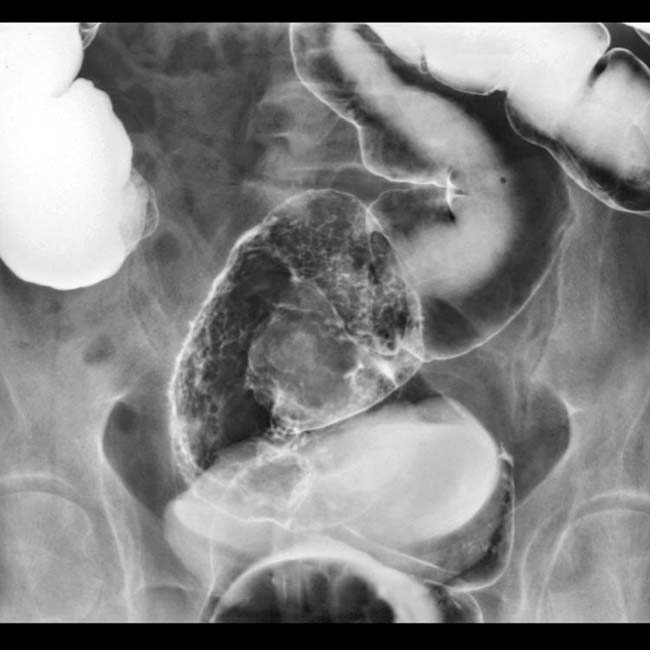
in the rectosigmoid colon having a shaggy, nodular surface, sometimes likened to the surface of a cauliflower.

image
(Right) Single contrast barium enema shows a large rectal mass image 
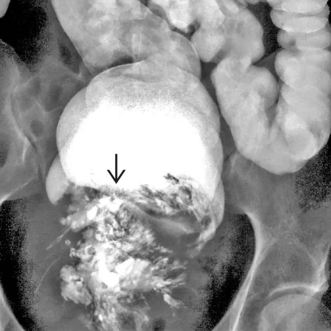
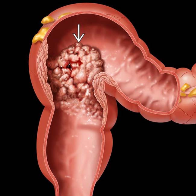
with a frond-like surface. Note the absence of a colonic obstruction, a typical feature of this soft and compressible tumor.
image
(Left) This 70-year-old man complained of frequent passage of watery stool, but had no symptoms of bowel obstruction. CT shows a large mass image 
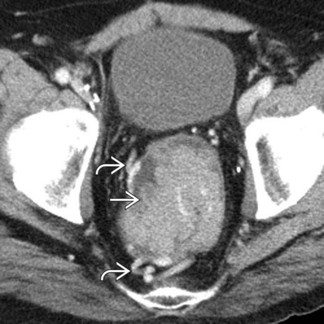
that fills the rectum. Note large vessels image within and draining the mass.

image
(Right) Coronal CT reformation in the same case shows the huge size of the mass image
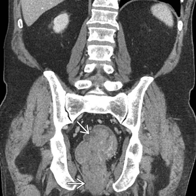
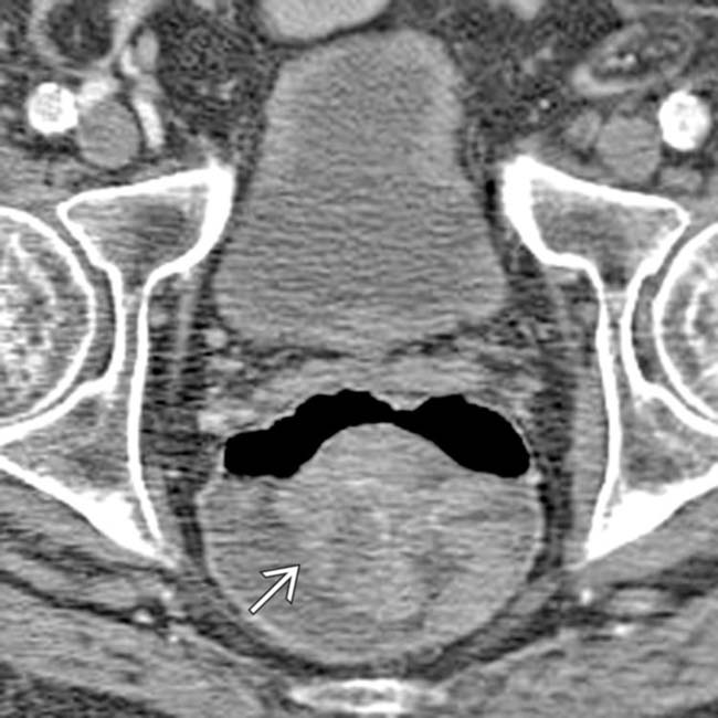
, but no definite signs of invasion through the rectal wall and no metastases. The resected villous adenoma had foci of frank carcinoma.

TERMINOLOGY

Synonyms

• Villous tumor

Definitions

• Adenomatous polyp that contains predominantly villous (“shaggy” surface) elements

IMAGING

General Features

• Best diagnostic clue

image Polypoid lesion with nodular or frond-like surface on barium enema or CT colonography
• Location

image Rectosigmoid > cecum > ascending colon > stomach > duodenum
• Size

image Range from < 1 to > 10 cm in diameter
image Giant villous tumor: 10-15 cm
• Morphology

image Cauliflower-like sessile growth with broad base or flat “carpet” lesion
• Other general features

image Villous adenoma is 1 histological type of adenomatous polyps (true neoplasms)

– Tubular adenoma: > 80% of neoplastic polyps
– Villous adenoma: 5-15% of colonic polyps, villous morphology in > 75% of lesion
– Tubulovillous adenoma: 5-15% of all colonic polyps
image As adenoma increases in size, degree of villous change usually increases
image Risk of cancer is related to size, location, and proportion of villous change in adenoma
image Greater risk of carcinoma in villous tumors of stomach and duodenum than colon

– Stomach: Carcinoma in 50% of lesions 2-4 cm and in 80% of lesions > 4 cm
– Duodenum: Carcinoma in 30-60% of villous tumors > 4 cm
– Colon: Carcinoma in situ in 10% and invasive carcinoma in up to 45% of cases

Radiographic Findings

• Fluoroscopic-guided double contrast barium enema

image 2 types of villous adenomas

– Polypoid mass
– “Carpet” lesion
image Polypoid mass

– May look cauliflower-like within colon
– Nodular, “lace,” or “soap bubble” pattern
– Due to trapping of barium between frond-like projections (interstices)
– Malignant transformation in bulky adenoma: Annular lesion with shelf-like, overhanging borders
image “Carpet” lesion

– Flat, lobulated lesion
– Localized or extensive
image Localized “carpet” lesion: Subtle alteration in surface texture
image Extensive “carpet” lesion: Involves large area of colon, encircling lumen

– En face: Fine nodular, reticular pattern with sharply demarcated border
– Profile: Irregular contour in contrast to smooth, fine contour of adjacent normal bowel
image Malignant transformation in “carpet” lesion (↑ risk)

– Radiolucent nodules surrounded by barium-filled grooves (produce fine nodular or reticular pattern)
– Polypoid carcinoma with surrounding mucosal change represents underlying adenoma
– Seen in rectum, cecum, ascending colon, stomach, and duodenum

CT Findings

• Large villous adenoma

image Low-attenuation, minimally enhancing, irregular polypoid mass
image Convolutional gyral enhancement pattern
image Corrugated, feathery appearance due to trapping of enteric contrast within interstices of villous adenoma
• CT colonography

image Appearance of villous tumors similar to that seen on barium enema
image CTC shows more polyps than barium enema

MR Findings

• Large villous adenoma

image T1WI: Low signal intensity mass with multiple frond-like projections and central cord-like structure
image T2WI: Frond-like projections will be more prominent
• Villous adenoma with more mucin-producing cells

image Short T1 and long T2 times
image Adenoma appears hyperintense on both T1- and T2WI

Ultrasonographic Findings

• Transrectal sonography

image Determines depth of invasion into colonic wall by adenoma

Imaging Recommendations

• CT colonography (or barium enema) for screening
• Transrectal US; plus CT or MR for staging

DIFFERENTIAL DIAGNOSIS

Colon Carcinoma

• Barium enema findings

image Early cancer: Sessile (plaque-like) lesion

– Typical early colon cancer
– Flat, protruding lesion with broad base and little elevation of mucosa (in profile view)
image Early cancer: Pedunculated lesion

– Short and thick polyp stalk
– Irregular or lobulated head of polyp
image Advanced cancer: Polypoid lesion (large)

– Dependent wall: Filling defect in barium pool
– Nondependent wall: Surface of tumor is etched in white
image Sessile and pedunculated polypoid cancers may be indistinguishable from villous adenoma
image Advanced cancer: Semiannular (“saddle”) lesion
image Advanced cancer: Annular (“apple core”) lesion

– Circumferential narrowing of bowel
– Shelf-like, overhanging borders (mucosal destruction)
• CT findings

image Asymmetric mural thickening ± irregular surface
image Extracolonic tumor extension

– Mass with irregular borders
– Extension from serosa to pericolic fat
– Loss of fat planes: Colon and adjacent muscles
image Metastases to regional mesenteric nodes
image Metastases to liver
• Diagnosis: Biopsy and histology

Fecal Mass

• Large, irregular colonic fecal impaction

image Most common location: Rectum
• Mimics large, cauliflower-like sessile polyp
• May cause bowel obstruction and proximal dilatation
• Usually seen in elderly, sedentary patients
• Diagnosis: Clinical history and colonoscopy

Intramural Benign Tumor, Colonic

• e.g., stromal tumors (leiomyoma, sarcoma, or GIST)
• Leiomyoma

image In profile

– Smooth surface, etched in white
– Borders: Right or obtuse angles with adjacent wall
image En face

– Seen as filling defect simulating polypoid type of villous adenoma
– Intraluminal surface: Abrupt, well-defined borders
• Leiomyosarcoma

image Bulky stromal tumors most frequently seen in rectum
image Broad-based mass simulating large villous adenoma
image Large tumors show ulceration or cavitation
image CT shows pericolonic extension (large extraluminal mass), liver, and peritoneal metastases
• Hypervascular on angiography
• Diagnosis: Biopsy

PATHOLOGY

General Features

• Etiology

image Villous adenoma or tumor

– Family history, idiopathic inflammatory disease
– Malignant potential: Lesions < 1 cm (5%), 1-2 cm (10%), > 2 cm (53%)

Gross Pathologic & Surgical Features

• Usually sessile

image May be polypoid, broad, flat, or carpet-like lesion
image Gray-tan lesion
• May have short, broad stalk and focal areas of hemorrhage or ulceration

Microscopic Features

• Frond-like papillary projections of adenomatous epithelium
• ± well-differentiated areas
• Carcinoma in situ, invasive cancer

CLINICAL ISSUES

Presentation

• Most common signs/symptoms

image Asymptomatic, diarrhea, pain, rectal bleeding, or melena
image Lesion closer to rectum: More likely to have diarrhea, electrolyte loss (hypokalemia)
• Lab data

image Guaiac positive stool
image Iron deficiency anemia
image Decreased serum protein, K+, Na+

– “McKittrick-Wheelock syndrome” if caused by rectal tumor, usually villous adenoma
image ± increased direct bilirubin levels (due to obstruction of ampulla of Vater by adenoma)
• Diagnosis: Endoscopy, biopsy, and histology

Demographics

• Age

image 60-70 years or older
• Gender

image M = F

Natural History & Prognosis

• Complications

image Malignant transformation or invasion; hemorrhage
• Prognosis

image Good: After removal of benign and carcinoma in situ adenoma
image Poor: Invasive carcinoma

Treatment

• Colonoscopic, endoscopic, or surgical resection

DIAGNOSTIC CHECKLIST

Consider

• Check for family history of colonic polyps and evaluate entire colon for synchronous lesions

Image Interpretation Pearls

• Cauliflower-like sessile mass with broad base or “carpet” lesion with reticular or “soap bubble” surface pattern
• Luminal obstruction is a late finding
image
(Left) Barium pool image from a double contrast barium enema shows a cauliflower-like mass image 
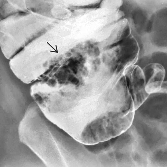
in the cecum, a typical appearance of a villous adenoma.

image
(Right) Spot film from an air contrast barium enema in the same patient shows the villous adenoma image 
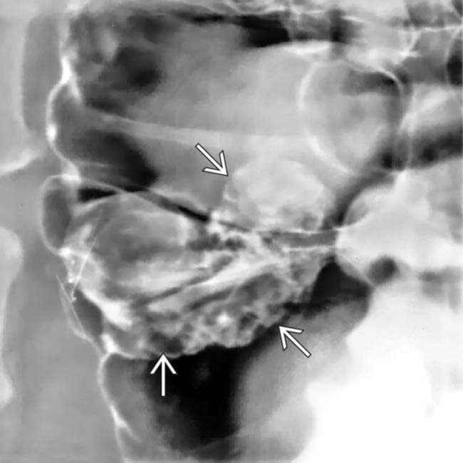
coated with barium and outlined by air on this view.
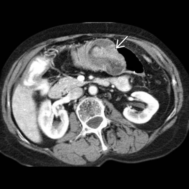
image
(Left) Axial CECT in an 85-year-old woman with acute abdominal pain shows a transverse colo-colonic intussusception due to a large lead point mass image.

image
(Right) More caudal CT section in the same patient shows the colonic mass image
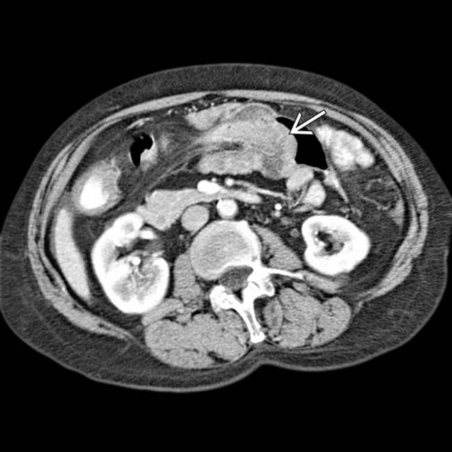
, a villous adenoma, as the lead point of the colonic intussusception.
image
(Left) Axial CECT in the same patient just proximal to the intussusception shows another smaller, lobulated, enhancing mass within the ascending colon image 
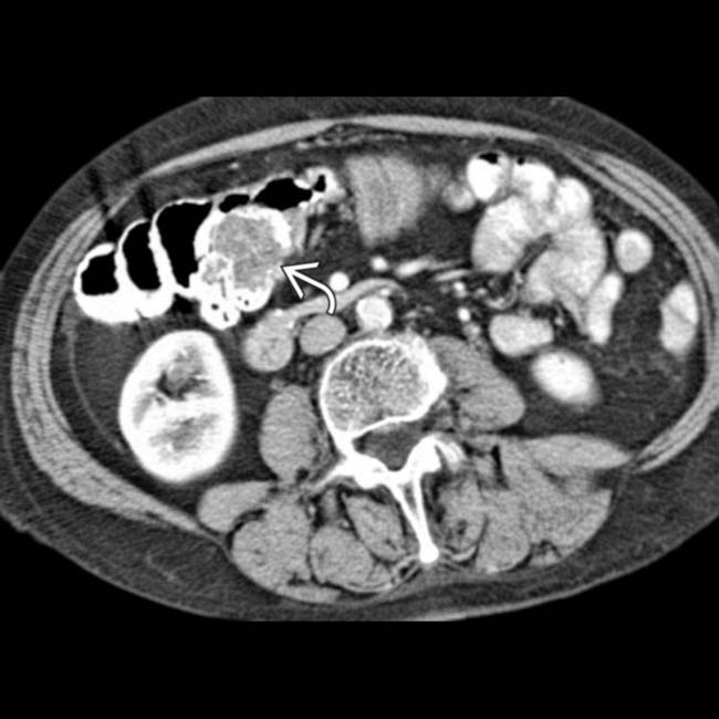
that proved to be a 2nd villous adenoma.

image
(Right) Single contrast BE in the same patient demonstrates temporary reduction of the intussusception, with the 2 villous adenomas image 
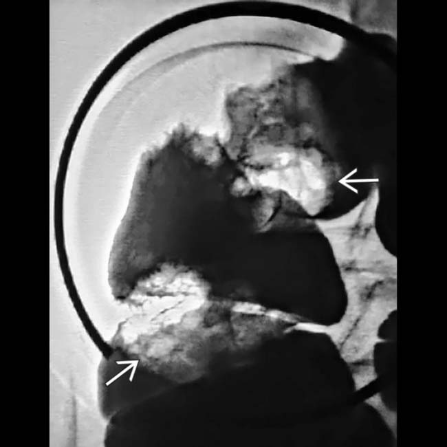
seen as masses with irregular surface contours within the barium pool. A right hemicolectomy and histopathology confirmed the diagnosis.
image
Single contrast BE shows a polypoid mass image 
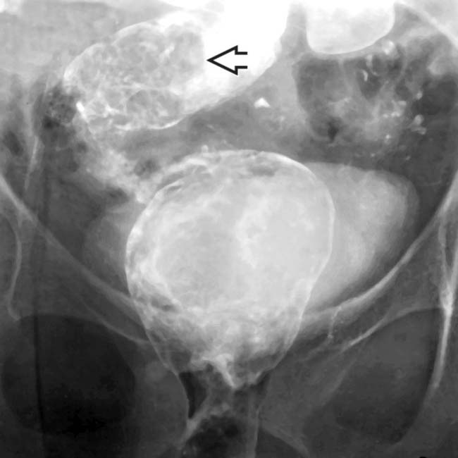
in the rectosigmoid colon with a very nodular surface. Barium within the rectum is diluted by mucus secreted by the tumor.
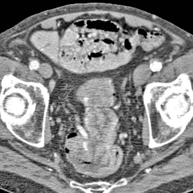
image
Axial CECT shows a large mass that fills the rectosigmoid colon with dilated stool-filled colon, noted more proximally.
image
Axial CECT shows a large polypoid mass image within the rectum.
image
Air contrast BE shows a rectal mass with a nodular surface that fills, but does not obstruct, the rectal lumen.

SELECTED REFERENCES

1. Serra-Aracil, X, et al. Transanal endoscopic surgery with total wall excision is required with rectal adenomas due to the high frequency of adenocarcinoma. Dis Colon Rectum. 2014; 57(7):823–829.

2. Bozkurt, N, et al. Adenoma with rectal villous diarrhoea and severe hypokalaemia (McKittrick-Wheelock syndrome). Br J Hosp Med (Lond). 2013; 74(11):648–649.

3. Sanchez Garcia, S, et al. Hypersecretory villous adenoma as the primary cause of an intestinal intussusception and McKittrick-Wheelock syndrome. Can J Gastroenterol. 2013; 27(11):621–622.

4. Sosna, J, et al. Critical analysis of the performance of double-contrast barium enema for detecting colorectal polyps > or = 6 mm in the era of CT colonography. AJR Am J Roentgenol. 2008; 190(2):374–385.

5. Ferrucci, JT. Double-contrast barium enema: use in practice and implications for CT colonography. AJR Am J Roentgenol. 2006; 187(1):170–173.

6. Taylor, SA, et al. Comparison of radiologists’ confidence in excluding significant colorectal neoplasia with multidetector-row CT colonography compared with double contrast barium enema. Br J Radiol. 2006; 79(939):208–215.

7. Johnson, CD, et al. Comparison of the relative sensitivity of CT colonography and double-contrast barium enema for screen detection of colorectal polyps. Clin Gastroenterol Hepatol. 2004; 2(4):314–321.

8. Smith, TR, et al. CT appearance of some colonic villous tumors. AJR Am J Roentgenol. 2001; 177(1):91–93.

9. Chung, JJ, et al. Large villous adenoma in rectum mimicking cerebral hemispheres. AJR Am J Roentgenol. 2000; 175(5):1465–1466.

Cunnane, ME, et al. Small flat umbilicated tumors of the colon: radiographic and pathologic findings. AJR Am J Roentgenol. 2000; 175(3):747–749.

Levine, MS, et al. Diagnosis of colorectal neoplasms at double-contrast barium enema examination. Radiology. 2000; 216(1):11–18.

Iida, M, et al. Endoscopic features of villous tumors of the colon: correlation with histological findings. Hepatogastroenterology. 1990; 37(3):342–344.