[level-membership-for-radiology-category] High-density material within gallbladder (GB)
– CT > 10x more sensitive than radiographs in detecting slight differences in density
– Mild to moderate opacification of GB bile on day after CECT or angiogram is a normal and expected finding
Does not necessarily imply abnormal renal function
– Dense opacification on CT indicates ↓ renal function
Increased attenuation of ascites fluid
– Visible a few hours after IV administration of contrast
– Results from hepatobiliary and peritoneal contrast excretion into ascites
– Mildly increased density of ascites fluid may not indicate renal impairment
– Significantly ↑ attenuation suggests ↓ renal function
Increased attenuation of pericardial fluid
– Most common in first few hours after IV contrast
– Mildly ↑ attenuation can be normal finding
• Radiographic findings
Opacification of bile within GB after IV contrast always implies impaired or delayed renal function
Mild to moderate vicarious excretion is visible on CT in normal patients, but is insufficient to be visible on radiographs
Often associated with prolonged nephrograms suggesting acute tubular necrosis or other causes of acute kidney injury
TOP DIFFERENTIAL DIAGNOSES
• Milk of calcium bile, gallstones, or sludge
• Iatrogenic high density bile due to cholangiography
• Hemobilia
• Exudative ascites or hemoperitoneum
DIAGNOSTIC CHECKLIST
• Mild to moderate ↑ density of bile or ascites on CT some hours following parenteral administration of contrast medium may be normal
Greater degrees of opacification of bile or ascites usually indicates renal dysfunction
Dense GB bile evident on plain films of abdomen usually indicates renal dysfunction
Dense GB bile with prolonged nephrograms suggests acute tubular necrosis or other cause of acute kidney injury
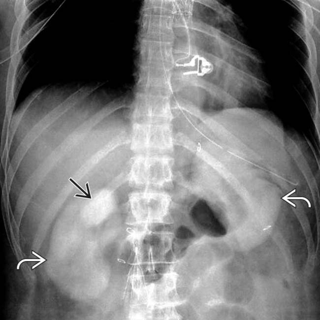
(Left) Radiograph in a patient with acute renal failure following coronary angiography shows dense opacification of the gallbladder bile and both kidneys many hours after the angiogram, compatible with vicarious excretion and shock- or contrast-induced nephropathy.
(Right) Axial NECT following embolization of a ruptured splenic artery aneurysm shows dense opacification of bile in the gallbladder and persistent nephrograms (left > right), the latter due to acute tubular necrosis.
(Left) Axial NECT in a patient with sickle cell disease who had a recent angiogram shows persistent opacification of the ureters due to slow excretion from the impaired kidneys. The bile is dense due to vicarious excretion.
(Right) Axial NECT a few hours after angiography shows persistent enhancement of the kidneys , compatible with shock- or contrast-induced nephropathy. The ascites measured 60 HU due to vicarious excretion of contrast medium from the peritoneum as a means of compensating for the failed renal excretion.
Axial NECT shows dense bile within the gallbladder due to vicarious excretion of the contrast medium that was used for a previous angiogram. The patient was in shock as a result of abdominal bleeding.
[/level-membership-for-radiology-category][not-level-membership-for-radiology-category] High-density material within gallbladder (GB)
– CT > 10x more sensitive than radiographs in detecting slight differences in density
– Mild to moderate opacification of GB bile on day after CECT or angiogram is a normal and expected finding
Does not necessarily imply abnormal renal function
– Dense opacification on CT indicates ↓ renal function
Increased attenuation of ascites fluid
– Visible a few hours after IV administration of contrast
– Results from hepatobiliary and peritoneal contrast excretion into ascites
– Mildly increased density of ascites fluid may not indicate renal impairment
– Significantly ↑ attenuation suggests ↓ renal function
Increased attenuation of pericardial fluid
– Most common in first few hours after IV contrast
– Mildly ↑ attenuation can be normal finding
• Radiographic findings
Opacification of bile within GB after IV contrast always implies impaired or delayed renal function
Buy Membership for Radiology Category to continue reading. Learn more here


 and both kidneys
  many hours after the angiogram, compatible with vicarious excretion and shock- or contrast-induced nephropathy.
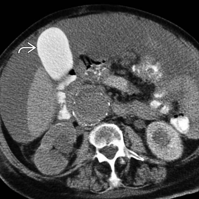
 and persistent nephrograms (left > right), the latter due to acute tubular necrosis.
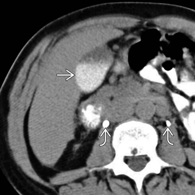
 due to slow excretion from the impaired kidneys. The bile is dense
  due to vicarious excretion.
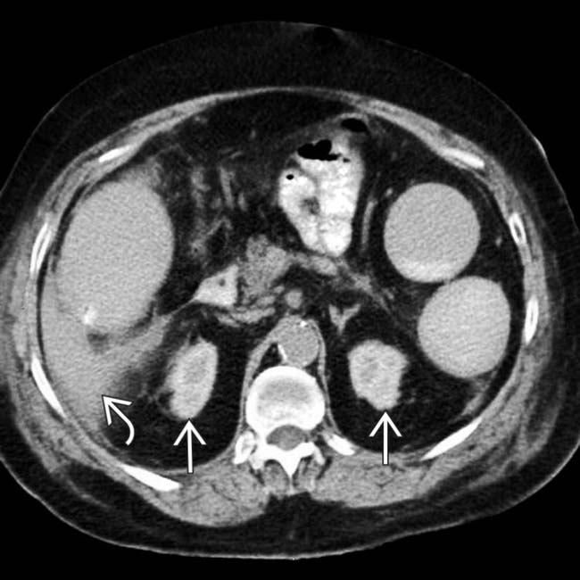
 , compatible with shock- or contrast-induced nephropathy. The ascites
  measured 60 HU due to vicarious excretion of contrast medium from the peritoneum as a means of compensating for the failed renal excretion.
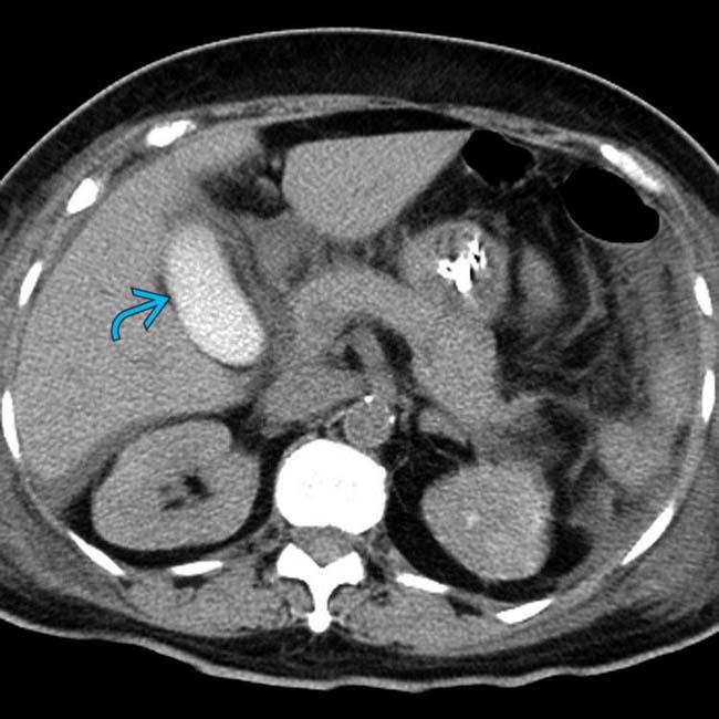
 due to vicarious excretion of the contrast medium that was used for a previous angiogram. The patient was in shock as a result of abdominal bleeding.
