1. Gao, H, et al. Definitive diagnosis of hepatic sinusoidal obstruction syndrome induced by pyrrolizidine alkaloids. J Dig Dis. 2012; 13(1):33–39.
2. Jevtic, D, et al. Veno-occlusive disease in pediatric patients after hematopoietic stem cell transplantation: relevance of activated coagulation and fibrinolysis markers and natural anticoagulants. J Pediatr Hematol Oncol. 2011; 33(3):227–234.
3. Lin, G, et al. Hepatic sinusoidal obstruction syndrome associated with consumption of Gynura segetum. J Hepatol. 2011; 54(4):666–673.
4. Mahgerefteh, SY, et al. Radiologic imaging and intervention for gastrointestinal and hepatic complications of hematopoietic stem cell transplantation. Radiology. 2011; 258(3):660–671.
5. Menke, J. Education and imaging. Hepatobiliary and pancreatic: sinusoidal obstruction syndrome during chemotherapy. J Gastroenterol Hepatol. 2011; 26(6):1083.
6. Sanei, MH, et al. Acute cellular rejection resulting in sinusoidal obstruction syndrome and ascites postliver transplantation. Transplantation. 2011; 92(10):1152–1158.
7. Sebagh, M, et al. Significance of isolated hepatic veno-occlusive disease/sinusoidal obstruction syndrome after liver transplantation. Liver Transpl. 2011; 17(7):798–808.
8. Rubbia-Brandt, L. Sinusoidal obstruction syndrome. Clin Liver Dis. 2010; 14(4):651–668.


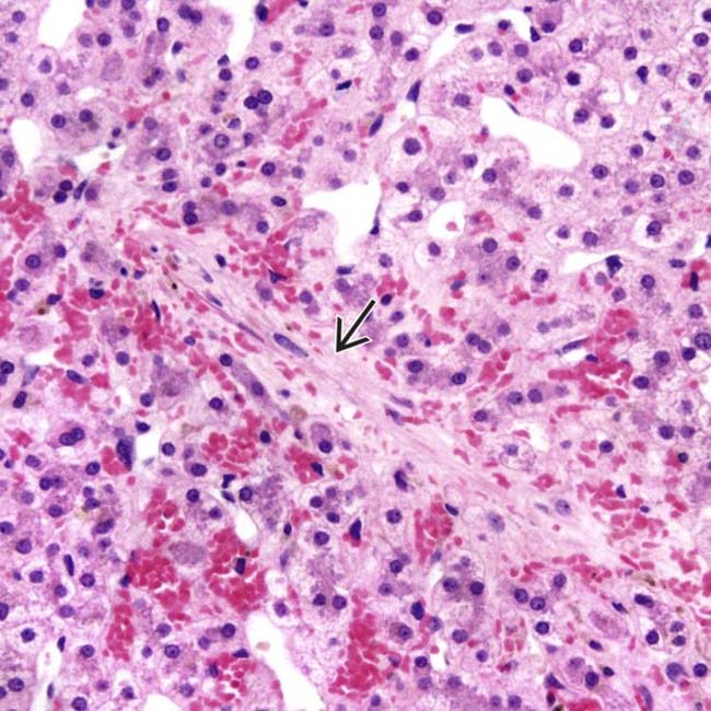
 accompanied by centrizonal sinusoidal congestion is seen in venoocclusive disease (VOD). (Courtesy S. Kakar, MD.)
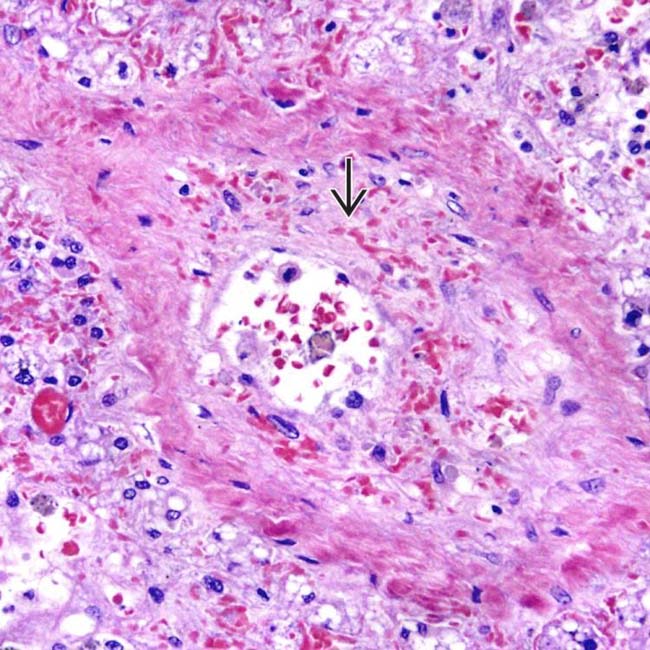
 led to partial occlusion of the lumen of a small hepatic vein in venoocclusive disease. (Courtesy S. Kakar, MD.)
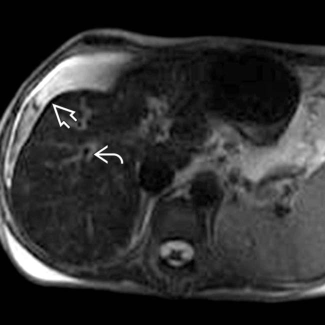
 and periportal edema
  , which are nonspecific findings. Liver biopsy showed sinusoidal congestion and fibrotic venules.
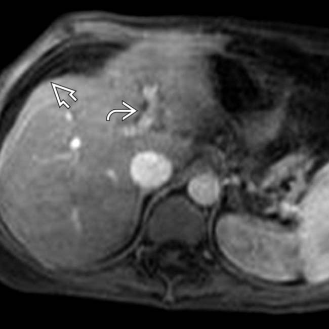
 and periportal edema
  .
.


