[level-membership-for-dermatology-category]Chapter 23
Vascular tumors
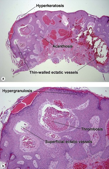
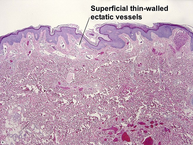
Angioma serpiginosum

Angioma serpiginosum presents as a progressive vascular lesion on a woman’s leg. The ectatic vessels begin as minute puncta in clusters, but merge to form a serpiginous array. They often bleed freely when traumatized.
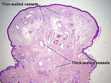
Venous lake
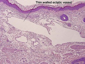
Venous lakes are common on the lips and ears of older patients. They may appear very dark, but blanch easily when compressed.
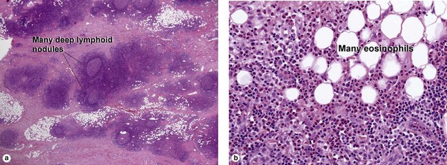
Angiolymphoid hyperplasia with eosinophilia
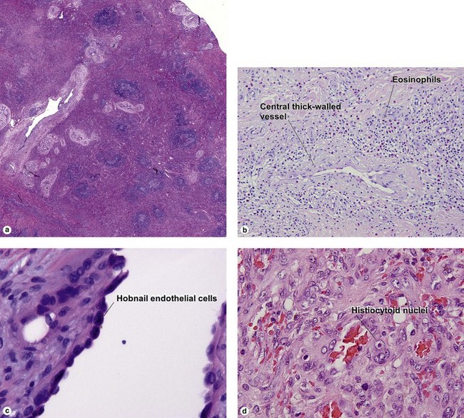
Subtypes include histiocytoid and epithelioid hemangioma. Histiocytoid endothelial cells are common.
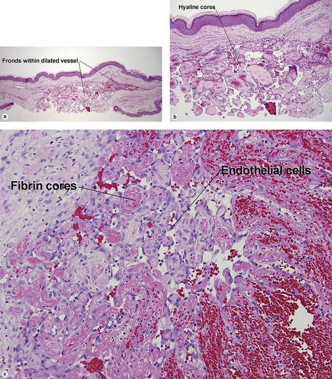
Intravascular papillary endothelial hyperplasia of Masson (IPEH)
IPEH can occur in any vascular space. It is common in angiokeratomas.
Targetoid hemosiderotic hemangioma (hobnail hemangioma)
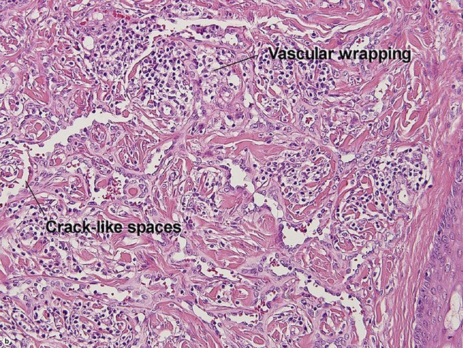
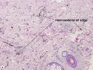
Targetoid hemosiderotic hemangiomas probably arise as a result of trauma to a pre-existing hemangioma.
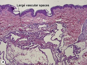
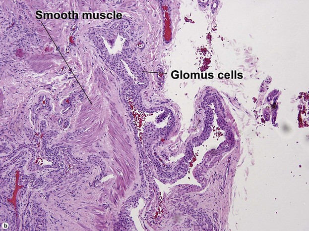
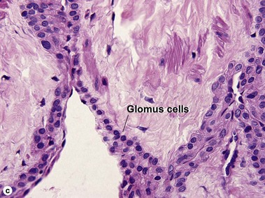
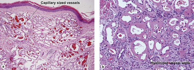
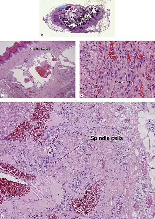
Eccrine angiomatous hamartoma
Usually a solitary bluish nodule. Often involves acral sites, although the example shown was on the back. May be tender.
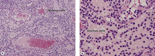
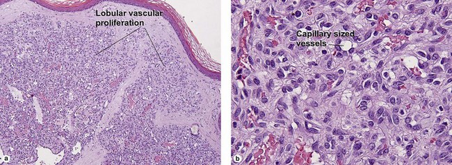
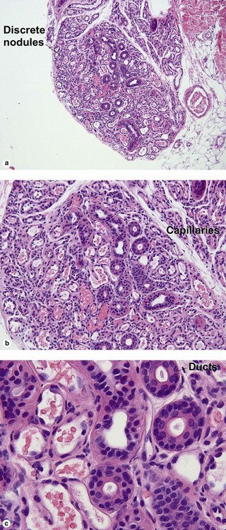
Glomeruloid hemangioma
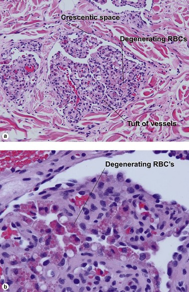
Glomeruloid hemangioma is associated with POEMS syndrome (Crow–Fukase syndrome, polyneuropathy, organomegaly, endocrinopathy, M protein, and skin changes) and Castleman’s disease. Two types of endothelial cell have been noted: cells with large vesicular nuclei, an open chromatin pattern, and large amount of cytoplasm, and a second population with small basal nuclei, a dense chromatin pattern, and scant cytoplasm. Lesions not associated with POEMS syndrome have been referred to as papillary hemangioma.
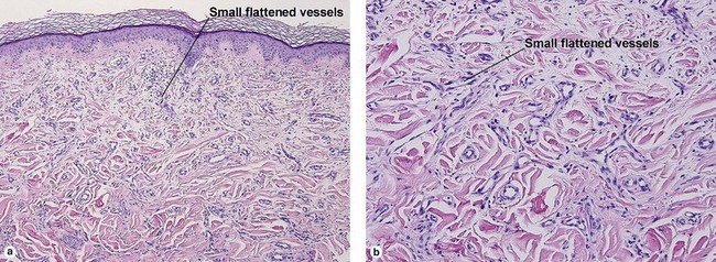
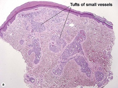
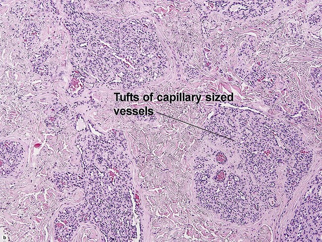
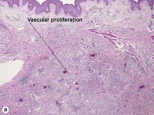
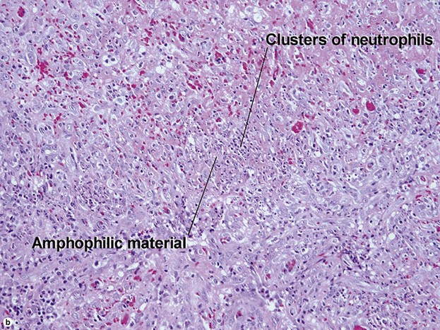
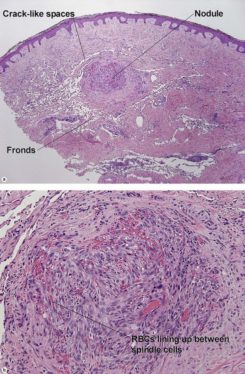
Kaposiform hemangioendothelioma
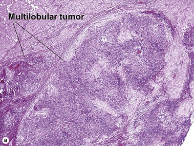
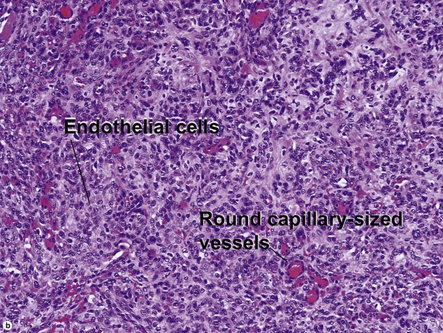
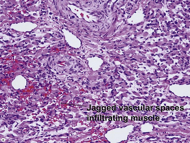
Kasabach–Merritt coagulopathy is usually associated with kaposiform hemangioendothelioma or tufted angioma, and hybrid tumors have been described.
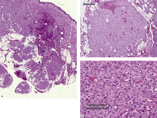
Hemangiopericytoma
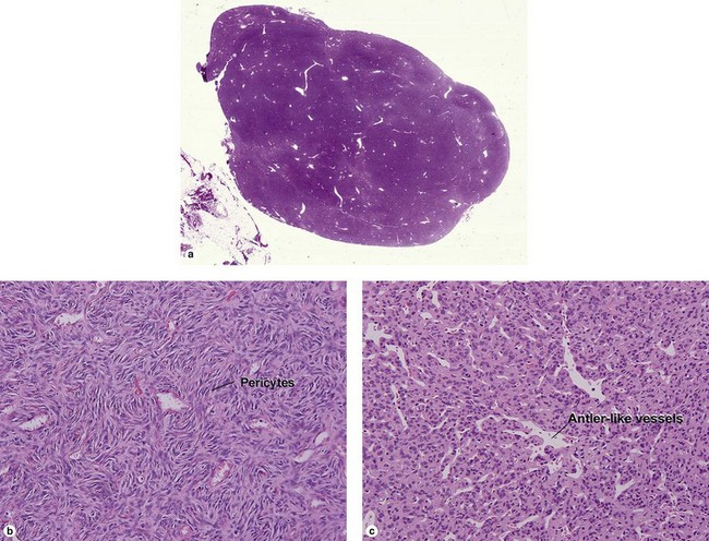
Hemangiopericytomas occur in the skin and soft tissues. It may be difficult to distinguish between benign and malignant hemangiopericytoma histologically. Large size and higher mitotic rate suggest a malignant potential. Infantile tumors are typically cutaneous, with a good prognosis.
Epithelioid hemangioendothelioma
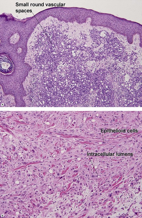
Epithelioid hemangioendotheliomas tend to occur on the extremities of young people. They are best regarded as low-grade malignancies.
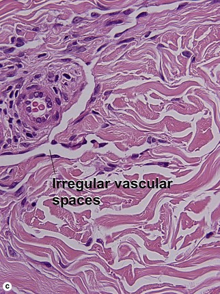
Retiform hemangioendothelioma
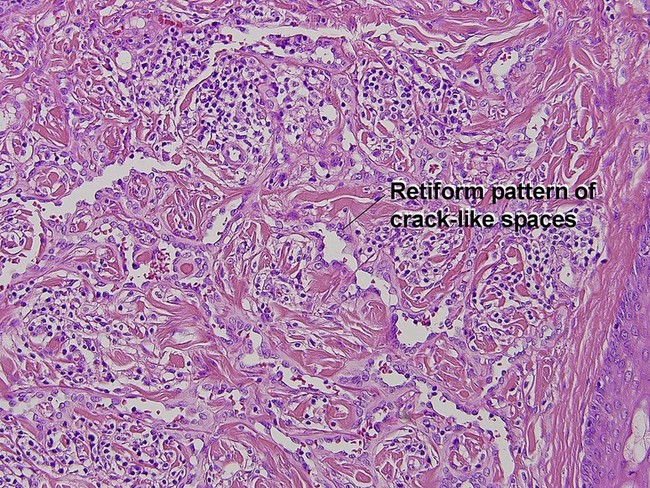
Retiform hemangioendothelioma is a low-grade angiosarcoma that occurs mostly on the extremities of young adults.
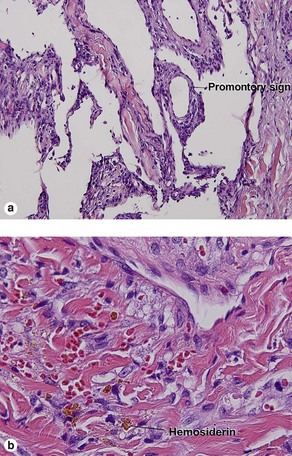
Angiosarcoma
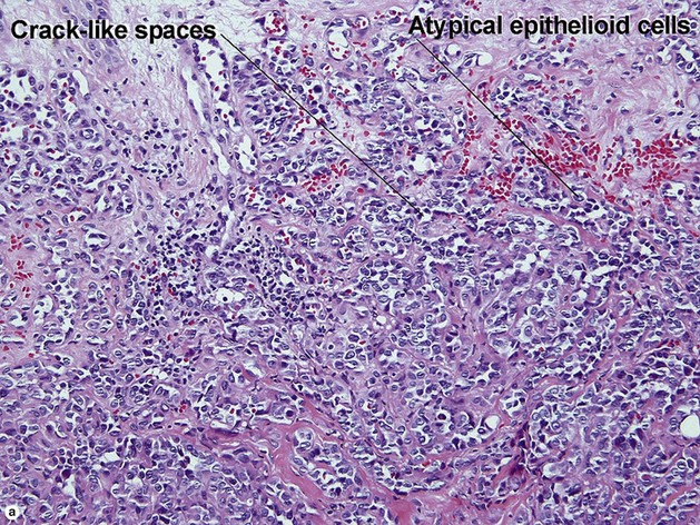

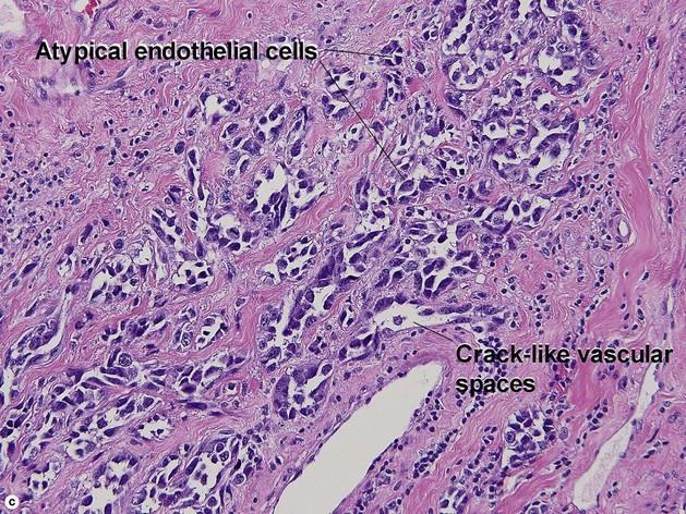
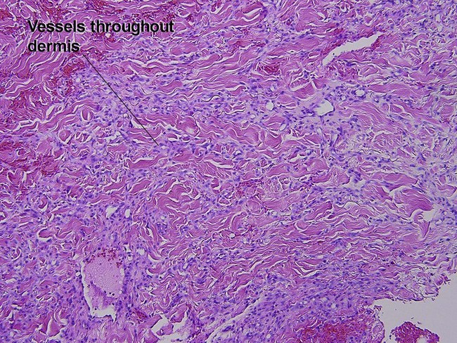
Angiosarcomas typically appear as bruise-like lesions on the forehead or scalp of an older patient. Epithelioid variants may be nodular. Stewart–Treves syndrome is angiosarcoma in the setting of a lymphedematous limb. Often, there is a history of radiation therapy.
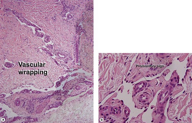
Kaposi’s sarcoma
Alvarez-Mendoza, A, Lourdes, TS, Ridaura-Sanz, C, et al. Histopathology of vascular lesions found in Kasabach-Merritt syndrome: review based on 13 cases. Pediatr Dev Pathol. 2000; 3(6):556–560.
Berenguer, B, Mulliken, JB, Enjolras, O, et al. Rapidly involuting congenital hemangioma: clinical and histopathologic features. Pediatr Dev Pathol. 2003; 6(6):495–510.
Billings, SD, Folpe, AL, Weiss, SW. Epithelioid sarcoma-like hemangioendothelioma. Am J Surg Pathol. 2003; 27(1):48–57.
Chu, CY, Hsiao, CH, Chiu, HC. Transformation between Kaposiform hemangioendothelioma and tufted angioma. Dermatology. 2003; 206(4):334–337.
Espat, NJ, Lewis, JJ, Leung, D, et al. Conventional hemangiopericytoma: modern analysis of outcome. Cancer. 2002; 95(8):1746–1751.
Kishimoto, S, Takenaka, H, Shibagaki, R, et al. Glomeruloid hemangioma in POEMS syndrome shows two different immunophenotypic endothelial cells. J Cutan Pathol. 2000; 27(2):87–92.
Mentzel, T, Partanen, TA, Kutzner, H. Hobnail hemangioma (“targetoid hemosiderotic hemangioma”): clinicopathologic and immunohistochemical analysis of 62 cases. J Cutan Pathol. 1999; 26(6):279–286.
Nayler, SJ, Rubin, BP, Calonje, E, et al. Composite hemangioendothelioma: a complex, low-grade vascular lesion mimicking angiosarcoma. Am J Surg Pathol. 2000; 24(3):352–361.
Pelle, MT, Pride, HB, Tyler, WB. Eccrine angiomatous hamartoma. J Am Acad Dermatol. 2002; 47(3):429–435.
Pellegrini, AE, Drake, RD, Qualman, SJ. Spindle cell hemangioendothelioma. J Cutan Pathol. 1995; 22:173.
Reis-Filho, JS, Paiva, ME, Lopes, JM. Congenital composite hemangioendothelioma: case report and reappraisal of the hemangioendothelioma spectrum. J Cutan Pathol. 2002; 29(4):226–231.
Requena, L, Sangueza, OP. Cutaneous vascular neoplasms. Part II. J Am Acad Dermatol. 1997; 37:887.
Requena, L, Sangueza, OP. Cutaneous vascular neoplasms. Part III. J Am Acad Dermatol. 1998; 38:143.
Requena, L, Kutzner, H. Hemangioendothelioma. Semin Diagn Pathol. 2013; 30(1):29–44.
Sangüeza, OP. Update on vascular neoplasms. Dermatol Clin. 2012; 30(4):657–665.
[/level-membership-for-dermatology-category][not-level-membership-for-dermatology-category]Chapter 23
Vascular tumors
Angioma serpiginosum
Angioma serpiginosum presents as a progressive vascular lesion on a woman’s leg. The ectatic vessels begin as minute puncta in clusters, but merge to form a serpiginous array. They often bleed freely when traumatized.
Venous lake
Venous lakes are common on the lips and ears of older patients. They may appear very dark, but blanch easily when compressed.
