Urinary tract
Methods of imaging the urinary tract
(a) CT for urological diagnosis and urological cancer staging
(b) CT for characterization of renal lesion
5. Magnetic resonance imaging (MRI):
6. Micturating cystography and cystourethrography
8. Retrograde pyeloureterography
9. Percutaneous renal procedures:
Intravenous excretion urography (IVU)
The technique is less frequently used than in the past and is now largely replaced by CT, MRI or US.
Contraindications
See Chapter 2 – general contraindications to intravenous (i.v.) water-soluble contrast media and ionizing radiation. In patients with contrast medium allergies alternative modalities such as ultrasound or MR can be considered. Patients with impaired renal function, particularly those with diabetes should be prepared with oral or intravenous hydration, or an alternative imaging modality considered. See Chapter 2.
Preliminary images
If necessary, the location of overlying opacities may be further determined by:
• supine AP film of the renal areas, in expiration. The X-ray beam is centred in the mid-line at the level of the lower costal margin or
• 35° posterior oblique views (side of interest towards the film) or
The examination should not proceed further until these images have been reviewed by the radiologist or radiographer and deemed satisfactory.
Images
1. Immediate film. AP of the renal areas. This film is exposed 10–14 s after the injection (approximate ‘arm-to-kidney’ time). It aims to show the nephrogram at its most dense, i.e. the renal parenchyma opacified by contrast medium in the renal tubules. Tomography may assist in evaluation of the renal outline or possible masses (or ultrasound if subsequently available).
2. 5-min film. AP of the renal areas. This film gives an initial assessment of pathology – specifically the presence or absence of obstruction before administering compression.
(a) after recent abdominal surgery
(c) if there is a large abdominal mass or aortic aneurysm
(d) when the 5-min film shows already distended calyces indicative of obstruction.
3. 10-min film. AP of the renal areas. There is usually adequate distension of the pelvicalyceal systems with opaque urine by this time. Compression is released when satisfactory demonstration of the pelvicalyceal system has been achieved. If the compression film is inadequate the compression should be checked and repositioned if necessary and a further 50 ml of contrast medium administered and the film after 5 minutes.
4. Release film. Supine AP abdomen taken immediately after release of compression. This film is taken to show the ureters. If this film is satisfactory, the patient is asked to empty their bladder.
5. After micturition film. Full-length supine AP abdomen. The aims of this film are to assess bladder emptying, to demonstrate drainage of the upper tracts, to aid the diagnosis of bladder tumours, to confirm ureterovesical junction calculi and, uncommonly, to demonstrate a urethral diverticulum in females.
Additional images
1. 35° posterior obliques of the kidneys, ureters or bladder – for equivocal collecting system lesions or localization of calculi.
2. Tomography – if renal outlines are not well seen.
3. Prone abdomen following the release film – may improve visualization of distal ureters by making them more dependent.
4. Delayed films at increasing (doubling of time intervals) up to 24 h after injection in renal obstruction.
Ultrasound of the urinary tract
Indications
Technique
1. Patient supine, right (RAO) and left anterior oblique (LAO) positions or lateral for kidneys. The kidneys are scanned longitudinally in an oblique coronal plane supplemented by transverse sections perpendicular to the axis. The right kidney may be scanned through the liver and posteriorly in the right loin. The left kidney is harder to visualize anteriorly, but can be visualized from a lateral approach. In difficult cases the patient should lie on their side with a pillow under the left loin to widen the space between the rib cage and pelvis.
2. The length of the kidney measured by US is 1–2 cm smaller than that measured at excretion urography because there is no geometric magnification. With US measurement, care must be taken to ensure that the true longitudinal lengthwise measurement is obtained. The range of lengths of the normal kidneys is 9–12 cm, and the difference between each kidney should be less than 1–2 cm.
3. The bladder is scanned suprapubically in transverse and longitudinal planes. Measurements taken of the three orthogonal diameters before and after micturition enable an approximate volume to be calculated by multiplying the three diameters and applying a conversion factor. (A conversion factor is usually pre-programmed into modern ultrasound machines.)
4. Renal transplants are usually located in the right or left iliac fossa. These usually lie fairly superficially and are easy to evaluate using oblique planes and gentle pressure to displace overlying bowel loops.
5. The native or transplant kidneys can be evaluated for vascular pathology using Doppler techniques.
• Renal artery stenosis is diagnosed by direct Doppler interrogation of the main renal arteries from a transabdominal approach. Elevated peak systolic velocities greater than 200 cm s–1 are suggestive of a >50% stenosis. Alternatively, as the main renal arteries in the native kidneys are often hard to visualize, the intrarenal arteries can be evaluated from a flank approach for downstream changes in waveform – the tardusparvus pattern;1 a slow rise (tardus) to a reduced peak (parvus) producing a prolonged acceleration time (a value >70 ms is indicative of a severe stenosis).
• Renal vein thrombosis is diagnosed by absent colour Doppler venous flow, direct visualization of thrombus within the distended vein, and a raised resistive index with reversal of arterial diastolic flow within the intrarenal arteries.
Computed tomography of the urinary tract
Indications
1. Renal colic/renal stone disease
5. Staging and follow-up of renal, collecting system or prostatic cancer (local staging of prostatic cancer is performed using thin-section MRI)
6. Investigation of renal tract obstruction
7. CT angiography may be used to assess renal vessels for suspected renal artery stenosis or arterio-venous fistula or malformation.
Techniques
Standard diagnostic CT
3. Scanogram of chest, abdomen and pelvis as appropriate
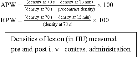
5. Scans obtained approximately 70 s (portal venous phase) after i.v. contrast (arterial phase scans of the liver (20–25 s after i.v. contrast) may be appropriate in those patients with suspected metastatic renal cancer who may have hypervascular liver metastases.)
Adrenal lesion characterization CT
Indication: adrenal mass is suspected or needs characterization.
Technique: Unenhanced CT of the abdomen to enable measurement of attenuation of any adrenal mass.
(Alternatively chemical shift MRI may be used for characterization.)
CT KUB
1. No intravenous or oral contrast is given.
2. Patient supine. (Some authorities advise prone scanning to differentiate if stones are impacted at the vesicoureteric junction or have passed into the bladder.)
3. A low-radiation-dose technique is used to scan from the top of the kidneys to include the bladder base with a slice thickness of 5 mm or less, as determined by CT scanner. (Due to the low-dose nature of the scan and the absence of i.v. and oral contrast, the scan has a very limited role in identifying pathology other than renal tract calculus disease and should not be used indiscriminately for investigation of non-specific abdominal pain.)
CT urogram (CTU)
1. An oral water load of 500–1000 ml 30–60 minutes before injection is recommended to ensure a diuresis and collecting system dilatation. No positive oral contrast
3. Initial low-dose unenhanced scans of urinary tract (CT KUB) to determine if renal tract calculus disease is present
4. Low osmolar contrast material (LOCM) 300 mg I ml–1 100 ml is given as bolus intravenously
5. Thin-section (usually 1 mm) scans are obtained from diaphragm to lower poles of kidneys during nephrographic/parenchymal enhancement phase (100 s following start of bolus injection). Alternatively, scan may instead be acquired during the portal venous phase (70 s) but normal corticomedullary differentiation may make small tumours difficult to appreciate
6. Delayed thin-section (1 mm) scans are acquired from upper pole of kidneys to bladder base 20 min after contrast injection, to examine collecting systems and ureters
7. Source images are reviewed along with multiplanar reconstructions. Post-processing with maximum-intensity projections and surface-shaded displays may be helpful, especially for demonstration.
CT angiography
Technique
1. No oral iodinated contrast used
2. Scan from the upper pole of the kidneys to the aortic bifurcation. Modern scanners are fast enough to produce high-quality studies of the whole abdomen
4. 100–150 ml i.v. contrast medium (LOCM 300) injected at 3–4 ml s–1
5. Use of bolus tracking/triggering devices or timing test injections is recommended to ensure appropriate timing. Otherwise scans are initiated after a preset empiric delay of 20–25 s from start of contrast material injection
6. Source axial scans are supplemented by multiplanar reconstructions and maximum intensity projection and volume-rendered surface-shaded display postprocessing.
Adrenal Lesion Characterization CT
Johnson, PT, Horton, KM, Fishman, EK. Adrenal imaging with multidetector CT: evidence-based protocol optimization and interpretive practice. RadioGraphics. 2009; 29:1319–1331.
Low, G, Dhliwayo, H, Lomas, DJ. Adrenal neoplasms. Clin Radiol. 2012; 67(10):988–1000.
O’Regan, KN, O’Connor, OJ, McLoughlin, P, et al. The role of imaging in the investigation of painless hematuria in adults. Semin Ultrasound, CT and MRI. 2009; 30(4):258–270.
Van Der Molen, AJ, Cowan, NC, Mueller-Lisse, UG, et al. CT urography working group of the European Society of Urogenital Radiology (ESUR). CT urography: definition, indications and techniques. A guideline for clinical practice. Eur Radiol. 2008; 18(1):4–17.
Magnetic resonance imaging of the urinary tract
Indications
1. Local staging of prostatic cancer
2. Local staging of bladder cancer
3. Staging of pelvic lymph nodes
5. Screening of patients with von Hippel–Lindau disease or their relatives, or other genetic conditions
6. MR urography where intravenous or CT urography contraindicated
7. MR angiography: potential living related donors, suspected renal artery stenosis.
Magnetic resonance imaging of the prostate
Technique/example protocol
1. Patient supine. Phased array body coil. The best images will be obtained with an endorectal coil, but many authorities do not use these. 1.5T or 3T scanners are both used. 3T scanners afford better signal-to-noise ratio but may be subject to more artefacts, notably susceptibility
2. Antiperistaltic drugs (hyoscine butyl-bromide or glucagon are recommended)
3. T1W and T2W axial scans whole pelvis
4. Thin-section (3–4 mm) small field of view T1-weighted spin echo (SE) scans in axial plane orthogonal to the axis of the prostate to evaluate for post-biopsy haemorrhage
5. Thin-section (3–4 mm) small field of view T2-weighted spin echo (SE) scans in transverse, sagittal and coronal planes orthogonal to the axis of the prostate
6. Multiparametric MRI – there is increasing use of the following functional studies:
Magnetic resonance urography
Indications
1. To demonstrate the collecting system/determine level of obstruction in a poorly functioning/obstructed kidney
2. Urinary tract obstruction unrelated to urolithiasis. Suspected renal colic from underlying calculus is better imaged with CT KUB
4. Renal transplant donor assessment (combined with MR angiography).
Technique
The two most common MR urographic techniques are:
• static fluid-sensitive urography using heavily T2-weighted MRI techniques to visualize fluid-filled structures (equivalent to magnetic resonance cholangio-pancreatography (MRCP))
• excretory MR urography using T1-weighted sequences post gadolinium enhancement.
1. Patient supine with an empty bladder for comfort. If the bladder is of interest, a moderately full bladder may be preferred.
3. Static MR urography may be performed prior to excretory urography. Thick-slab, single-shot, fast-spin echo or a similar thin-section technique, e.g. half-Fourier rapid acquisition with relaxation enhancement; single-shot, fast-spin echo; single-shot, turbo-spin echo. 3D respiratory triggered sequences may be used to obtain thin-section data sets that may be further post-processed.
4. Oral or intravenous hydration, compression or diuretics may be used to enhance collecting system distension.
5. Excretory MR urography: a gadolinium-based contrast agent is administered i.v. using a dose of 0.1 mmol gadolinium kg–1 body weight. The collecting systems are imaged during the excretory phase (10–20 min) using a breath-hold, three-dimensional gradient echo, T1-weighted sequence. Fat suppression will improve the conspicuity of the ureters. T2* effects from a high concentration of contrast agent may reduce the signal intensity of urine and potentially obscure small masses within the collecting system. This can be overcome by using a lower volume of i.v. contrast but may compromise soft-tissue imaging.
Magnetic resonance imaging of the adrenals
Magnetic resonance renal angiography
Technique
1. Three-dimensional spoiled gradient echo sequences before and following i.v. injection (2 ml s–1) of 0.1 mmol kg–1 gadolinium.
2. A saline flush (20 ml) at the same rate is given.
3. Accurate timing of injection is determined by bolus tracking with ROI (region of interest) positioned over abdominal aorta.
4. The scan is triggered when signal intensity at the ROI reaches a preset value.
5. Source imaging is supplemented by post processing with multiplanar reconstructions and maximum-intensity projections.
Micturating cystourethrography
Technique
To demonstrate vesico-ureteric reflux (this indication is almost exclusively confined to children):
1. Using aseptic technique the bladder is catheterized. Residual urine is drained.
2. Contrast medium (LOCM 150 mg I ml–1) is slowly injected or dripped in with the patient supine and bladder filling is observed by intermittent fluoroscopy. It is important that early filling is monitored by fluoroscopy in case the catheter is malpositioned, e.g. in the distal ureter or vagina.
3. Intermittent monitoring is also necessary to identify transient reflux. Any reflux should be recorded.
4. The catheter should not be removed until the radiologist is confident that the patient will be able to micturate, the patient does not tolerate further infusion or until no more contrast medium will drip into the bladder.
5. Older children and adults are given a urine receiver but smaller children should be allowed to pass urine onto absorbent pads on which they can lie. Children can lie on the table but adults will probably find it easier to micturate while standing erect. In infants and children with a neuropathic bladder, micturition may be accomplished by suprapubic pressure.
6. Spot images are taken during micturition and any reflux recorded. A video recording may be useful. The lower ureter is best seen in the anterior oblique position of that side. Boys should micturate in an oblique or lateral projection, so that spot films can be taken of the entire urethra.
7. Finally, a full-length view of the abdomen is taken to demonstrate any undetected reflux of contrast medium that might have occurred into the kidneys and to record the post-micturition residue.
8. Lateral views are helpful when fistulation into the rectum or vagina are suspected.
Aftercare
1. No special aftercare is necessary, but patients and parents of children should be warned that dysuria, possibly leading to retention of urine, may rarely be experienced. In such cases a simple analgesic is helpful and children may be helped by allowing them to micturate in a warm bath.
2. Most children will already be receiving antibiotics for their recent urinary tract infection – the dose will usually be doubled for 3 days, starting on the day prior to the procedure. Children not already on antibiotics will also usually be prescribed a 3-day course (often trimethoprim).
Ascending urethrography in the male
Technique
2. The catheter is connected to a 50 ml syringe containing contrast medium and flushed to eliminate air bubbles
3. Using aseptic technique the tip of the catheter is inserted so that the balloon lies in the fossa navicularis (i.e. immediately proximal to the meatus within the glans) and its balloon is inflated with 2–3 ml of water to anchor the catheter and occlude the meatus
4. Contrast medium is injected under fluoroscopic control and steep (30–45°) oblique films taken. Gentle traction on the catheter is used to straighten the penis over the ipsilateral leg and prevent urethral overlap or foreshortening from obscuring pathology.
Depending on the clinical indication, ascending urethrography may be followed by descending micturating cystourethrography to demonstrate the proximal urethra and bladder, assuming there is no contraindication to bladder catheterization, e.g. false passage, stricture. It may be possible to fill the bladder retrogradely via the urethral catheter if the patient is able to relax the bladder neck (and thus avoid bladder catheterization).
Retrograde pyeloureterography
Technique
In the X-ray department
1. With ureteric catheter(s) in situ, the patient is transferred from the operating theatre to the X-ray department.
2. Urine is aspirated and, under fluoroscopic control, contrast medium is slowly injected. Care should be taken to eliminate air bubbles before injection (as these may mimic pathology such as tumour or calculus. About 3–5 ml is usually enough to fill the pelvis but if the patient complains of pain or fullness in the loin, the injection should be terminated before this.
3. Images are taken as the catheter is withdrawn. These should include frontal and oblique projections.
Conduitogram
Technique
1. The patient should be advised to bring a spare stoma bag or one should be available.
2. A urinary catheter is chosen tailored to the size of the stoma. (Although larger catheters may pass more easily, a smaller catheter may be needed.)
3. The catheter is flushed with contrast medium and a syringe or giving set with contrast medium is connected. It is very important to exclude air bubbles which can be confused for upper tract tumours.
4. The conduit is then catheterized using a sterile technique.
5. The catheter balloon is inflated and minimal traction applied to the catheter to occlude the stoma.
6. Contrast is instilled under fluoroscopic control into the lumen of the conduit.
7. AP and oblique views are taken as appropriate, of sufficient number to demonstrate the whole pelvicalyceal systems and ureters.
Percutaneous renal cyst puncture and biopsy
Technique
Insertion of the needle can be controlled by either ultrasonography or CT:
1. The patient is placed in the prone position, or as appropriate depending on patient habitus and position of lesion.
2. The kidney, mass or cyst is located directly with US or CT or indirectly, after opacification of the kidneys with i.v. contrast medium. The optimum site for puncture is marked on the skin. For renal biopsy in the investigation of parenchyma disease the lower pole of the left kidney is often preferred.
3. The skin and subcutaneous tissues are infiltrated with 1% lidocaine.
4. The needle is passed directly into the lesion during suspended respiration. US or CT are used to monitor the path of the needle. For cyst puncture the stilette is removed and the cyst contents are aspirated and examined.
5. For biopsy, the biopsy needle is deployed following confirmation of needle position with imaging.
Percutaneous antegrade pyelography and nephrostomy
This is the introduction of a drainage catheter into the collecting system of the kidney.
Technique
Identifying the collecting system prior to the definitive procedure
1. Freehand or with a biopsy needle attachment; US guidance is the most common method for localizing the kidney and guiding the initial needle puncture into the collecting system.
2. Excretion urography, if adequate residual function and a non-dilated system using a parallax technique.
3. Occasionally retrograde injection through an ileal conduit or a ureteric catheter may be used to demonstrate the target collecting system.
Techniques of puncture, catheterization
The skin and soft tissues are infiltrated with local anaesthetic using a spinal needle.
Puncture may then be made using one of the following systems (depending on preference):
1. An 18G sheathed needle, or Kellett needle, using the Seldinger technique for catheterization. Contrast injection is used to confirm successful siting of the needle and for preliminary demonstration of the pelvicalyceal system. On occasion air is used as a negative contrast medium to enable targeting of a posterior non-dependent calyx. Upon successful puncture a J-guidewire is inserted and coiled within the collecting system; the sheath is then pushed over the wire, which may be exchanged for a stiffer wire. Dilatation is then performed to the size of the drainage catheter, which is then inserted. Care must be taken not to kink the guidewire within the soft tissues. Sufficient guidewire should be maintained within the collecting system, ideally with the wire in the upper ureter to maintain position and, if kinking does occur, the kinked portion of the wire can be withdrawn outside the skin.
2. Coaxial needle puncture systems using a 22/21G puncturing needle that takes a 0.018 guidewire. This affords a single puncture with a fine needle, with insertion of a three-part coaxial system to allow insertion of 0.035 guidewire and then proceed as in (1) above.
3. The trocar-cannula system, in which direct puncture of the collecting system is made with the drainage catheter already assembled over a trocar. On removal of the trocar the drainage catheter is advanced further into the collecting system.
Having successfully introduced the catheter, it is securely fixed to the skin and drainage commenced.
Percutaneous nephrolithotomy
Equipment
Patient preparation
1. Full discussion between radiologist/urologist concerning indications, etc.
2. Imaging (IVU, CT KUB, CTU) to demonstrate position of calculus and relationship to calyces
5. Two units of blood cross matched
8. Bladder catheterization, as large volumes of irrigation fluid will pass down the ureter during a prolonged procedure.
Technique
Methods of opacification of the collecting system
1. Retrograde ureteric catheterization for demonstration and distension of the collecting system may be achieved. In addition, a retrograde occlusion balloon catheter in the ureter will prevent large fragments of stone passing down the ureter
2. Intravenous excretion urography
3. Antegrade pyelography; this also enables distension of the collecting system.
Complications
Immediate
1. Failure of access, dilatation or removal
2. Perforation of the renal pelvis on dilatation
3. Inadvertent access to renal vein and IVC
4. Haemorrhage. Less than 3% of procedures should require transfusion. Rarely, balloon tamponade of the tract or embolization may be required
5. Damage to surrounding structures, i.e. diaphragm, colon, spleen, liver and lung
Renal arteriography
Indications
1. Renal artery stenosis prior to angioplasty or stent placement. Diagnostic arteriography has been replaced generally by MR or CT angiography (MRA or CTA).
2. Assessment of living related renal transplant donors – replaced generally by MRA or CTA.
3. Embolization of vascular renal tumour prior to surgery.
4. Haematuria particularly following trauma, including biopsy. This may precede embolization.
5. Prior to prophylactic embolization of an angiomyolipoma (AML) or therapeutic embolization of a bleeding AML.
Technique
The catheter is placed proximal to the renal vessels (i.e. approximately T12) and AP and oblique runs are performed (the oblique run demonstrating the renal origins). Selective catheterization as required is used for optimal demonstration of intrarenal vessels, and prior to interventional procedures.
Static renal radionuclide scintigraphy
Indications
1. Assessment of individual renal function
2. Investigation of urinary tract infections, particularly in children for scarring
3. Assessment of reflux nephropathy for scarring
4. Identification of horseshoe, solitary or ectopic kidney
5. Differentiation of a pseudotumour due to a hypertrophied column of Bertin from a true tumour.
Additional techniques
1. Single photon emission computed tomography (SPECT) – in the assessment of scarring and renal masses/‘pseudotumours’.
2. 99mTc-mercaptoacetyltriglycine (MAG-3) (max. 100 MBq, 0.7 mSv ED) may be considered as a possible alternative to DMSA; it gives inferior kidney visualization, but has the advantage of additional dynamic assessment of excretion in the same study and a lower radiation dose.
3. Fast dynamic frames with motion correction may be useful to reduce movement artefact, particularly in young children.
Dynamic renal radionuclide scintigraphy
Indications
Radiopharmaceuticals
1. 99mTc-MAG-3 (mercaptoacetyltriglycine), 100 MBq max (0.7 mSv ED); (200 MBq max (1 mSv ED) for first-pass blood flow imaging). Highly protein-bound, it is mainly cleared by tubular secretion (80%), but with around 20% glomerular filtration. It is now the radiopharmaceutical of choice owing to better image quality, particularly in patients with impaired renal function (compared with 99mTc-diethylene triaminepentacetic acid (DTPA)).
2. 99mTc-DTPA, 150 MBq typical (1 mSv ED), 300 MBq max (2 mSv ED); (800 MBq max (5 mSv ED) for first-pass blood flow imaging). This is cleared by glomerular filtration. There is a lower kidney/background ratio than MAG-3 resulting in poorer image quality and noisier clearance curves.
3. 123I-orthoiodohippurate (hippuran). This is almost entirely cleared by tubular secretion. It is not in common clinical use now.
Technique
1. The patient lies supine or sits reclining with their back against the camera.
2. The radiopharmaceutical is injected i.v. and image acquisition is started simultaneously.
3. Perform dynamic acquisition with 10–15 s frames for 30–40 min. (For quantitative perfusion studies, e.g. in the transplanted kidney, 1–2 s frames over the first minute are acquired.)
4. If poor drainage is seen from one or both kidneys after 10–20 min, a diuretic (furosemide 40 mg) is administered slowly during imaging. Imaging should be continued for at least a further 15 min. Since maximum diuresis does not occur until 15 min after administration of furosemide, as an alternative it may be given 15 min before the radiopharmaceutical (the so-called ‘F – 15’ renogram), which can be useful after equivocal standard ‘F + 20’ studies.
5. If significant retention in the kidneys is apparent at the end of the imaging period, the patient is asked to void and walk around for a minute, then a further short image is taken.
Analysis
The following information is produced using standard computer analysis:
Additional figures are sometimes calculated:
Additional techniques
1. Pre- and 1 h post-captopril (25–50 mg) study for diagnosis of renal artery stenosis (RAS). Radionuclide techniques have the advantage of showing the functional effect of a stenotic renal artery, as opposed to the anatomical demonstration as provided by angiographic techniques. This, therefore, helps identify patients in whom RAS is the cause of hypertension (renovascular). The patient should, ideally, stop diuretic and ACE inhibitor medication 3–5 days prior to the test. The renogram curves post-captopril are compared with the baseline study to look for a deterioration.
2. In direct micturating cystography following renography to demonstrate vesicoureteric reflux. The bladder must not have been emptied and the kidneys should be reasonably clear of activity. Continuous dynamic 5-s images are acquired for 2 min before and up to 3 min after micturition, with generation of bladder and kidney time–activity curves.
3. Glomerular filtration rate (GFR) measurement and individual kidney GFR can be performed with DTPA studies by taking blood samples for counting. GFR is measured using a non-imaging technique with 51Cr EDTA.
Aftercare
1. The patient should be warned that the effects of diuresis may last a couple of hours. The patient may experience postural hypotension when standing erect at the end of the procedure.
2. If captopril has been administered, blood pressure should be monitored until it has returned to baseline level.
Blaufox, MD. Procedures of choice in renal nuclear medicine. J Nucl Med. 1991; 32:1301–1309.
Hilson, AJ. Functional renal imaging with nuclear medicine. Abdominal Imaging. 2003; 28:176–179.
Taylor, A, Nally, J, Aurell, M, et al. Consensus report on ACE inhibitor renography for detecting renovascular hypertension. J Nucl Med. 1996; 37:1876–1882.
Woolfson, RG, Neild, GH. The true clinical significance of renography in nephro-urology. Eur J Nucl Med. 1997; 24(5):557–570.
Direct radionuclide micturating cystography
Johnson, PT, Horton, KM, Fishman, EK. Optimizing detectability of renal pathology with MDCT: Protocols, pearls, and pitfalls. Am J Roentgenol. 2010; 194(4):1001–1012.
Mandell, GA, Eggli, DF, Gilday, DL, et al. Procedure guideline for radionuclide cystography in children. J Nucl Med. 1997; 38:1650–1654.