Undifferentiated Sarcoma

Published on 19/07/2015 by admin

Filed under Radiology

Last modified 22/04/2025

Print this page

rate 1 star rate 2 star rate 3 star rate 4 star rate 5 star
Your rating: none, Average: 0 (0 votes)

This article have been viewed 1714 times

 Undifferentiated embryonal sarcoma

• Definition

image Primary malignant tumor of liver composed of primitive mesenchymal cells with partial, divergent differentiation

IMAGING

• Large, encapsulated, spherical mass
• May have peripheral rim of viable, hypervascular tumor
• Often has large complex, cystic spaces with focal hemorrhage
• May show signs of vascular invasion

image Hepatic, portal vein, or inferior vena cava (IVC) invasion
• Large subcapsular tumors may rupture
• Nodal or distant metastases are common

image Lung and osseous mets are most common

TOP DIFFERENTIAL DIAGNOSES

• Metastases and lymphoma, hepatic
• Hydatid (echinococcal) disease
• Angiosarcoma, liver

CLINICAL ISSUES

• Majority of patients are children (ages 6-10)
• Comprises 6-13% of primary hepatic neoplasms in children
• Rare reports in adults of all ages
• Generally grim prognosis; 5-year survival averages ∼ 15%
• Resection ± adjuvant or neoadjuvant therapy have recently improved prognosis

DIAGNOSTIC CHECKLIST

• Check for primary extrahepatic tumor
• Metastases are much more common cause of complex cystic hepatic tumors
image
(Left) Axial T2WI MR in a 57-year-old woman shows a huge hepatic mass image 
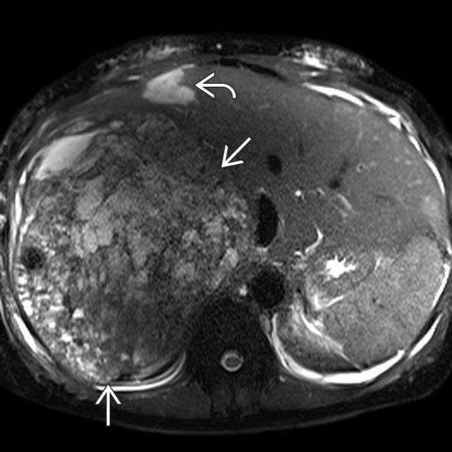
that replaces most of the right lobe. The mass is heterogeneously hyperintense, though not as bright as another lesion image that proved to be a cavernous hemangioma.

image
(Right) Axial T1WI C+ MR in the same case shows nodular peripheral enhancement of the hemangioma image
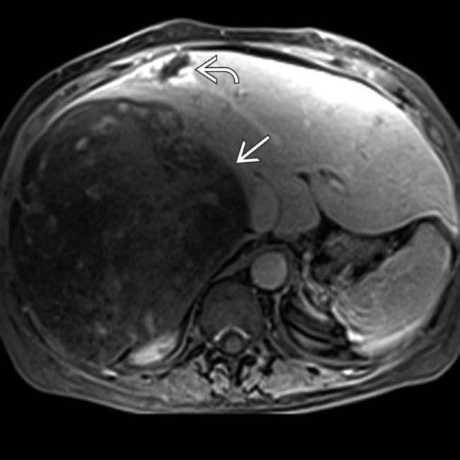
, whereas most of the sarcoma image shows no enhancement. The tumor is an undifferentiated sarcoma, which is largely necrotic and hemorrhagic as suggested by MR.
image
(Left) Axial arterial phase CECT in a 73-year-old man shows a large, encapsulated mass image 
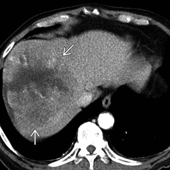
that has central necrosis and a periphery of hypervascular solid tumor, a common feature of sarcomas in general, though not specific for a primary hepatic sarcoma.

image
(Right) Coronal CT reconstruction in the same case shows the large, centrally necrotic hepatic sarcoma image
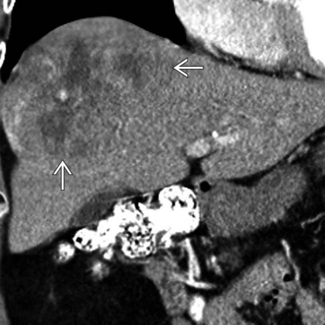
. Absence of immunohistochemical evidence of muscle, epithelial, or vascular differentiation led to the final diagnosis of undifferentiated sarcoma.

TERMINOLOGY

Synonyms

• Undifferentiated embryonal sarcoma
• Malignant mesenchymoma

Definitions

• Primary malignant tumor of liver composed of primitive mesenchymal cells with partial, divergent differentiation

IMAGING

General Features

• Best diagnostic clue

image Large, spherical, encapsulated necrotic or complex cystic mass
• Location

image Right lobe predilection
• Size

image Usually large (9-30 cm) at time of diagnosis
• Morphology

image Spherical

CT Findings

• Large, encapsulated, spherical mass
• May have peripheral rim of viable, hypervascular tumor
• Often has large complex, cystic spaces with focal hemorrhage
• May show signs of vascular invasion

image Hepatic, portal vein, or inferior vena cava (IVC) invasion
• Large subcapsular tumors may rupture
• Nodal or distant metastases are common

image Lung and osseous mets are most common

MR Findings

• Large, encapsulated, spherical mass
• Central necrosis, hemorrhage, cystic degeneration

image These areas are heterogeneously bright on both T1- and T2WI

Ultrasonographic Findings

• Large, complex, cystic-appearing mass

Imaging Recommendations

• Best imaging tool

image Multiplanar, multiphasic CECT or MR

DIFFERENTIAL DIAGNOSIS

Metastases and Lymphoma, Hepatic

• Metastases from primary cystic tumor (e.g., ovarian cystadenocarcinoma)
• Metastases from other tumors, with spontaneous or therapy-induced necrosis

image e.g., metastatic sarcoma, gastrointestinal stromal tumor (GIST)
image Immunohistochemical differentiation (e.g., C-Kit for GIST)

Hydatid (Echinococcal) Disease

• Several cases have been reported in which hepatic sarcomas were misdiagnosed as hydatid cysts

image Presence of large, encapsulated, complex cystic mass with internal debris

Angiosarcoma, Liver

• May be indistinguishable by imaging and light microscopy
• Immunohistochemical analysis may be definitive

PATHOLOGY

Gross Pathologic & Surgical Features

• Solitary, well demarcated, large
• Some solid areas, but mostly gelatinous, cystic areas with focal hemorrhage and necrosis

Microscopic Features

• Spindled, oval, or stellate tumor cells in myxoid stroma
• Marked nuclear pleomorphism and mitotic activity
• Foci of extramedullary hematopoiesis (in up to 50% of cases)
• Immunohistochemistry

image Tumor cells stain positive for α-1-antitrypsin and α-1-antichymotrypsin
image Characteristic PAS(+) cytoplasmic and extracellular eosinophilic globules

CLINICAL ISSUES

Presentation

• Most common signs/symptoms

image Vague right upper quadrant (RUQ) discomfort
image Subcapsular tumor may rupture → severe pain and hypotension
• Other signs/symptoms

image Jaundice, dyspnea (with IVC compression or invasion)

Demographics

• Majority of patients are children (ages 6-10)

image Comprises 6-13% of primary hepatic neoplasms in children
• Rare reports in adults of all ages
• Equal gender distribution

Natural History & Prognosis

• Generally grim prognosis; 5-year survival averages ∼ 15%

Treatment

• Resection ± adjuvant or neoadjuvant therapy

image Recent studies suggest improved survival
• Liver transplantation has been used in children with encouraging short-term success

DIAGNOSTIC CHECKLIST

Consider

• Check for primary extrahepatic tumor

image Metastases are much more common cause of complex cystic hepatic tumors

SELECTED REFERENCES

1. Ismail, H, et al. Treatment of undifferentiated embryonal sarcoma of the liver in children—single center experience. J Pediatr Surg. 2013; 48(11):2202–2206.

Kalra, N, et al. Undifferentiated embryonal sarcoma of liver in an adult masquerading as complicated hydatid cyst. Ann Hepatol. 2011; 10(1):81–83.

Celikbilek, M, et al. Primary hepatic carcinosarcoma. Hepatobiliary Pancreat Dis Int. 2011; 10(1):101–103.

Gasljevic, G, et al. Undifferentiated (embryonal) liver sarcoma: synchronous and metachronous occurrence with neoplasms other than mesenchymal liver hamartoma. Ann Diagn Pathol. 2011; 15(4):250–256.

Oral, A, et al. A case of undifferentiated embryonic liver sarcoma mimicking cystic hydatid disease in an endemic region of the world. J Pediatr Surg. 2011; 46(11):e5–e9.

Schoofs, G, et al. Hepatic rhabdomyosarcoma in an adult: a rare primary malignant liver tumor. Case report and literature review. Acta Gastroenterol Belg. 2011; 74(4):576–581.

Zacherl, M, et al. Surgery for liver metastases originating from sarcoma-case series. Langenbecks Arch Surg. 2011; 396(7):1083–1091.

Goto, H, et al. Carcinosarcoma of the liver. Intern Med. 2010; 49(23):2577–2582.

Massani, M, et al. Hepatic cystic lesion in adult: a challenging diagnosis of undifferentiated primary embryonal sarcoma. G Chir. 2010; 31(5):225–228.

Tsukada, A, et al. Embryonal sarcoma of the liver in an adult mimicking complicated hepatic cyst: MRI findings. J Magn Reson Imaging. 2010; 31(6):1477–1480.

Upadhyaya, M, et al. Primary hepatic sarcomas in children—a single-center experience over 19 years. J Pediatr Surg. 2010; 45(11):2124–2128.

Huang, YJ, et al. Education and imaging. Hepatobiliary and pancreatic: Huge hepatic carcinosarcoma. J Gastroenterol Hepatol. 2009; 24(5):929.

Kullar, P, et al. Primary hepatic embryonal sarcoma masquerading as metastatic ovarian cancer. World J Surg Oncol. 2009; 7:55.

Matthaei, H, et al. Long-term survival after surgery for primary hepatic sarcoma in adults. Arch Surg. 2009; 144(4):339–344. [discussion 344].