[level-membership-for-critical-care-medicine-category]50
Ultrasound imaging in space flight
Between the 1970s and mid-1990s, eight different ultrasound imagers were flown on National Aeronautics and Space Administration (NASA) and Russian spacecraft and successfully operated for research purposes. These successes justified the installation of the first permanent ultrasound system (HDI-5000, ATL/Philips, Andover, MA) for the ISS Human Research Facility (2002). After serving for 10 years, the ISS Ultrasound-1was replaced by a new unit with advanced research and clinical capabilities (Figure 50-1).
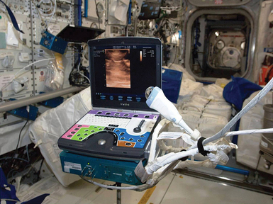
Figure 50-1 The International Space Station (ISS) Ultrasound System (Vivid Q, General Electric, Waukesha, WI) is ready for use. The box underneath the machine is a data and power interface. Note the oversized water drop on the 12-MHz linear array transducer, to be used for acoustical coupling instead of gel. Also note the multicolor keyboard overlay specially designed for remote guidance purposes. (Image courtesy National Aeronautics and Space Administration.)
Significant efforts were needed before space-based ultrasound imaging could be recognized for clinical use, with no medical expertise onboard and little evidence on the imaging representations of disease in microgravity.1 The successful implementation of clinical ultrasound in space flight is a great testament to the essential universality and versatility of ultrasound imaging. We hope this chapter will motivate more physicians to seek and promote imaging solutions in their areas of practice.
Scope of diagnostic ultrasound in space
Space medicine depends on ultrasound imaging more than most other clinical disciplines because of the absence of other diagnostic imaging modalities and the operational nature of the setting with limited resources and the very limited ability to safely and quickly evacuate the ill or injured crew member. Depending on the clinical and operational circumstances, a focused diagnostic examination in space with a single binary clinical question (emergency medicine model) can evolve into a broader, multitarget assessment and monitoring (critical care model) or into a comprehensive and specific imaging application (radiology model). The current medical requirements for the International Space Station foresee a possibility of advanced life support of a seriously ill or injured crew member for up to 72 hours, which, regardless of the initial offending factors, essentially includes close monitoring of the hemodynamic parameters, pulmonary physiology, airway management, intracranial pressure, and so forth, without the trained personnel and resources taken for granted in any terrestrial hospital. Therefore space medicine experts have great interest in both established and emerging ultrasound applications, and they monitor the recently accelerating implementation of bedside ultrasound approaches and techniques in terrestrial emergency medicine and intensive care.
Implications of microgravity
Patient and operator positioning
In the absence of gravity, the patient must often be physically restrained by using elastic cords or fabric belts to ensure positional stability; in general, the mutual positioning of the operator, patient, and imaging hardware must be globally compatible with the medical equipment setup for emergency medical treatment and life support activities. Thus a seriously ill patient would be scanned on the special electrically isolated restraint system designed for advanced life support procedures (Figure 50-2). The operator should also be restrained in a comfortable and sustainable position to consistently exert a contact force on the transducer and have both hands available for the imaging procedure.
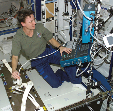
Figure 50-2 Astronaut Peggy Whitson (International Space Station [ISS]-5 expedition, 2002) developing the optimal setup for ultrasound examinations on the ISS by using the Crew Medical Restraint System (CMRS). Note the use of foot restraints. This position was found unacceptable because applying force on the transducer would make the operator position unstable. The CMRS was moved closer and was used for restraining both the subject (using straps) and the operator (inserting one knee into the space under CMRS). (Image courtesy National Aeronautics and Space Administration.)
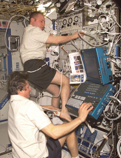
Self-scanning is also possible with minimal foot restraint, except for patients in distress or when a more experienced operator is available. Furthermore, some crews use creative ways of positioning in microgravity for specific applications (Figure 50-3).
Normal and pathologic anatomy
In the lack of gravity, the position of an object or the shape and distribution of a fluid collection are determined by the combined effect of weaker physical forces, such as properties of the fluid; surface interaction forces; tissue and organ compliance; random pressure fluctuations and gradients, such as peristalsis; and small accelerations. The absence of gravity may thus require significant modifications of imaging techniques as well as data interpretation. A terrestrial “gold standard” imaging procedure may not work in microgravity, whereas a previously unexplored technique may be the method of choice. Animal models of internal bleeding2–4 in simulated microgravity strongly suggest high diagnostic accuracy of ultrasonography if performed and interpreted using microgravity-based evidence and considerations. Intrathoracic hemorrhage and pneumothorax, maxillary sinusitis, lung abscess, ileus with dilatated bowel loops, and urinary calculi are examples of the many imaging situations influenced by the gravity vector or its absence.
Lack of imaging expertise onboard
Even with a comprehensive set of standardized scanning protocols, the variability of normal and affected structures and random factors, such as imaging conditions and acoustical artifacts, require knowledge and experience. Space medicine experts agree that the expertise necessary to independently perform an ultrasound examination in space cannot be expected because most crew members have no medical background and receive only basic medical training. To make the crew ultrasound operators, three measures are currently used: (1) limited preflight training in equipment use and general scanning technique, (2) preprocedure multimedia-based performance enhancement, and (3) real-time remote guidance by an expert from the ground.
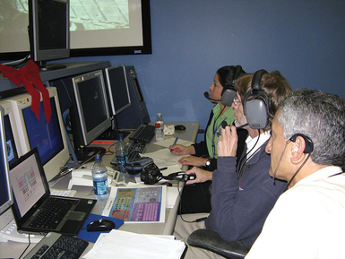
A very important component of remote guidance is the convention among all participants on the exact language used in training, multimedia materials, and real-time discourse, including transducer positioning and manipulation terms, anatomic landmarks, and instrument controls. As of the end of 2012, NASA medical personnel and the research community had conducted hundreds of successful remote guidance sessions in all areas of adult ultrasound imaging (Figure 50-4).
Selected medical problems and ultrasound imaging solutions
Urolithiasis, urinary obstruction, and retention are very common emergencies terrestrially and are among the recognized risks for human space flight. In some crew members, renal colic has been observed during space flight and shortly after landing. Urinary tract imaging was conducted on at least eight healthy crew members in space for procedure development purposes by using terrestrial techniques, which retain validity in microgravity5,6; notwithstanding a usually favorable prognosis for small stones, it is easy to foresee a scenario leading to patient evacuation, especially if urinary tract infection is present. Prognostic determination would be a prime focus of imaging in such cases because the size and location of the calculus help forecast the course of the obstruction.
Peritoneal fluid and gas are principal imaging targets in abdominal trauma and in a number of systemic and localized pathologies. Free blood in microgravity does not localize to the focused assessment with sonography in trauma (FAST) sites as readily as on Earth.3 Small quantities of blood remain in the place of origin, slowly spreading over adjacent mesothelial surfaces by means of surface interaction and capillary action. As bleeding continues, localized collections form and slowly spread according to the peritoneal anatomy. Although the basic FAST locations remain valid, additional non-FAST locations must be added to the FAST examination.5,7,8
Decompression sickness (DCS) is caused by rapid transition to a lower ambient pressure. The rate of gas bubbles in the lower extremity venous return is easy to assess but not as important as the “bubble crossover” (e.g., penetration of the bubbles into the left circulation).9 Although the ISS program invested primarily in the prevention of DCS during extravehicular activity with low in-suit pressure, the capability is still important for accidents resulting in the drop of ambient pressure inside the vehicle.
Eye trauma can be caused by airborne objects, cluttered environment, elastic cords, and pressurized gases; it can be assessed in the field by scanning through closed eyelids. A comprehensive ocular protocol has been tested in several space crew members.10 In addition, a novel method of ultrasound pupillometry was also proposed and tested on the ISS, with a subsequent terrestrial validation.11
Intracranial hypertension may develop in a small subset of crew members if the overall adaptive capacity to microgravity-related cephalad fluid redistribution is saturated. Responding to this concern, since early 2010, ISS astronauts undergo eye and orbit ultrasonography before, during, and after flight as part of an occupational monitoring program. Eye and orbit ultrasonography has thus become the single most practiced clinical imaging modality in human space flight (Figure 50-5). The quantitative and qualitative parameters include the optic nerve sheath diameter (ONSD), globe flattening, optic disk protrusion, and others. Eye and orbit ultrasonography is thoroughly addressed in Chapter 6.
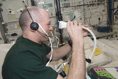
Figure 50-5 International Space Station (ISS) Flight Engineer Dr. Donald Pettit (ISS-31 expedition, 2012) is about to begin an eye and orbit ultrasound procedure required for all crew members as part of the occupational surveillance program. Note the small amount of water placed over the orbit area for acoustical coupling purposes. The use of water allows conserving gel (an available but expensive commodity in space) and gaining additional advantages of convenient, pressure-free standoff, time saving, and ease of cleanup. (Image courtesy National Aeronautics and Space Administration.)
Pneumothorax (PT) is either idiopathic or associated with chest trauma, positive-pressure lung damage, or other identifiable causes. NASA investigators studied the potential of ultrasonography in PT first in a microgravity animal model12 and then in a prospective human trial; ultrasonography was 98% sensitive and 100% specific.13 In September 2002, for the first time in history of space flight, NASA scientist Peggy A. Whitson, assisted from the Mission Control Center (author A.S.), demonstrated the normal pleural interface in microgravity.5 The same procedure was routinely repeated in several subsequent expeditions.
Free pleural fluid would present a diagnostic challenge in space even to a skilled physician, primarily because of its unusual distribution. Animal studies in parabolic aerial flight (20- to 25-second microgravity periods) have shown pleural separation by fluid throughout the pleural cavity, rather than in dependent locations only. Microgravity ultrasound reliably detected as little as 50 mL of pleural blood in a 50-kg porcine model by using then-current multipurpose equipment.3 Even higher sensitivity is expected in human pleura in continuous microgravity when using modern equipment and proper imaging technique.
Pulmonary pathology, in the absence of radiographic and meaningful auscultation capability may be easily overlooked. Evidence corroborates the relevance of ultrasound imaging in chemical pneumonitis or infectious processes, congestive lungs, pulmonary embolism, or atelectasis in previously healthy lungs. In 2011, evidence-based consensus statements were published to guide implementation, development, and standardization of lung ultrasound in all relevant clinical settings.14
Bone fractures: The clinical utility of ultrasonography in fractures is widely recognized and of interest to space medicine. Besides identifying a fracture, ultrasonography reveals mutual mobility of fragments, proximity and condition of vascular trunks, tendons and nerves, and can aid in reposition and monitoring of healing. NASA investigators have reported a high accuracy of ultrasonography in long-bone fractures in an emergency room setting.15
Pearls and highlights
• Ultrasound imaging is a permanent research capability aboard the International Space Station, as well as the sole diagnostic imaging capability for crew medical support.
• The space program–affiliated experts contribute to the development and promotion of novel diagnostic solutions, including pleural and lung ultrasound, trauma imaging, and others.
• Imaging representations of normal states and especially disease in microgravity may differ from those on Earth, and may require modifications of both imaging technique and interpretation.
• Patient positioning in microgravity is used only for convenient scanning setup and to ensure stability upon transducer pressure.
• The current paradigm of ultrasonography in space includes real-time ultrasound video downlink with verbal remote guidance of the crew-member operator. Telemedicine solutions developed by the space program have important terrestrial applications.
• Future missions outside the low Earth orbit will eliminate the ability for real-time guidance and rapid evacuation to Earth, requiring increased medical autonomy. Ultrasound expertise will have to reside aboard the vehicle, along with automated image recognition and other solutions.
• Medical results of the space program can improve the well-being of people on Earth, including expansion of ultrasound applications and development of advanced telemedicine techniques.
References
1. Sargsyan, AE, Hamilton, DR, Melton, S, et al. The International Space Station ultrasound imaging capability overview for prospective users, TP-2006-213731, S-989, NASA Technical Publication. Houston, TX: NASA Johnson Space Center; 2006.
2. Kirkpatrick, AW, Nicolaou, S, Campbell, MR, et al. Percutaneous aspiration of fluid for management of peritonitis in space. Aviat Space Environ Med. 2002; 73:925–930.
3. Hamilton, DR, Sargsyan, AE, Kirkpatrick, AW, et al. Sonographic detection of pneumothorax and hemothorax in microgravity. Aviat Space Environ Med. 2004; 75:272–277.
4. Kirkpatrick, AW, Nicolaou, S, Rowan, K, et al, Thoracic sonography for pneumothorax: the clinical evaluation of an operational space medicine spin-off. Acta Astronau. 2005; 56:831–838.
5. Sargsyan, AE, Hamilton, DR, Jones, JA, et al, FAST at MACH 20: clinical ultrasound aboard the International Space Station. J Trauma 200. 2005; 58:35–39.
6. Jones, JA, Sargsyan, AE, Barr, YR, et al, Diagnostic ultrasound at MACH 20: retroperitoneal and pelvic imaging in space. Ultrasound Med Bio. 2009; 35:1059–1067.
7. Kirkpatrick, AW, Nicolaou, S, Sargsyan, AE, et al. Focused assessment with sonography for trauma in weightlessness. J Am Coll Surg. 2003; 196:833–844.
8. Sargsyan, AE, Hamilton, DR, Jones, JA, et al, FAST at MACH 20: clinical ultrasound aboard the International Space Station. J Traum. 2005; 58:35–39.
9. Pilmanis, AA, Meissner, FW, Olson, RM. Left ventricular gas emboli in six cases of altitude-induced decompression sickness. Aviat Space Environ Med. 1996; 67:1092–1096.
10. Chiao, L, Sharipov, S, Sargsyan, AE, et al. Ocular examination for trauma; clinical ultrasound aboard the International Space Station. J Trauma. 2005; 58:885–889.
11. Sargsyan, AE, Hamilton, DR, Melton, SL, et al. Ultrasonic evaluation of pupillary light reflex. Crit Ultrasound J. 2009; 1:53–57.
12. Dulchavsky, SA, Hamilton, DR, Diebel, LN, et al. Thoracic ultrasound diagnosis of pneumothorax. J Trauma. 1999; 47:970–971.
13. Dulchavsky, SA, Schwarz, KL, Kirkpatrick, AW, et al, Prospective evaluation of thoracic ultrasound in the detection of pneumothorax. J Trauma 200. 2001; 50:201–205.
14. Volpicelli, G, Elbarbary, M, Blaivas, M, et al. International evidence-based recommendations for point-of-care lung ultrasound. Intensive Care Med. 2012; 38:577–591.
15. Marshburn, TH, Legome, E, Sargsyan, A, et al. Goal-directed ultrasound in the detection of long-bone fractures. J Trauma. 2004; 57:329–332.
[/level-membership-for-critical-care-medicine-category][not-level-membership-for-critical-care-medicine-category]50
Ultrasound imaging in space flight
Between the 1970s and mid-1990s, eight different ultrasound imagers were flown on National Aeronautics and Space Administration (NASA) and Russian spacecraft and successfully operated for research purposes. These successes justified the installation of the first permanent ultrasound system (HDI-5000, ATL/Philips, Andover, MA) for the ISS Human Research Facility (2002). After serving for 10 years, the ISS Ultrasound-1was replaced by a new unit with advanced research and clinical capabilities (Figure 50-1).
Figure 50-1 The International Space Station (ISS) Ultrasound System (Vivid Q, General Electric, Waukesha, WI) is ready for use. The box underneath the machine is a data and power interface. Note the oversized water drop on the 12-MHz linear array transducer, to be used for acoustical coupling instead of gel. Also note the multicolor keyboard overlay specially designed for remote guidance purposes. (Image courtesy National Aeronautics and Space Administration.)
Significant efforts were needed before space-based ultrasound imaging could be recognized for clinical use, with no medical expertise onboard and little evidence on the imaging representations of disease in microgravity.1 The successful implementation of clinical ultrasound in space flight is a great testament to the essential universality and versatility of ultrasound imaging. We hope this chapter will motivate more physicians to seek and promote imaging solutions in their areas of practice.
Scope of diagnostic ultrasound in space
Space medicine depends on ultrasound imaging more than most other clinical disciplines because of the absence of other diagnostic imaging modalities and the operational nature of the setting with limited resources and the very limited ability to safely and quickly evacuate the ill or injured crew member. Depending on the clinical and operational circumstances, a focused diagnostic examination in space with a single binary clinical question (emergency medicine model) can evolve into a broader, multitarget assessment and monitoring (critical care model) or into a comprehensive and specific imaging application (radiology model). The current medical requirements for the International Space Station foresee a possibility of advanced life support of a seriously ill or injured crew member for up to 72 hours, which, regardless of the initial offending factors, essentially includes close monitoring of the hemodynamic parameters, pulmonary physiology, airway management, intracranial pressure, and so forth, without the trained personnel and resources taken for granted in any terrestrial hospital. Therefore space medicine experts have great interest in both established and emerging ultrasound applications, and they monitor the recently accelerating implementation of bedside ultrasound approaches and techniques in terrestrial emergency medicine and intensive care.
Implications of microgravity
Patient and operator positioning
In the absence of gravity, the patient must often be physically restrained by using elastic cords or fabric belts to ensure positional stability; in general, the mutual positioning of the operator, patient, and imaging hardware must be globally compatible with the medical equipment setup for emergency medical treatment and life support activities. Thus a seriously ill patient would be scanned on the special electrically isolated restraint system designed for advanced life support procedures (Figure 50-2). The operator should also be restrained in a comfortable and sustainable position to consistently exert a contact force on the transducer and have both hands available for the imaging procedure.
Figure 50-2 Astronaut Peggy Whitson (International Space Station [ISS]-5 expedition, 2002) developing the optimal setup for ultrasound examinations on the ISS by using the Crew Medical Restraint System (CMRS). Note the use of foot restraints. This position was found unacceptable because applying force on the transducer would make the operator position unstable. The CMRS was moved closer and was used for restraining both the subject (using straps) and the operator (inserting one knee into the space under CMRS). (Image courtesy National Aeronautics and Space Administration.)
Self-scanning is also possible with minimal foot restraint, except for patients in distress or when a more experienced operator is available. Furthermore, some crews use creative ways of positioning in microgravity for specific applications (Figure 50-3).
Normal and pathologic anatomy
In the lack of gravity, the position of an object or the shape and distribution of a fluid collection are determined by the combined effect of weaker physical forces, such as properties of the fluid; surface interaction forces; tissue and organ compliance; random pressure fluctuations and gradients, such as peristalsis; and small accelerations. The absence of gravity may thus require significant modifications of imaging techniques as well as data interpretation. A terrestrial “gold standard” imaging procedure may not work in microgravity, whereas a previously unexplored technique may be the method of choice. Animal models of internal bleeding2–4 in simulated microgravity strongly suggest high diagnostic accuracy of ultrasonography if performed and interpreted using microgravity-based evidence and considerations. Intrathoracic hemorrhage and pneumothorax, maxillary sinusitis, lung abscess, ileus with dilatated bowel loops, and urinary calculi are examples of the many imaging situations influenced by the gravity vector or its absence.
