Chapter 635 Tumors of the Ear and Temporal Bone
Benign tumors of the external canal include osteomas and monostotic and polyostotic fibrous dysplasia. Osteomas manifest as bony masses in the canal and require removal only if hearing is impaired or external otitis results; osteomas may be confused clinically with exostoses (Chapter 495.2). Masses occurring over the mastoid bone, such as first branchial cysts, dermoid cysts, and lipomas, may be confused with primary mastoid tumors; imaging can help with the diagnosis and treatment plan.
Eosinophilic granuloma, which can occur in isolation or as part of the systemic Langerhans cell histiocytosis (Chapter 501), should be suspected in patients with otalgia, otorrhea (sometimes bloody), hearing loss, abnormal tissue within the middle ear or ear canal, and roentgenographic findings of a sharply delineated destructive lesion of the temporal bone. Definitive diagnosis is made by biopsy. Treatment depends on the site of the lesion and histology. Depending on the site, it may be treated by surgical excision, curettage, or local radiation. If the lesion is part of a systemic presentation of Langerhans’ cell histiocytosis, chemotherapy in addition to local therapy (surgery with or without radiation) is indicated. Long-term follow-up is necessary whether the temporal bone lesion is a single isolated lesion or part of a multisystem disease.
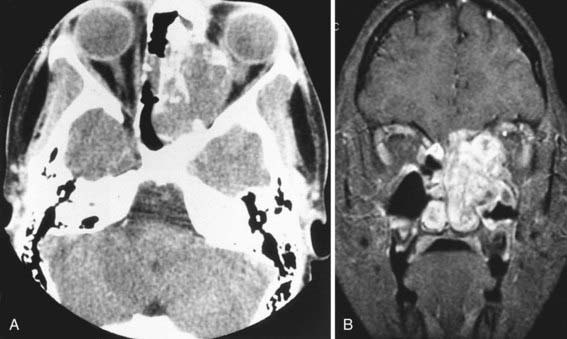
Symptoms and signs of rhabdomyosarcoma (Chapter 494) originating in the middle ear or ear canal include a mass or polyp in the middle ear or ear canal, bleeding from the ear, otorrhea, otalgia, facial paralysis, and hearing loss. Other cranial nerves also may be involved. Diagnosis is based on biopsy, but the extent of disease is determined by both CT and MRI of the temporal and facial bones, skull base, and brain (Fig. 635-1). Management usually involves a combination of chemotherapy, radiation, and surgery.
Non-Hodgkin lymphoma (Chapter 490.2) and leukemia (Chapter 489) also occur rarely in the temporal bone. Although primary neoplasms of the middle ear are very uncommon in children, they include adenoid cystic carcinoma, adenocarcinoma, and squamous cell carcinoma. Benign tumors of the temporal bone include glomus tumors. The initial signs and symptoms of the more common nasopharyngeal neoplasms (angiofibroma, rhabdomyosarcoma, epidermoid carcinoma) may be associated with insidious onset of chronic otitis media with effusion (often unilateral). A high index of suspicion is needed for diagnosing these tumors early.


