Tumors of the Cervical Spine

Published on 11/04/2015 by admin

Filed under Orthopaedics

Last modified 22/04/2025

Print this page

rate 1 star rate 2 star rate 3 star rate 4 star rate 5 star
Your rating: none, Average: 0 (0 votes)

This article have been viewed 2549 times

32 Tumors of the Cervical Spine

Introduction

Tumors of the vertebral axis are usually described and grouped based upon their location: intramedullary (IM), intradural-extramedullary (IDEM), and extradural (ED). IM lesions are found within the spinal cord parenchyma, comprise only 5% of all spinal lesions, and 50% of these tumors are located in the cervical spine. Ependymomas account for 60% to 70% of all IM tumors found in adults, followed by astrocytomas and hemangioblastomas. Extramedullary-intradural lesions comprise 40% of all spine tumors and may commonly extend to the spinal roots. IDEM tumors include meningiomas and nerve sheath tumors (neurofibromas and schwannomas); each occurs with similar frequency. Only 15% of meningiomas are located in the cervical spine, while nerve sheath tumors have an equal distribution throughout the cervical, thoracic, and lumbar spine. Extradural lesions arise in vertebral bodies or epidural space and comprise 55% of all spine lesions. Primary ED tumors are relatively uncommon, with the vast majority of tumors in this region being metastases. Primary ED tumors include osteoid osteomas, osteblastomas, osteochondromas, hemangiomas, osteosarcomas, chordomas, and chondrosarcomas. Metastatic ED tumors to the spine include breast, lung, prostate, gastrointestinal, and renal cell carcinomas; myeloma; and lymphoma; and they often invade the vertebral column. The cervical spine is the region least often involved by spinal metastases (10%).

In this chapter, we discuss tumors of the three compartments of the spine: IM, IDEM, and ED. We provide the basic presentation, imaging findings, details of surgery, and management of tumors with adjuvant therapies. Additional attention is placed on describing the more common cervical tumors found in adults as well as the most challenging tumors of the cervical spine. Specifically, there are more detailed discussions and case examples of the surgical management of intramedullary ependymomas, cervical schwannomas, and cervical chordomas.

Intramedullary Spinal Tumors

General Information, Clinical Presentation, and Imaging

Patients with intramedullary glial spinal cord tumors may present with pain, sensory dysfunction, and weakness referable to the level of the lesion. A characteristic presentation of an intramedullary tumor is a central, dull aching of a gradual onset that is not referable to a specific neurological location. The symptoms can be of a longer duration for lesions that grow slowly. As the lesion increases in size, the symptoms may progress from the vague ones described above to more neurologically localizing sensory, and even later, motor changes. In contrast, in patients with malignant astrocytomas, symptoms occur rapidly with a mean duration of less than 6 months before diagnosis.

The diagnostic imaging modality of choice, to differentiate the three most common IM lesions (ependymomas, astrocytomas, and hemangioblastomas), to evaluate the possibility of malignancy, and for preoperative evaluation and planning, is gadolinium-enhanced magnetic resonance imaging (MRI). An MRI of the entire spine axis is indicated to evaluate for other lesions or metastasis. IM lesions commonly result in widening of the spinal cord. Also, MRI provides evaluation of associated findings commonly seen with IM lesions, such as edema, hemorrhage, cyst, syringomyelia, and cord atrophy. Intramedullary tumors may be difficult to see on T1-weighted images as they appear isointense to the adjacent spinal cord. T2-weighted images are the most diagnostic, as these tumors often appear hyperintense to the surrounding spinal cord. Although ependymomas often display homogeneous enhancement on MRI, astrocytomas are often much less uniform. This is caused by inconsistent contrast uptake as a result of their irregular margins and associated necrosis. Whereas ependymomas result in symmetric expansion of the spinal cord, astrocytomas are more infiltrative, have margins that are less sharp, and are eccentrically located. Hemangioblastomas can be differentiated from the other tumors by their origin and location on the posterior surface of the spinal cord with the tumor nodule on the pial surface and more intense enhancement than that of ependymomas.

Ependymomas

Ependymomas account for the majority of all intramedullary spinal cord tumors found in adults. They comprise about 60% of all IM spinal cord tumors and are the most common glial tumors of the spinal cord. IM ependymomas arise from the ependymal cells lining the central canal. Ependymomas are benign and slow-growing, centrally located, well-circumscribed and sometimes encapsulated, and they cause symmetric expansion of the cord without infiltration into the surrounding neural tissue. Their arterial supply is most often derived from the anterior spinal artery. Ependymomas appear as reddish or purple-gray masses with many small blood vessels. Associated hemorrhage at the outer margins of the tumor and reactive cysts occur with many IM ependymomas.

Intramedullary ependymomas most commonly occur in the cervical and cervicothoracic regions of the spinal cord. The mean age at presentation is 42 years, and there is a slight female predominance. The most common presenting symptom is neck pain localized to the region of the spine, but patients may also present with dysesthetic pain or numbness and, with larger tumors, with symptoms from neural compression. Given the slow growth and well-circumscribed quality of these tumors, symptoms generally progress slowly, and patients often have a long history prior to diagnosis.

On T1-weighted images, ependymomas appear isointense relative to the neural parenchyma although, infrequently, they may appear hypointense. Heterogeneity and hyperintensity on T1-weighted images may reflect a hemorrhagic component. On T2-weighted images, ependymomas are commonly hyperintense relative to the normal spinal cord. Ependymomas are homogeneously and intensely enhancing with a well-defined border of enhancement. Hemorrhage at the cranial or caudal margin of ependymomas is common and T2-weighted images may demonstrate a low-signal-intensity rim. About half of ependymomas have nonenhancing reactive cysts with similar signal intensity to cerebrospinal fluid. Also, half of cervical ependymomas are associated with a syrinx. Cervical lesions average 4.2 vertebral segments in length.

Astrocytomas

Astrocytomas of the spinal cord are rare in adults and arise from glial cells in the spinal cord. They are less common than ependymomas in the adult population. The majority of IM astrocytomas are low-grade tumors, but approximately 25% of adult spinal cord astrocytomas have anaplastic features. IM astrocytomas are commonly found in the cervical region of the spinal cord.

There is slight male predominance of IM astrocytomas. In adults, the incidence of spinal cord astrocytomas peaks between the third and fifth decades of life, but these tumors may occur in individuals of any age. As with ependymomas, patients often present with symptoms near the level of the tumor. The most common signs and symptoms of spinal cord tumors include localized pain, numbness and paresthesias, unilateral or bilateral weakness, bowel or bladder dysfunction, spasticity, and gait difficulties. Patients with malignant astrocytomas are more likely to present with neurological deficit, given the rapid growth of the tumor and subsequent neural compression.

On imaging, intramedullary astrocytomas of the spinal cord vary in size and length, with 6 vertebral-body segments being the average length. Spinal astrocytomas have variable and heterogeneous enhancement patterns. The tumor margin is often not well defined. Tumor cysts are a common finding, and reactive cysts may be observed at the cranial and caudal ends. IM astrocytomas may be associated with a syrinx. Spinal astrocytomas are more infiltrative, have less defined margins than the other IM lesions, and are more eccentric in location. Spinal astrocytomas are less prone to have extratumoral hemorrhage. MRI may demonstrate significant cord edema, drop metastasis, and leptomeningeal spread with malignant astrocytomas.

Operative Techniques (See Figures 32-1 and 32-2)

Intramedullary Tumors

The patient is positioned prone on gel rolls in a Mayfield head holder with the arms tucked to the side, with care taken to pad all pressure points. We use intraoperative neurophysiologic monitoring with somatosensory evoked potentials, transcranial motor evoked potentials, and continuous electromyography for all IM cervical spine tumors. The use of total intravenous anesthesia allows for effective monitoring during tumor resection. We maintain the mean arterial pressure at a normal range to ensure adequate spinal cord perfusion. A lateral x-ray with skin markers in place allows for identification of the relevant level of the spine and a precise skin exposure. A standard dorsal midline approach is used. A subperiosteal dissection of the paraspinal musculature exposes the lamina and spinous processes. Prior to laminectomy, the level is confirmed again with radiograph. Generally, we perform a joint-sparing laminectomy at the vertebral level above and below the area of the lesion. If there is a suspicion of a high-grade astrocytoma and we obtain biopsy confirmation of this lesion, we target the location of the greatest size of the mass, and may not require a laminectomy overlying the entire lesion. Prior to dural opening, the tumor is precisely visualized with ultrasound to confirm adequate exposure. A durotomy is made in the midline, and the dural edges are sutured to the soft tissues laterally, exposing the arachnoid overlying the spinal cord. Using an operating microscope, the arachnoid is opened and tacked laterally to the dural edges with vascular clips. Care is taken to avoid any blood rundown into the operative field by placing sponges along the outer dural margin. For ependymomas, or lesions not extending to the dorsal surface, we prefer approaching the tumor, if possible, with a midline myelotomy between the dorsal columns. For more lateral lesions, a myelotomy dorsal to the dorsal root entry zone approach is performed. We use neurophysiological monitoring to define the dorsal columns and midline sulcus, as the midline may be difficult to identify due to the tumor growth. A bipolar cautery is used on the pial surface, and it is sharply incised. The myelotomy is extended beyond both poles of the tumor to facilitate a gross total resection. We make the best attempt to preserve all veins and arteries. The dorsal columns are dissected apart. Commonly, we enhance exposure with retraction of the cord with a pial suture.

Demarcating and establishing a plane between the parenchyma and tumor is key to achieving a gross total or more aggressive resection. Regardless of the suspected tumor pathology, we always send tumor specimens early in the dissection for evaluation with frozen pathology. The bulkiest component of the lesion is often in the center, and thus a good place to begin the dissection, as there is decreased potential for harm to surrounding neural structures. As mentioned earlier, it is also important to have complete exposure to the poles of the tumor. For ependymomas, we generally can identify and develop a clear plane between cord and tumor, and the tumor capsule is dissected circumferentially from the spinal cord parenchyma. We recommend en bloc resection of the tumor whenever possible, as it reduces the potential for tumor spillage, avoids encountering excessive bleeding from the tumor during dissection, and thus allows maintenance of better surgical planes. Dissection along the anterior median raphe is usually the most difficult portion of the surgery, as the tumor tends to adhere to this thin segment of the cord. Because small branches of the anterior spinal artery penetrate the neoplasm along the anterior aspect of the cord, great care must be taken to avoid disrupting the anterior spinal artery. The final step in removing the ependymoma after dissection from the ventral surface is the separation from the anterior spinal artery.

For large tumors or ones with indistinct planes, it may be necessary to debulk the mass with an ultrasonic aspirator. When en bloc resection is not feasible due to an inability to clearly visualize the dissection planes, internal debulking may be performed to decrease the amount of traction on the cord. The major disadvantage of extensive debulking is fragmentation of the tumor, thereby destroying the correct dissection planes. For large tumors, after debulking, it may be possible to remove further tumor capsule and delineate the tumor margins from the cord.

Generally, for an ependymoma or low-grade astrocytoma, we will always attempt a total resection if there are no persistent changes on neurophysiological monitoring. If there are neurological changes on monitoring, we typically elect to perform a second staged resection at a later date. For biopsy-confirmed anaplastic astrocytomas we resect as much abnormal appearing tissue as possible and only attempt a total resection if there is no evidence of significant changes on monitoring, and the tumor is easily resectable with a visible cord-to-tumor interface and is not disseminated. The extent of resection may be confirmed via intraoperative ultrasonography. We do not perform more than a biopsy on a glioblastoma multiforme or a disseminated anaplastic astrocytoma. Gross total resection is the acceptable surgical option for a hemangioblastoma.

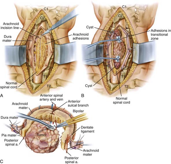
After tumor resection, we achieve hemostasis, avoiding the temptation to coagulate any of the surface vessels. The tacked sutures on the pia and dura are removed. The dura is closed primarily, in a watertight fashion. The subarachnoid space is irrigated to remove any blood prior to final closure, and a Valsalva maneuver confirms lack of CSF egress. Fibrin glue is placed over the dural closure. The wound is closed in the standard fashion and we leave a subfascial drain until there is limited output. We allow the patient to ambulate and sit up immediately after surgery.

In the authors’ experience with intramedullary cervical tumor resection, patients presenting with myelopathic motor symptoms or those undergoing three or more levels of cervical laminectomy were found to have an increased likelihood of developing subsequent symptomatic instability requiring fusion, and we elect to fuse these patients with standard lateral mass and pedicle screws at the time of tumor resection. In the absence of myelopathy and/or the need for three-or-more-level laminectomies, we prefer laminoplasty or laminectomy if the majority of the facet is maintained.

Postsurgical Management

The best current treatment of ependymomas consists of gross total resection without need for adjuvant therapy. The implementation of radiotherapy for ependymomas is warranted in cases of malignancy or disseminated tumor. In the case of a subtotal resection, we generally consider a secondary stage resection. The senior author (ZG) resected 26 spinal cord ependymomas, 11 of which had previous treatment with surgery and/or radiation therapy. A gross total resection was achieved in 88% of patients. Only one patient developed a recurrence over a mean follow-up period of 31 months. This study demonstrated that radical surgical resection of spinal cord ependymomas can be safely achieved in the majority of patients. A trend toward neurological improvement from a postoperative deficit can be expected between 1 and 3 months after surgery and continues for up to 1 year. The best predictor of outcome is the preoperative neurological status.

In the setting of residual ependymoma or low-grade astrocytoma that is not surgically resectable, radiotherapy is advocated. The dose is generally lower for these IM lesions compared to the adjuvant treatment of anaplastic astrocytomas (AAs) or glioblastoma multiforme. Higher grade lesions receive higher doses, and disseminated disease requires complete craniospinal radiation. There are no well-defined chemotherapeutic or radiosurgery regimens for IM tumors, but they remain possible treatment options for the future. In general, most authors only recommend biopsy for high-grade astrocytomas, followed by radiotherapy, but others have advocated for more aggressive surgical treatment of nondisseminated anaplastic astrocytomas. For example, in one large series of 35 high-grade spinal astrocytomas, radical resection of AAs was associated with a trend of increased overall survival in nondisseminated AA cases. The overall survival rate with these high-grade lesions remains poor.

Intradural-Extramedullary Spinal Cord Tumors

General Information, Clinical Presentation, and Imaging

The two most common IDEM tumors, occurring with approximately equal incidence, are meningiomas and nerve sheath tumors, including neurofibromas and schwannomas. Meningiomas are more common in females and occur with the highest frequency in the thoracic spine, but approximately 20% present in the cervical spinal cord, with most localizing to the upper cervical spine near the foramen magnum. In comparison, schwannomas show no preference with respect to sex or region of the spinal cord. Whereas meningiomas and schwannomas commonly result in local pain or signs from mass effect on the spinal cord including myelopathy, nerve sheath tumors cause radicular pain from their involvement of the spinal roots.

Gadolinium-enhancing MRI is the imaging modality of choice for IDEM spinal tumors to differentiate between these two types of lesions and to identify the origin, location, and extent of the lesion. An MRI of the entire spine should be obtained to ensure there are not other lesions elsewhere. Preoperative imaging also is important to evaluate the tumor’s relation to major vessels, including the vertebral artery (VA). Furthermore, a vascular study including CT angiography or MR angiography is indicated to evaluate whether a tumor extends into the transverse foramen or is adjacent to major vessels, including the VA.

Nerve Sheath Tumors

The vast majority (85%) of nerve sheath tumors are comprised of schwannomas, with only 15% consisting of neurofibromas. Schwannomas may occur sporadically or as part of neurofibromatosis (NF) 2, while neurofibromas occur in NF1. Typically, schwannomas arise from Schwann cells of the dorsal sensory spinal roots (77%), and therefore often present localized to the posterolateral region of the spinal canal. The schwannoma often arises from a single nerve fascicle, with the remaining fascicles either displaced to one side or located around the tumor. Schwannomas appear grossly as a smooth globoid mass attached to a nerve, do not produce nerve enlargement, and are suspended eccentrically from the nerve. They are firm, encapsulated neoplasms and can be cystic, hemorrhagic, or fat-containing. Schwannomas can be localized completely intradurally, may extend intraforaminally with or without an extradural component, or may be entirely extradural. Dumbbell tumors are schwannomas with contiguous intraspinal, foraminal, and extraforaminal components. In general, 49% to 84% of schwannomas are intradural, 8% to 32% are completely extradural, 1% to 19% are both intradural and extradural, 6% to 23% are dumbbell, and 1% are intramedullary. While purely intradural schwannomas are more common in the thoracic and lumbar regions, dumbbell tumors occur predominantly in the cervical spine. The frequency of schwannomas occurring in the cervical spine is similar to the occurrence in the lumbar or thoracic regions.

Sporadic spinal schwannomas usually present between the fifth and sixth decades of life. Men and women are affected equally. Typically, patients present with local pain and signs of compression of adjacent neural structures, with neurological deficits developing late in the course of the illness. The symptoms are vague in the beginning and worsen gradually, primarily because of the slow growth of the lesions. Most patients present initially with segmental pain followed by local pain; gait ataxia, motor weakness, bladder paresis, and dysesthesia are less common. Patients usually report an average duration of symptoms from 2 to 3 years, but some may have had symptoms for more than 15 years.

On MRI, schwannomas can be differentiated from meningiomas by their heterogeneous and less avid enhancement. Also, schwannomas often appear isointense on T1-weighted images, but they can be distinguished from meningiomas by their increase in signal intensity seen in T2-weighted images. Schwannomas may be associated with an expansion of the intervertebral foramen of the involved nerve root, and thus CT images are valuable to evaluate the bony anatomy. Smooth bony expansion at the foramen with lack of significant erosion suggests this benign, slow-growing lesion. Overall, it is important to evaluate the degree of extraspinal extension of a tumor to help with surgical approach and preoperative planning.

Meningiomas

Typically, spinal meningiomas are located in the intradural extramedullary space, grow slowly, and spread laterally in the subarachnoid space until they result in symptoms. Spinal meningiomas are typically IDEM (83% to 94%), but rarely (5 to 14%), tumors have an extradural component or are entirely extradural (3% to 9%). Most meningiomas are located laterally and a posterolateral location is more common than anterolateral location. Only a small proportion of meningiomas (<20%) are located in the cervical spine, but in younger populations the cervical location is more common. When located in the cervical spine, spinal meningiomas are commonly located in the upper cervical spine, are located more anterolateral and often result in motor dysfunction, especially in the hand and distal arm.

Meningiomas typically occur between the ages of 49 and 62 years. With their slow growth, there is commonly a significant delay between the onset of symptoms and diagnosis. The mean duration of symptoms prior to presentation is usually 1 to 2 years, but patients may have noted pain symptoms beginning 15 to 20 years prior to diagnosis. Patients often present with pain, sensorimotor deficits, and sphincter disturbances. Typically, back or radicular pain precedes the weakness and sensory changes; the sphincter dysfunction is always a late finding. Additionally, signs of myelopathy are present in most patients. Weakness is present in 64%, and 32% of patients are nonambulatory at the time of presentation.

MRI is the best imaging technique for diagnosing spinal meningiomas. It clearly delineates the level of the tumor and its relation to the cord, which is useful in planning surgery. Typically, spinal meningiomas are isointense to the normal spinal cord parenchyma on T1- and T2-weighted images, and they display intense enhancement after gadolinium injection. The characteristic sign of a meningioma is a “dural tail” of enhancement.

Operative Techniques

Intradural-Extramedullary Tumors

Many spinal meningiomas and schwannomas present eccentrically, dorsolateral to the spinal cord; therefore, they are accessible via a posterior or posterolateral approach and are easily seen after the dura is opened. Unilateral laminectomy, with or without facetectomy, may be used for eccentrically located ventral tumors. Ventrolaterally located tumors often require dentate ligament sectioning to obtain further ventral exposure and visualization, but some ventral tumors may provide the necessary spinal cord retraction to provide access via the standard posterior exposure. A divided dentate ligament or noncritical nerve root may be retracted to provide further ventral exposure. Intraoperative ultrasonography is used to establish the location of the tumor before dural opening. Tumor resection is performed with intraoperative electrophysiological monitoring, and additionally, for schwannomas, with intraoperative stimulation with motor-evoked potentials of nerve fascicles (at levels responsible for limb function) prior to sectioning.

IDEM tumors in the cervical area may present surgical challenges because of the need for extensive bony exposure, which results in the potential for spinal instability. In addition, large tumors of the cervical spine may compress adjacent neurovascular structures and cause tight adhesion between the tumor capsule and spinal cord, adjacent nerve roots, vertebral artery, cervical plexus, or carotid sheath, all of which may be injured with tumor resection. The majority of IDEM tumors are approached posteriorly; some midline ventral tumors may be approached anteriorly with a corpectomy and fusion. Some of the arguments against the use of the anterior approaches to ventrally located spinal tumors include inadequate access to the tumor because of a deep and constrained operative field, excessive bleeding from the epidural venous plexus, the need for spinal reconstruction, and the risk of postoperative cerebrospinal fluid leak. Access to ventrally located tumors by a posterior approach may not always be possible, however, because of the need to place undue retraction on the spinal cord. Thus a purely ventral approach or a combined anterior and posterior approach is indicated.

Spinal Schwannomas

Many schwannomas that are located completely intradurally, with or without a small foraminal component, may be approached through a laminectomy, but very large tumors, tumors that are located extradurally, or those with an ED component often require an additional or different surgical approach to achieve gross total resection. For the standard midline exposure of an ID schwannoma, the schwannoma is exposed and the plane of dissection on the tumor surface must be identified. An arachnoid membrane often adheres to the tumor and must be incised and reflected off the tumor surface. Next, the tumor and its capsule are cauterized to decrease the size of the tumor and its vascularity. The normal proximal and distal aspects of the involved nerve are exposed, and the attachment to the involved nerve root is identified. Internal debulking may be performed with an ultrasonic aspirator. The schwannoma is then separated from the nerve. Although it is often necessary to section the fascicle involved with the tumor, in the majority of cases, it is possible to preserve all other fascicles of the nerve root. Functioning nerve fascicles often can be dissected free and swept circumferentially off the surface of an underlying schwannoma, thus preserving their function. Interestingly, sectioning of nerve fascicles at levels relevant for arm function often does not result in a deficit, as many fascicles are already nonfunctional.. Some proximally located schwannomas may be embedded in the pia, and resection of these tumors may require resection of a segment of the pia.

For dumbbell cervical schwannomas, one may perform a single-stage, modified posterior midline exposure. First, the ID component is resected as earlier, but a complete unilateral facetectomy is performed. Then the dural incision is extended laterally over the nerve root sleeve to gain access to the foraminal and extraforaminal tumor extension. The exposure extends up to 4 cm from the lateral dural margin; tumor extension beyond these limits often requires an additional anterolateral approach. If it is possible to preserve the nerve root, an intrafascicular tumor dissection is performed. If it is necessary to sacrifice the nerve root, the lateral dural incision is extended around the dural root sleeve to disarticulate it from the dural tube. Next, the dural tube is reconstructed to be watertight. Removal of the foraminal and extraforaminal tumor components depends on their size and relation to the nerve root and nerve root sleeve. Generally, the tumor is followed distally along the nerve root to the lateral margin. Detaching the levator scapulae and posterior and middle scalene muscle attachments from the posterior tubercles of the transverse processes allows the exposure to extend 3.5 to 4 cm from the lateral dural margin. The rostral and caudal tumor margins are then defined. The VA is displaced ventromedially and separated from the dumbbell tumors by the tumor capsule or nerve sheath, the periosteum, and a perivertebral venous plexus. If necessary, spinal stabilization can be performed after tumor removal. After this approach in patients with significant ED components, it may be necessary to resect residual tumor with an additional anterior approach or a more lateral posterior approach.

Postsurgical Management

The primary treatment for spinal schwannomas and meningiomas is surgical resection; gross total resection is the goal. Complete resection of spinal schwannomas and meningiomas conveys an excellent prognosis. In general, the rate of gross total resection for these tumors is over 85%. For schwannomas, it may be more difficult to achieve a gross total resection of tumors with extensive ED involvement and those occurring in patients with neurofibromatosis type 2. For meningiomas, potential challenges for total resection include anterior location, en plaque meningiomas, recurrent tumors with arachnoid scarring, tumors with epidural components, and calcified meningiomas. Recurrences are rare when a gross total resection has been achieved, and range from 5% to 10%. Thus the benefit of complete resection needs to always be considered in terms of risk of spinal cord damage, given that these are benign lesions. For IDEM tumors, complications are rare and typically occur in less than 5% of cases. Surgical morbidity is related to cerebrospinal fluid leakage, wound healing, postoperative hematomas, and instability. In general, 80% of patients demonstrate neurological improvement postoperatively, and neurological deterioration is seen in less than 10%. New motor and sensory deficits usually improve over time. The most common late symptoms are mild or intermittent radiating pain, cystic myelopathy, spinal deformities, and arachnoiditis. Although the optimal treatment for spinal meningioma is total microsurgical resection, some authors advocate adjunctive radiotherapy in cases of recurrent tumors.

Extradural Spinal Cord Tumors

General Information, Clinical Presentation, and Imaging

Primary tumors of the vertebral column are relatively infrequent, with the vast majority of extradural tumors being spinal column metastases. Metastatic tumors are the most common neoplastic lesions of the spine, and the vertebral column is the most common site of bone metastasis, but metastasis to the cervical spine (10%) occurs less often than to the thoracic (70%) and lumbar regions (20%). Nearly 5% to 10% of patients with systemic cancer suffer spinal metastases, and approximately 30% to 70% of patients with solid tumors have spinal metastatic disease on autopsy. Breast, lung, prostate, and renal cell carcinomas; lymphoma; and sarcoma account for 70% of all sources of spinal metastasis. The metastases occur in the vertebral body (60%), posterior elements (30%), or both (10%). The most common symptom is neck pain (90%); the pain is usually local with tenderness on palpation, but there can also be a radicular component. More than 50% of patients who present with symptomatic epidural spinal cord compression may be nonambulatory, may have bowel or bladder dysfunction, and may present with severe deficits, including acute weakness that may progress to quadriplegia. It is important to assess for mechanical pain secondary to instability, as this is common in the cervical spine, specifically with tumors with significant bone destruction associated with pathological fractures and deformity.

Although epidural spinal cord compression occurs in only 5% to 10% of patients with metastasis, it has a great impact on the quality of a patient’s remaining life and his or her overall survival. There has been an increased trend with epidural compression to prolong neurological function, ability to ambulate, and survival with more aggressive surgical management, including circumferential decompression with or without spondylectomy and stabilization with spinal reconstruction followed by radiotherapy. Studies like the one by Patchell and colleagues have demonstrated that direct decompressive surgery plus postoperative radiotherapy is superior to treatment with radiotherapy alone for patients with spinal cord compression caused by metastatic cancer. For example, they demonstrated that surgical resection followed by radiotherapy compared to radiotherapy alone for treatment of spinal cord compression resulted in significantly more patients retaining the ability to ambulate after treatment (84% compared to 57%), and for significantly longer periods of time (122 compared to 13 days).

With respect to primary spinal cord tumors, the cervical region is unique in that malignant lesions are more common than benign. Primary benign tumors of the spinal column include osteoid osteomas, osteoblastomas, osteochondromas, giant cell tumor, aneurysmal bone cyst, and hemangiomas, and primary malignant tumors of the spinal column include plasmacytoma/multiple myeloma (most common), osteosarcomas, chondrosarcomas, chordoma, lymphoma, and malignant fibrous histiocytoma. Generally, benign primary spine tumors are more common in younger individuals, 35% have a neurological deficit on presentation, and they often involve the posterior elements. Malignant primary spinal tumors are far more common in middle-aged individuals (40 to 60 years), more often result in neurological deficit or spinal canal compression (55%), and more often involve the vertebral body (80%).

Imaging modalities used for identification and evaluation of epidural spinal column tumors including primary and metastatic lesions are plain x-ray, CT, and MRI. Interestingly, 80% to 90% of patients with symptomatic spinal metastasis have abnormalities on plain radiograph including osteolytic, or less often, osteoblastic changes of the pedicle or body. Plain x-rays are helpful in determining the stability of the spine and clearly delineate pathological fractures, abnormal alignment, and deformities. CT further demonstrates bony involvement, including the extent of osseous destruction, and is also useful in evaluating spinal stability. As with all types of tumors of the spine, an MRI of the entire neuroaxis is vital to evaluate for tumors at other sites and other levels of epidural compression. MRI with contrast clearly demonstrates the extent of tumor and its relation to the spinal canal and other relevant anatomy including the lower cervical roots, important for arm function, and the VA. If there is tumor involvement extending to vascular structures, a dedicated vascular imaging study is indicated. Bone or PET scans may be helpful to evaluate for other sites of skeletal or systemic involvement.

Biopsy for histological diagnosis of the tumor is performed once the lesion has been fully investigated with imaging studies. In the absence of known diagnosis of primary tumors, a clear diagnosis based on characteristic imaging, a more accessible primary or metastatic lesion, or catastrophic neurological deficit, we perform a CT-guided biopsy of the spinal tumor for diagnosis and for surgical decision-making. During biopsy, it is very important to provide meticulous attention to prevent the seeding of neoplastic cells to healthy tissue. Thus, the route taken during the biopsy should coincide with a region that can be adequately excised during the surgical resection, and imaging studies help dictate the biopsy approach. This detail is very important for tumors in which an en bloc resection provides superior local control or a surgery-related cure or impacts survival, including chordomas and sarcomas. Finally, diagnosis of a radiosensitive tumor may eliminate the need for surgical decompression. After tissue diagnosis, highly vascular tumors including renal cell carcinoma, melanoma, and thyroid carcinoma may be treated with preoperative embolization to limit operative blood loss.

It is beyond the scope of this chapter to discuss the surgical management of every type of primary and metastatic spine tumor, and thus we provide a general overview of this topic. Also, as benign spinal tumors are rarer in adults, we limit any further discussion to malignant ones, which are relevant to the adult and aging population. Below, we discuss the specific management of a primary malignant tumor, a cervical chordoma.

Operative and Postoperative Management

Spinal Metastatic Tumors

Generally, indications for surgery include focal spinal metastasis with cord compression, failed radiotherapy, unknown pathology, pathological fracture with or without deformity, or progressive or rapid neurological decline. Surgery may not be indicated if there has been paralysis for over 24 to 48 hours; with anticipated survival of less than 3 months; with radiosensitive tumors including lymphoma, multiple myeloma, or prostate; if there is diffuse metastatic involvement or diffuse epidural compression; or in the presence of significant comorbidities.

The goals of a surgical intervention should be clearly identified before surgery, based on knowledge of the patient’s pathology, predicted survival, extent of spinal disease and other systemic metastasis, control of the primary tumor, associated comorbidities, stability created by the tumor or by potential resection, and the tumor’s ability to respond to other modalities including chemotherapy and radiation. Generally, the treatment options for cervical metastasis vary from laminectomy with or without stabilization for a mostly dorsal tumor compressing the cord, anterior cervical decompression with corpectomy and reconstruction for tumors involving mostly the vertebral body, to a two-staged anterior and posterior decompression with reconstruction. It is far more difficult to achieve a complete spondylectomy in the cervical spine than in the thoracic and lumbar regions, due to the need to preserve the lower cervical roots for arm function as well as the vertebral arteries. Although it may be possible to perform a complete intralesional resection or an en bloc spondylectomy, both with circumferential spinal reconstruction, these procedures, specifically the latter, have high morbidity and mortality and are rarely associated with increased long-term survival. En bloc spondylectomy requires extensive instrumentation to achieve difficult fusions, and requires extensive exposure of neurovascular structures that poses additional risk of nerve root and vascular injury; thus more limited resections in the cervical spine may reduce these risks. A curative surgery may only be useful and justified with an isolated metastasis to the spine, particularly one of breast or renal cell origin, in patients who have proven themselves fit for long-term survival. Generally, palliative treatment is indicated for cervical spinal metastasis, for treatment of pain and to promote neurological function. In addition to decompression and stabilization, other palliative treatment options may include balloon kyphoplasty; however, it is used far less often in the cervical spine due to its pedicles being smaller and more difficult to approach.

As most metastatic lesions originate in the vertebral body, an anterior cervical corpectomy offers the most direct approach for tumor excision, neurological decompression, and effective reconstruction of the weight-bearing vertebral column. This approach is especially appropriate in patients with significant vertebral body destruction resulting in neck pain or symptomatic spinal cord compression. When choosing spinal reconstructive materials and techniques, multiple biomechanical factors must be considered to achieve anatomical restoration of sagittal and coronal plane deformity and physiological load bearing. Stabilization and reconstruction of the cervical vertebral body defect after corpectomy can be performed with bone allograft, cement, pins or Silastic tubes, or titanium or PEEK interbody spacers and cages. Stabilization is then achieved with anterior instrumentation with cervical plate fixation, to prevent distraction failure and to provide increased rigidity. Additionally, posterior instrumentation with or without bone grafting may be necessary to supplement the anterior construct. As mentioned above, some tumors can also be addressed with anterior followed by posterior decompression or posterior decompression alone with laminectomy or more extensive bony decompression, with stabilization with lateral mass or pedicle screws.

Generally, surgery is followed by standard radiotherapy. As mentioned previously, some tumors, such as those of lymphoreticular origin, are very radiosensitive, and some cervical tumors with epidural spinal compression and even instability can be effectively treated with radiotherapy without surgical intervention. For example, a plasmacytoma with pathological fracture will reossify and regain stability after radiotherapy treatment. Some tumors are radioresistant, including renal cell, melanoma, and sarcoma, but even these often show some response to radiation treatment. There is also an increased primary role of radiosurgery for some of these typically radioresistant tumors. It may result in less neurological complications, and improve local tumor control by delivering a high dose of radiation in a single fraction to a focal area. Also, radiosurgery may be used in addition to standard radiotherapy. Although a variety of treatments are employed based on tumor histology and location, a common spine tumor radiotherapy dose is 3000 rads over 10 fractionated treatments 24 hours apart. The target volume usually encompasses 1 or 2 vertebral bodies above and below the lesion, including paravertebral and epidural disease.

Overall, effective treatment is surgery that decompresses the spinal cord circumferentially, reconstruction and immediate stabilization, followed by radiation therapy. Patient’s who undergo this treatment are 1.3 times more likely to be ambulatory after treatment compared to radiation alone and twice as likely to regain ambulatory function. Also, 70% to 90% of patients have significant relief of pain, and 60% to 100% improve or retain ambulatory status. Primary pathology is the principal factor determining survival — some primary tumors including breast and renal cell carcinoma have longer survival after diagnosis of spinal metastasis — and overall, the median survival with metastatic spine disease is 10 months. Patients with preoperative and postoperative ambulatory function have significantly longer survival than nonambulatory patients.

Primary Malignant Tumors

A CT-guided biopsy is invaluable in the surgical planning for cervical primary tumors. There are classification scales employed for primary spinal tumors for oncologic staging (Enneking) or for planning surgical treatment (Weinstein, Boriani, and Biagini: WBB). For example, malignant lesions in the Enneking system are divided into low-grade (I), high-grade (II), or high-grade with distant metastases (III). Low-grade lesions are further subdivided into those confined to vertebra (IA) or those with extension into paravertebral compartment (IB). These tumors are treated by a wide en bloc resection followed by radiation therapy. High-grade lesions (II) are further subdivided into intracompartmental without capsule (IIA) or invasion of surrounding structures with extensive bone destruction or fracture (IIB). The treatment of high-grade lesions is usually with multiple treatment modalities including wide resection, chemotherapy, and radiation therapy; results are still poor.

In the WBB system, the vertebra, surrounding soft tissue, and spinal canal are divided into 12 zones to allow accurate staging and surgical decision-making regarding operative approach. Tumors can extend to involve soft tissue, bone, spinal canal, epidural space, dura or neural elements, or the VA. The highest chance of curing a primary spine tumor is with wide resection with negative margins, and conversely, the presence of positive margins increases the probability of local recurrence and disease progression. A decision is made based on tumor location, involvement of adjacent structures, size of the tumor, and response to other treatment modalities to determine whether it is necessary or possible to perform an en bloc resection compared to an intralesional resection (with or without negative margins). For some malignant primary spine tumors, en bloc spondylectomy with or without wide margins is not always feasible or necessary for oncological control. For example, the senior author (ZG) effectively treated a C6 primary osteogenic sarcoma with a total cervical spondylectomy with intralesional resection followed by neoadjuvant chemotherapy. Overall, en bloc procedures can be divided into intralesional, wide, and marginal.

En Bloc Resection and Treatment of Cervical Chordoma (See Figures 32-3 through 32-5)

Chordomas are one of the most common primary malignant tumors of the mobile spine; only plasmacytomas occur at a higher incidence. They account for 2% to 4% of primary malignant bone tumors and arise from the notochord remnants from the clivus to the coccyx. The majority of lesions occur in the sacrum, but 10% to 15% of cases occur in the mobile spine with the cervical region most common among these (50%). C2 and C3 are the most common cervical locations, and there may be significant epidural spread and extensions to the retropharyngeal space. They are slow-growing but locally aggressive and have significant propensity for local bone destruction, neural compression, and recurrence. The extension of the disease to the surrounding vital anatomy may ultimately result in neurological compromise, instability, and death. They arise in the vertebral body and may grow posteriorly to compress the spinal canal or extend anteriorly into the paraspinal muscles. Metastases are initially infrequent, but with later stage disease, they occur in up to 65% of cases.

The median age at diagnosis is 58 years, and 80% occur in patients over 40 years. They occur almost twice as frequently in men. The mean duration of symptoms is 14 months, and patients initially present with local pain. Patients with chordomas of the spine are more likely to present with neurological deficit including weakness, sensory change, or bowel and bladder dysfunction compared to those in the sacrum, which affords more room for tumor growth prior to neurological compromise. Cervical chordomas may result in dysphagia, airway obstruction, or an oropharyngeal mass.

On CT, there is lytic bone destruction as well as a large soft tissue mass, and frequently calcifications. On MRI, it is often possible to see extension or infiltration of tumor across the intervertebral disc space. On T1-weighted images, chordomas are isointense or slightly hypointense compared with muscle, and on T2-weighted images, they are hyperintense. They enhance with contrast, and have foci of low signal in areas of calcifications. A vascular study is performed if there is involvement of the VA or, if there is any concern that one of them must be sacrificed for tumor resection, to confirm patency of the opposite VA. Bone scan may show low or normal isotope uptake. The entire spine and body should be evaluated for other lesions. As mentioned previously, it is very important for treatment of chordomas to select a biopsy tract that does not traverse a major cavity which could seed the area with tumor cells, and that the tract can be clearly defined/marked for future resection.

Chordomas are generally considered poor responders to standard radiation therapy and chemotherapy, and intralesional resection is associated with a high local recurrence. Currently, the best initial treatment includes radical en bloc resection with or without high energy photon or proton beam radiation. Protons allow improved sparing of critical structures because the dose deposition of protons is limited mainly by the Bragg peak, where the dose is low, but the treatment is extremely conformal to the target volume. Residual or recurrent disease is treated with surgical resection followed by these radiation modalities. Mobile spine chordomas treated with intralesional resection and radiation have a high recurrence rate (approximately 66% at 37 months). Overall, 5-year and 10-year survival are 50% to 68% and 28% to 40%, respectively.

Thus, en bloc spondylectomy is an ideal treatment for chordomas, but it is very difficult in the cervical and upper cervical spine because of difficulties with adequate exposure, the need to preserve the VAs and cervical nerve roots, and difficulties with cutting the cervical pedicles. Following is a description of the senior author’s (ZG) en bloc resection and reconstruction of a multilevel cervical spine chordoma involving C2 with total spondylectomy of C2-4 and sacrifice of the right C2-4 nerve roots and a segment of the right VA. The patient, a 54-year-old man, presented with 2 years of progressive dysphagia, numbness in hands, and unsteadiness. Imaging revealed a large retropharyngeal mass extending from C2-4 involving the C2-4 vertebra with epidural extension and encasement of the right VA. The treatment of cervical chordomas often involves a multidisciplinary approach with otolaryngologists and plastic surgeons to assist in access and exposure.

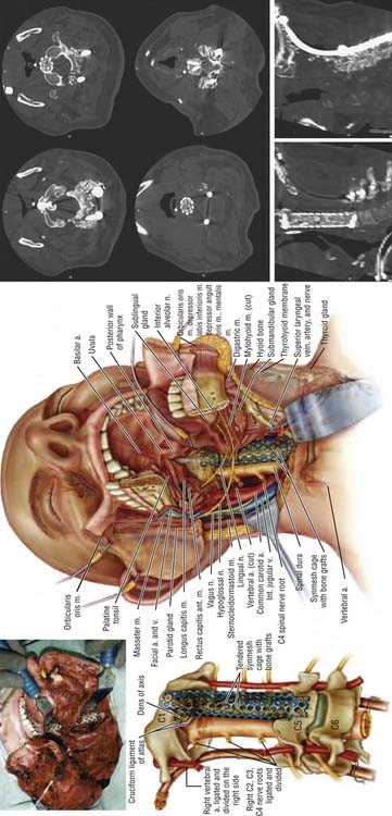
The operation was performed in two stages. The first stage, in the prone position, involved the resection of the posterior elements of the involved vertebrae, freeing neural elements that could be spared, completing the dissection around the VAs and the posterior margins of the tumor, and stabilizing the spine. After a tracheostomy was placed, the patient was placed in the prone position with head fixation, and lateral films confirmed the neutral position of the head and neck. Bilateral laminectomies and facetectomies of C2-4 (and a partial C1 right laminectomy) were performed to allow wide exposure of the exiting nerve roots. The right C2-4 roots were ligated and transected as they entered the tumor mass. Next, the right VA was exposed from C2-4 with drilling both rostral and caudal to the tumor. An initial plane of dissection was created on the right lateral side of the tumor, and a Silastic sheet was placed between the tumor and the ventral thecal sac to protect the neural structures during the subsequent anterior procedure. Occipito-cervicothoracic fixation was performed. At this point, prior to the second stage, MRA was performed to evaluate patency of the left VA to ensure safe sacrifice of the right VA for tumor resection.

Next, the patient was positioned in the supine position. The second stage, an anterior approach, was designed to complete the en bloc resection and reconstruct and stabilize the ventral spinal defect. A right lateral neck dissection accompanied by a transmandibular, circumglossal, retropharyngeal exposure was performed. Subsequently, a C4-5 discectomy was performed, the uncovertebral joints were drilled, and the posterior longitudinal ligament was resected for visualization of the ventral dura. Soft tissue free of tumor was freed from the anterior arch of C1.

On the left, the longus colli insertion on the transverse process of C2-4 was released to allow the transverse process to be drilled away, completing the circumferential exposure of the left VA from C2-5. The surgical margin on the left was thus freed from all structures. On the right, the longus colli muscles were mobilized above and below the tumor and the VA was dissected above C2 and below C4 so as not to violate the tumor. Additional dissection was performed around the lateral aspect of the tumor, medial to the carotid sheath, until this met with the dissection plane from the posterior approach. Temporary aneurysm clips were placed on the right VA and SSEPs were noted to remain stable for 30 minutes. The vessel was ligated and transected at both ends beyond the tumor, freeing the specimen along the right lateral aspect. Finally, a high-speed drill was used to cut across the base of the dens, and rongeurs were used to resect the ligamentous complex behind the dens. This established a superior margin for the resection. The entire tumor mass, including the C2-4 vertebral bodies, the right VA segment, and the right C2-4 nerve roots were removed en bloc, but the resection was marginal at the dura.

A fibular allograft was then cut to size and fashioned to form a sharp spike that could be embedded into the residual dens. The inferior end of the graft rested firmly against the superior endplate of the first remaining vertebra. A cervical plate was then fashioned to C1 and the most superior remaining vertebra (C5) The screws fixing the plate superiorly were placed with tricortical purchase, penetrating the anterior arch of C1 and engaging the residual dens. The posterior pharyngeal nerve was evaluated to determine if it is still intact (if it is not, a free flap should be placed from an external location that has remained prepped during the surgery).

The patient required several weeks of ventilatory support and needed a gastrostomy tube for difficulties with swallowing. Common complications after resection of cervical primary malignant tumors include failure of stabilization, swallowing difficulties, hoarseness, Horner syndrome, and hypoglossal injury; often tracheostomy and gastrostomy tubes are required after surgery. At a year from surgery, the patient is fully ambulatory, is able to swallow a regular diet, had his tracheostomy and gastrostomy tubes removed, his spinal construct remains stable, and he has no clinical or radiologic evidence of tumor recurrence. Radiation therapy has not been administered.

References

1. Cohen Z., Fourney D., Marco R., Rhines L., Gokaslan Z. Total cervical spondylectomy for primary osteogenic sarcoma. J. Neurosurg. Spine. 2002;97:386-392.

2. Gottfried O., Gluf W. Quinones-Hinojosa, Kan P, Schmidt M: Spinal meningiomas: surgical management and outcome. Neurosurg. Focus. 2003;14:1-7.

3. Gottfried O., Binning M., Schmidt M. Surgical approaches to spinal schwannomas. Contemp. Neurosurg.. 2005;27:1-8.

4. Hanbali F., Fourney D., Marmor E., Suki D., Rhines L., Weinberg J., McCutcheon I., Suk I., Gokaslan Z. Spinal cord ependymoma: radical surgical resection and outcome. Neurosurgery. 2002;51:1162-1174.

5. McGirt M., Goldstein I., Chaichana K., Tobias M., Kothbauer K., Jallo G. Extent of surgical resection of malignant astrocytomas of the spinal cord: outcome analysis of 35 patients. Neurosurgery. 2008;63:55-60.

6. Patchell R., Tibbs P., Regine W., Payne R., Saris S., Kryscio R., Mohiuddin M., Young B. Direct decompressive surgical resection in the treatment of spinal cord compression caused by metastatic cancer: a randomized trial. Lancet. 2005;366:643-648.

7. Rhines L., Fourney D., Siadati A., Suk I., Gokaslan Z. En bloc resection of multilevel cervical chordoma with C-2 involvement. J. Neurosurg. Spine. 2005;2:199-205.

8. Sciubba D., Chi J., Rhines L., Gokaslan Z. Chordoma of the spinal column. Neurosurg. Clin. N. Am.. 2008;19:5-15.

9. Vincent F., Fehlings M. Spinal column tumors. In Bernstein M., Berger M., editors: Neuro-oncology: the essentials, ed 2, New York: Thieme Medical Publishers, 2008.