Treatment of Rheumatic Diseases

Published on 22/03/2015 by admin

Filed under Pediatrics

Last modified 22/04/2025

Print this page

rate 1 star rate 2 star rate 3 star rate 4 star rate 5 star
Your rating: none, Average: 0 (0 votes)

This article have been viewed 1637 times

Chapter 148 Treatment of Rheumatic Diseases

The rheumatic diseases of childhood are complex chronic illnesses that present management challenges to both primary care and subspecialty providers. Optimal disease management requires family-centered care delivered by a multidisciplinary team of health care professionals providing medical, psychologic, social, and school support. Rheumatologic conditions, such as juvenile idiopathic arthritis (JIA) and systemic lupus erythematosus (SLE), most often follow a disease course marked by flares and periods of remission, but some children have unremitting disease. Treatment is aimed at achieving and maintaining clinical remission while minimizing medication toxicities. Disease management includes surveillance for potential complications of disease, such as inflammatory eye disease in JIA and early nephritis in SLE.

The goals of treatment are to avert disability, maximize the physical function and quality of life of affected children, relieve discomfort, prevent or reduce organ damage, and avoid or limit drug toxicity. Nonpharmacologic therapy is an important adjunct to medical management of rheumatic diseases. A key predictor of long-term outcome consists of early recognition and referral to a rheumatology team experienced in the specialized care of children with rheumatic diseases. Significant differences in outcome are seen at 10 yr after onset in patients with JIA depending on whether referral to a pediatric rheumatology center was accomplished within 6 mo of onset.

Pediatric Rheumatology Teams and Primary Care Physicians

The multidisciplinary pediatric rheumatology team (Table 148-1) offers coordinated services for children and their families. General principles of treatment include: early recognition of signs and symptoms of rheumatic disease with timely referral to rheumatology for prompt initiation of treatment; monitoring for disease complications and adverse effects of treatment; coordination of subspecialty care and rehabilitation services with communication of clinical information; and child and family–centered chronic illness care, including self-management support, alliance with community resources, partnership with schools, resources for dealing with the financial burdens of disease, and connection with advocacy groups. Planning for transition to adult care providers needs to start in adolescence. Central to effective care is partnership with the primary care provider, who helps coordinate care, monitor compliance with treatment plans, ensure appropriate immunization, monitor for medication toxicities, and identify disease exacerbations and concomitant infections. Communication between the primary care provider and subspecialty team permits timely intervention when needed.

Table 148-1 MULTIDISCIPLINARY TREATMENT OF RHEUMATIC DISEASES IN CHILDHOOD

Accurate diagnosis and education of family

Physical medicine and rehabilitation Consultant team Nephrology Orthopedics Dermatology Gastroenterology Physical and psychosocial growth and development Peer group relationships Individual and/or family counseling Coordination of care Involvement of patient and family as active team members Communication among health care providers Involvement of school (school nurse) and community (social worker) resources

Therapeutics

A key principle of pharmacologic management of rheumatic diseases is that early disease control, striving for induction of remission, leads to less tissue and organ damage with improved short- and long-term outcomes. Medications are chosen from broad therapeutic classes on the basis of diagnosis, disease severity, anthropometrics, and adverse effect profile. Many of therapeutics used do not have U.S. Food and Drug Administration (FDA) indications for pediatric rheumatic diseases. The evidence base may be limited to case series, uncontrolled studies, or extrapolation from use in adults. The exception is JIA, for which there is a growing body of randomized control trial evidence, particularly for newer therapeutics. Therapeutic agents used for treatment of childhood rheumatic diseases (Table 148-2) have various mechanisms of action, but all suppress inflammation. Both biologic and nonbiologic disease-modifying antirheumatic drugs (DMARDs) directly affect the immune system. DMARDs should be prescribed by specialists. Live vaccines are contraindicated in patients taking immunosuppressive glucocorticoids or DMARDs. A negative test result for tuberculosis (purified protein derivative [PPD]) should be verified and the patient’s immunization status updated, if possible, before such treatment is initiated. Killed vaccines are not contraindicated, and annual injectable influenza vaccine is recommended in children with rheumatic diseases.

Nonsteroidal Anti-Inflammatory Drugs

Nonsteroidal anti-inflammatory drugs (NSAIDs) are prescribed to decrease both pain symptoms and the acute and chronic inflammation associated with arthritis, pleuritis, pericarditis, uveitis, and cutaneous vasculitis, but they are not disease modifying. NSAID anti-inflammatory effects require regular administration at adequate doses based on weight (mg/kg) or body surface area (mg/m2), for longer periods than needed for analgesia alone. The mean time to achieve anti-inflammatory effect in JIA is 4-6 wk of consistent administration. NSAIDs work primarily by inhibiting the enzyme cyclo-oxygenase (COX), which is critical in the production of prostaglandins, a family of substances that promote inflammation. Two types of COX receptors have been demonstrated; selective COX-2 inhibitors (such as celecoxib and meloxicam) inhibit receptors responsible for promoting inflammation with potential for fewer gastrointestinal (GI) adverse effects. Clinical trials in children with JIA found celecoxib and meloxicam to be similar, in effectiveness and tolerability, to the nonselective NSAID naproxen.

The most frequent adverse effects of NSAIDs in children are nausea, decreased appetite, and abdominal pain. Gastritis or gastric or duodenal ulceration occurs less frequently in children. Less common adverse effects, occurring in ≤ 5% of children undergoing long-term NSAID therapy, include mood change, concentration difficulty that can simulate attention deficit disorder, sleepiness, irritability, headache, tinnitus, alopecia, anemia, elevated liver enzyme values, proteinuria, and hematuria. Certain agents (indomethacin) have a higher risk of toxicity than others (ibuprofen); naproxen has an intermediate risk. These NSAID-associated adverse effects reverse quickly once the medication is stopped. Several NSAID-specific adverse reactions may also occur. Ibuprofen can induce aseptic meningitis, primarily affecting patients with lupus. Naproxen is more likely than other NSAIDs to cause a unique skin reaction called pseudoporphyria, which is characterized by small hypopigmented depressed scars occurring in areas of minor skin trauma, such as fingernail scratches, or after small spontaneous vesicular lesions. Pseudoporphyria is more likely to occur in fair-skinned individuals and on sun-exposed areas. If pseudoporphyria develops, the inciting NSAID should be discontinued because scars can persist for years or be permanent. NSAIDs should be used cautiously in patients with dermatomyositis or systemic vasculitis because of an increased frequency of GI ulceration with these disorders. Salicylates have been supplanted by other NSAIDs owing to the relative frequency of salicylate hepatotoxicity and these agents’ association with Reye syndrome.

The response to NSAIDs varies greatly among individual patients, but overall, 40-60% of children with JIA experience improvement in their arthritis with NSAID therapy. Patients may try several different NSAIDs for 6-wk trials before finding one that demonstrates clinical benefit. NSAIDs with longer half-lives or sustained-release formulations allow for once- or twice-daily dosing and improve compliance. Laboratory monitoring for toxicity includes a complete blood count (CBC), serum creatinine, liver function tests (LFTs), and urinalysis every 6-12 mo, though guidelines for frequency of testing are not established.

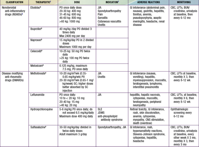
Nonbiologic Disease-Modifying Antirheumatic Drugs

Methotrexate

Methotrexate (MTX), an antimetabolite, is a cornerstone of therapy in pediatric rheumatology because of its sustained effectiveness and relative low toxicity over prolonged periods of treatment. The mechanism of action of low-dose methotrexate for arthritis is uncertain. MTX inhibits folate-dependent metabolic steps, including dihydrofolate reductase activity. MTX metabolites further inhibit other folate-dependent enzymes, including 5-aminoimidazole-4-carboxamide ribonucleotide (AICAR) transformylase, important in de novo biosynthesis of inosine monophosphate, and results in accumulation of 5-aminoimidazole-4-carboxamide. As a result, purine nucleotide biosynthesis is blocked, which may be immunosuppressive. 5-Aminoimidazole-4-carboxamide and its metabolites may themselves be immunotoxic. MTX may also work by other mechanisms, including increasing adenosine, which inhibits lymphocyte proliferation.

MTX has a central role in the treatment of arthritis, especially in children with polyarticular JIA. The response to oral MTX (10 mg/m2 once a week) is better than the response to placebo (63% vs 36%). Children who show no response to standard doses of MTX often do show response to higher doses (15 or 30 mg/m2/week). Patients with JIA who demonstrate clinical response to MTX also have improvements in growth rate, functional ability in daily tasks, and radiologic appearance of joint damage. Subcutaneous administration of MTX is similar in absorption and pharmacokinetic properties to intramuscular injection, with less pain. MTX is commonly used in treatment of juvenile dermatomyositis as a steroid-sparing agent, with efficacy in 70% of patients. It has also been used successfully at a dosage of 10-20 mg/m2/week in patients with SLE to treat arthritis, serositis, and rash.

MTX is well tolerated by children. Because of the lower dose and alternative mechanisms of action, MTX toxicity in the rheumatic diseases is milder and qualitatively different from that observed with treatment of neoplasms with MTX. In eight published studies including 288 patients with JIA taking MTX, adverse effects included GI toxicity (13%), stomatitis (3%), elevated liver enzyme values (15%), headache (1-2%), and leukopenia, interstitial pneumonitis, rash, and alopecia (<1%). Hepatotoxicity observed among adults with rheumatoid arthritis treated with MTX has raised concern about similar problems in children. Analysis of liver biopsy specimens in children with JIA undergoing long-term MTX treatment has revealed occasional mild fibrosis and no evidence of even moderate liver damage. Children receiving MTX should be counseled to avoid alcohol, smoking, and pregnancy. Folic acid is given as an adjunct to minimize adverse effects. Lymphoproliferative disorders have been reported in adults treated with MTX, primarily in association with Epstein-Barr virus infection. Regression of lymphoma may follow withdrawal of MTX.

Monitoring laboratory tests for MTX toxicity include CBC and LFTs at regular intervals, initially every 4 wk for the first 3 mo of treatment, then every 8-12 wk, with more frequent intervals after dosing adjustments or in response to abnormal values.

Glucocorticoids

Glucocorticoids are given through oral, intravenous, ocular, topical, and intra-articular administration as part of treatment of rheumatic disease. Oral steroids are foundational treatment for moderate to severe lupus, dermatomyositis, and most forms of vasculitis; their long-term use is associated with a long list of well-described, dose-dependent complications, including linear growth suppression, cushingoid features, osteoporosis, avascular necrosis, hypertension, impaired glucose tolerance, mood disturbance, and increased infection risk. Glucocorticoids should be tapered to the lowest effective dose over time, and DMARDs introduced as steroid-sparing agents.

Intravenous steroids have been used to treat severe, acute manifestations of systemic rheumatic diseases such as SLE, dermatomyositis, and vasculitis. The intravenous route allows for higher doses to obtain an immediate, profound anti-inflammatory effect. Methylprednisolone, 10-30 mg/kg/dose up to a maximum of 1 g, given over 1 hr daily for 1 to 5 days, is the intravenous preparation of choice. Although generally associated with fewer adverse effects than oral steroids, intravenously administered steroids are associated with significant and occasionally life-threatening toxicities, such as cardiac arrhythmia, acute hypertension, hypotension, hyperglycemia, shock, pancreatitis, and avascular necrosis.

Ocular steroids are prescribed only by ophthalmologists as ophthalmologic drops or injections into the soft tissue surrounding the globe (sub–Tenon capsule injection) for active uveitis. Long-term ocular steroid use leads to cataract formation and glaucoma. Current ophthalmologic management has significantly decreased the frequency of blindness as a complication of JIA-associated uveitis.

Intra-articular steroids are utilized with increasing frequency as initial therapy for children with pauciarticular JIA or when one or several affected joints have not responded to standard parenteral drug therapy in polyarticular disease. Most patients have significant clinical improvement within 3 days. Duration of response depends on steroid preparation used, joint affected, and arthritis subtype, with the anticipated response rate to knee injection between 60% and 80% at 6 mo. Intra-articular administration may result in subcutaneous atrophy and hypopigmentation of the skin at the injection site, as well as subcutaneous calcifications along the needle track.

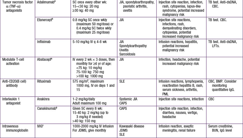
Biologic Disease-Modifying Antirheumatic Drugs

Biologic response–modifying drugs that target specific components of the immune system are becoming increasingly available for treatment of various rheumatic diseases. The initial biologic DMARDs targeted tumor necrosis factor-α (TNF-α), a proinflammatory cytokine implicated in inflammatory arthritis. Subsequently, biologic therapeutics have been developed to target specific cytokines such as interleukin-1 (IL-1) and IL-6 or to interfere with specific immune cell function through depletion of B cells or suppression of T-cell activation (Table 148-3).

Table 148-3 SUMMARY OF BIOLOGIC THERAPIES STUDIED IN JUVENILE IDIOPATHIC ARTHRITIS AND THEIR METHOD OF ACTION

DRUG METHOD OF ACTION
Etanercept Soluble TNF p75 receptor fusion protein that binds to and inactivates TNF-α
Infliximab Chimeric human/mouse monoclonal antibody that binds to soluble TNF-α and its membrane-bound precursor, neutralizing its action
Adalimumab A humanized IgG1 monoclonal antibody that binds to TNF-α
Abatacept Soluble, fully human fusion protein of the extracellular domain of (CTLA-4, linked to a modified Fc portion of the human IgG1. It acts as a co-stimulatory signal inhibitor by binding competitively to CD80 or CD86, where it selectively inhibits T-cell activation
Tocilizumab A humanized anti–human IL-6 receptor monoclonal antibody
Anakinra An IL-1 receptor antagonist (IL-1 RA)

CTLA, cytotoxic T lymphocyte–associated antigen; Ig, immunoglobulin; IL, interleukin; TNF, tumor necrosis factor.

From Beresford MW, Baildam EM: New advances in the management of juvenile idiopathic arthritis— 2: the era of biologicals, Arch Dis Child Educ Pract Ed 94:151–156, 2009.
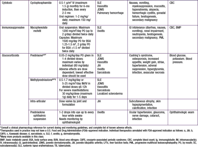
Tumor Necrosis Factor-α Antagonists

Five TNF-αantagonists are currently approved by the FDA for treatment of rheumatoid arthritis in adults, two of which also have an FDA indication for treatment of children with moderate to severe polyarticular JIA (etanercept and adalimumab). Etanercept is a genetically engineered fusion protein consisting of two identical chains of the recombinant extracellular TNF receptor monomer fused with the Fc domain of human immunoglobulin (Ig) G1. Etanercept binds both TNF-α and lymphotoxin-α (formerly called TNF-β) and inhibits their activity. Three quarters of children with active polyarticular JIA that fails to respond to MTX demonstrate response to etanercept after 3 mo of therapy. Dosing is 0.8 mg/kg subcutaneous weekly (maximum 50 mg/dose) or 0.4 mg/kg subcutaneously twice a week (maximum 25 mg/dose). Adalimumab is a fully human anti-TNF monoclonal antibody used alone or in combination with MTX. In a placebo-controlled withdrawal-design study, children continuing to receive adalimumab were less likely to experience disease flares (43% vs. 71%) even if they were also taking MTX (37% vs 65%). Adalimumab is administered subcutaneously every other week at a dose of 20 mg for children weighing 15-30 kg and 40 mg for those weighing >30 kg.

Infliximab, a chimeric mouse-human monoclonal antibody, was tested in a randomized controlled clinical trial for use in JIA but did not achieve study end points. However, it is FDA approved for pediatric Crohn disease and has been used “off label” for treatment of polyarticular JIA, uveitis, Behçet syndrome, and sarcoidosis. Two additional anti-TNF agents—golimumab, a human monoclonal antibody against TNF, and certolizumab pegol, a pegylated humanized antibody against TNF—have been approved by the FDA for rheumatoid arthritis in adults but are without pediatric indications.

The most common adverse reactions are injection site reactions that diminish over time. TNF blockade is associated with an increased frequency of serious systemic infections, including sepsis, dissemination of latent tuberculosis, and invasive fungal infections in endemic areas. TNF blockade should not be initiated in subjects with history of chronic or frequent recurrent infections. Tuberculosis should be tested for prior to initiation of therapy with TNF antagonists. If test results are positive, antituberculous treatment must be administered before anti-TNF treatment can be started. There is a theoretically increased risk of malignancy with TNF-α antagonists, and there have been reports of development of lupus-like syndromes, demyelinating syndromes, antibody formation to the drug, rashes, cytopenias, and other reactions. The benefit-risk profile appears favorable after a decade of experience with this therapeutic class; the safety of longer-term suppression of TNF function is unknown.

Cytotoxics

Cyclophosphamide

Cyclophosphamide requires metabolic conversion in the liver to its active metabolites, which alkylate the guanine in DNA, leading to immunosuppression by the inhibition of the S2 phase of mitosis. The subsequent decrease in numbers of T and B lymphocytes result in diminished humoral and cellular immune responses. Cyclophosphamide infusions (500-1,000 mg/m2) given monthly for 6 mo, and then every 3 mo for 12-18 mo, have been shown to reduce the frequency of renal failure in patients with lupus and diffuse proliferative glomerulonephritis. Open trials suggest efficacy in severe CNS lupus. Oral cyclophosphamide (1-2 mg/kg/day) is effective as induction treatment of severe Wegener granulomatosis and other forms of systemic vasculitis as well as interstitial lung disease or pulmonary hemorrhage associated with rheumatic disease. Cyclophosphamide is a potent cytotoxic drug associated with significant toxicities. Potential short-term adverse effects include nausea, vomiting, anorexia, alopecia, mucositis, hemorrhagic cystitis, and bone marrow suppression. Long-term complications include an increased risk for sterility and cancer, especially leukemia, lymphoma, and bladder cancer. Thirty percent to 40% of adult women with lupus treated with intravenous cyclophosphamide become infertile; the risk of ovarian failure appears to be significantly lower in adolescent and premenarchal girls. Ovarian suppression with an inhibitor of gonadotropin-releasing hormone to preserve fertility is currently being studied.

Other Drugs

Azathioprine is sometimes used to treat Wegener granulomatosis following induction therapy or to treat SLE. Cyclosporine has been used occasionally in the treatment of dermatomyositis on the basis of uncontrolled studies and is helpful in the treatment of macrophage activation syndrome complicating systemic JIA (Chapter 149). There are case reports describing the successful use of thalidomide, or its analog lenalidomide, as treatment for systemic JIA, inflammatory skin disorders, and Behçet disease. Several drugs commonly used in the past to treat arthritis are no longer part of standard treatment, including salicylates, gold compounds, and D-penicillamine.

Bibliography

Ardoin S, Schanberg LE. The management of pediatric systemic lupus erythematosus. Nat Clin Pract Rheumatol. 2005;1:1-11.

Beresford MW, Baildam EM. New advances in the management of juvenile idiopathic arthritis—2: the era of biologicals. Arch Dis Child Educ Pract Ed. 2009;94:151-156.

Burgos-Vargas R, Vazquez-Mellado J, Pacheco-Tena C, et al. A 26 week randomized, double blind, placebo controlled exploratory study of sulfasalazine in juvenile onset spondyloarthropathies. Ann Rheum Dis. 2002;61:941-942.

Cleary AG, Murphy HD, Davidson JE. Intra-articular corticosteroid injections in juvenile idiopathic arthritis. Arch Dis Child. 2003;88:192-196.

. Clinical pharmacology [online database]. Tampa, FL, Gold Standard, 2009 http://www.clinicalpharmacology.com

Feldman BM, Rider LG, Reed AM, et al. Juvenile dermatomyositis and other idiopathic inflammatory myopathies of childhood. Lancet. 2008;371:2201-2212.

Gedalia A, Shetty AK. Chronic steroid and immunosuppressant therapy in children. Pediatr Rev. 2004;25:425-434.

Gürcan HM, Ahmed AR. Efficacy of various intravenous immunoglobulin therapy protocols in autoimmune and chronic inflammatory disorders. Ann Pharmacother. 2007;41:812-823.

Hashkes PJ, Laxer RM. Medical treatment of juvenile idiopathic arthritis. JAMA. 2005;294:1671-1684.

Lovell DJ, Ruperto N, Goodman S, et al. Adalimumab with or without methotrexate in juvenile rheumatoid arthritis. Pediatric Rheumatology Collaborative Study Group; Pediatric Rheumatology International Trials Organisation. N Engl J Med. 2008;359:810-820.

McGonagh JE. Young people first, juvenile idiopathic arthritis second: transitional care in rheumatology. Arthritis Rheum. 2008;59:1162-1170.

Morgan SL, Oster RA, Lee JY, et al. The effect of folic acid and folinic acid supplements on purine metabolism in methotrexate-treated rheumatoid arthritis. Arthritis Rheum. 2004;50:3104-3111.

Navaneethan SD, Viswanathan G, Strippoli GF. Treatment options for proliferative lupus nephritis: an update of clinical trial evidence. Drugs. 2008;68:2095-2104.

Ruperto N, Murray KJ, Gerloni V, et al. A randomized trial of parenteral methotrexate comparing an intermediate dose with a higher dose in children with juvenile idiopathic arthritis who failed to respond to standard doses of methotrexate. for the Pediatric Rheumatology International Trials Organization. Arthritis Rheum. 2004;50:2191-2201.

Ruperto N, Lovell DJ, Quartier P, et al. Abatacept in children with juvenile idiopathic arthritis: a randomised, double-blind, placebo-controlled withdrawal trial. Paediatric Rheumatology International Trials Organization; Pediatric Rheumatology Collaborative Study Group. Lancet. 2008;372(9636):383-391.

Smolen JS, Aletaha D, Koeller M, et al. New therapies for treatment of rheumatoid arthritis. Lancet. 2007;370:1861-1874.

Tolleson-Rinehart S, Morgan DeWitt E, Massie S, Pediatric Education for Drug Safety. Pharmacological treatment of juvenile idiopathic arthritis: a pediatric education for drug safety (PEDS) continuing education module. http://pedscme.unc.edu. Accessed May 1, 2008