TREATMENT OF ESOPHAGEAL INJURY

Published on 10/03/2015 by admin

Filed under Critical Care Medicine

Last modified 22/04/2025

Print this page

rate 1 star rate 2 star rate 3 star rate 4 star rate 5 star
Your rating: none, Average: 0 (0 votes)

This article have been viewed 1785 times

CHAPTER 42 TREATMENT OF ESOPHAGEAL INJURY

The treatment of esophageal perforations remains controversial, with uncertainty surrounding several aspects of surgical management: the preferred method of surgical repair, the management of injuries recognized late, and the role of cervical esophagostomy for diversion, among others. Much of this controversy stems from the uncommon nature of these injuries. The relative infrequency of esophageal injury may be attributed to the deep location of this collapsed structure that is surrounded by vital organs. Many penetrating wounds injure the heart or aorta and result in death at the scene. Blunt injuries to the esophagus are very unusual. Anatomic features, with the lack of serosa, contribute to the difficulty in treatment. Given the frequency of delay in diagnosis and the complexity of operative treatment, injuries of the esophagus remain among the most difficult injuries to manage without major morbidity or mortality. Even the busiest trauma centers often encounter fewer than five esophageal injuries per year.

INCIDENCE

The incidence of esophageal injuries is low with most resulting from penetrating trauma. Esophageal trauma received little notice until the completion of World War II, with only 18 esophageal injuries recorded in the military records reviewed from that war and the Korean and Vietnam Wars combined. Numerous reports in the literature document the incidence of esophageal trauma to be less than 1% with the most common cause being penetrating injuries sustained from stab and gunshot wounds. The cervical esophagus represents the most common site of injury followed by thoracic and abdominal esophageal injuries. While these wounds occur relatively infrequently, they continue to be associated with high mortality ranging from 20% to 30%. Virtually all patients who sustain an esophageal injury also incur associated injuries to other respiratory, gastrointestinal, and vascular injuries. As a result, surgeons must maintain a high index of suspicion so that untoward diagnostic delays may be avoided.

Penetrating injuries of the esophagus far outnumber blunt esophageal injuries. The predominant mechanism of esophageal trauma or injury is gunshot wounds (70%–80%) followed by stab wounds (15%–20%) and shotgun wounds (3%–5%). Esophageal injuries resulting from blunt trauma account for less than 1% of all esophageal injuries. These injuries are most often located in the cervical esophagus as the result of an anterior blow with the neck in a hyperextended position. An acute blow to a distended stomach may produce tears of the distal esophagus.

DIAGNOSIS

Diagnostic delays are often cited as significant factors in the high morbidity and mortality associated with injuries to the esophagus. Several factors contribute to this diagnostic delay including the uncommon occurrence of these injuries. Because associated injuries are common, delays may occur before the initiation of specific diagnostic tests to evaluate the esophagus. All the while, even a “simple” perforation elicits a massive inflammatory response with mediastinal tissue destruction that may further complicate the repair.

Esophageal injuries must be suspected in penetrating neck injuries that violate the platysma, in transmediastinal gunshot wounds, and with significant chest trauma with associated tracheobronchial injuries. The clinical findings most commonly associated with cervical esophageal injuries include neck pain and dysphagia. Tenderness to palpation and with passive motion, dyspnea, and/or hoarseness may be present. Hematemesis, hemoptysis, or bloody nasogastric tube aspirate in the absence of obvious oral or pharyngeal trauma should suggest the possibility of esophageal injury. Expanding cervical hematoma is certainly a cause for concern as are the subsequent development of fever, cough, and stridor. Palpable crepitus or air within the soft tissues or a wide prevertebral shadow on neck or cervical spine radiographs may be the initial suggestion of an esophageal injury. Computed tomography examination may demonstrate subcutaneous emphysema within the soft tissues or in the upper mediastinum.

The clinical findings associated with thoracic esophageal injuries may be nonspecific and initially absent. They may include abdominal tenderness and/or rigidity, cervical crepitation from tracking of mediastinal emphysema, and Hamman’s sign (mediastinal crunch on auscultation). The presence of mediastinal emphysema and pleural effusion in the face of penetrating thoracic trauma should elevate awareness for the possibility of a thoracic esophageal injury.

Subdiaphragmatic esophageal injuries often present with abdominal tenderness and/or rigidity. Patients frequently complain of abdominal pain and may progress to signs of frank peritonitis. Upright chest radiographs or computed tomography scans may demonstrate pneumoperitoneum.

While penetrating neck or thoracic wound with hemodynamic instability will often necessitate immediate exploration for associated injuries, the hemodynamically stable patient often presents a diagnostic challenge. In the past, all penetrating neck wounds that violated the platysma were routinely explored. However, many trauma centers now practice selective management of neck wounds. Selective management necessitates some type of study to exclude esophageal injury. We recommend a water-soluble contrast esophagogram in stable patients. If no injury is seen with this technique, addition of dilute barium adds a measure of safety in excluding an injury. Because contrast studies yield a false-negative rate of up to 25%, esophagoscopy may be added in patients regarded as high risk for injury. The specificity of a negative esophagogram accompanied by negative esophagoscopy approaches 100%. Even in hemodynamically stable patients with cervical hematomas who will undergo exploration, we advocate esophagoscopy, as the injury may be localized by the appreciation of blood or hematoma within the esophagus. It is often difficult to identify an esophageal injury during exploration due to extensive blood staining of the tissues. All studies should be obtained in an expeditious manner as prolonged time to diagnosis has been widely correlated to increased morbidity and mortality. Once an esophageal injury has been diagnosed, all oral intake is held, nasogastric tube decompression is performed, and intravenous fluid resuscitation and broad-spectrum antibiotics are initiated before prompt surgical intervention.

SURGICAL TREATMENT

While some penetrating cervical wounds may be managed nonoperatively, all confirmed esophageal injuries should be managed operatively in expedient fashion. The preferred surgical management of esophageal injuries is dictated by the location of the injury, stability of the patient, time to diagnosis, and associated injuries.

In our opinion, all esophageal injuries should be treated by general unifying principles regardless of location. These principles include (1) attempted closure of all defects by some method; (2) the use of onlay flaps, preferably muscular, as a buttress or for primary closure; and (3) tube drainage near the repair. Given the lack of a serosa, primary healing is not uniform. Therefore, the use of a buttress often enhances healing without fistula development. Local muscle flaps, in particular, are useful for either buttress or as a primary onlay repair.

Injuries to the cervical esophagus may be approached either by a collar incision or by an incision anterior to the sternocleidomastoid. An anterior unilateral incision should be made for unilateral cervical and single injuries, whereas a collar incision is indicated for midline, multiple, or bilateral cervical injuries. The esophagus is located deep to the trachea and placement of a nasogastric tube often facilitates localization by palpation. Throughout the dissection, great care must be taken to identify and avoid injury to the recurrent laryngeal nerves which are located in the tracheoesophageal groove. If further exposure is needed, the omohyoid muscle may be divided. After blunt dissection, the esophagus should be encircled by a Penrose drain in order to further facilitate the dissection.

Thoracic esophageal injuries are best approached through thoracotomy incisions based on the suspected level of the injury. Following initial studies, the decision for the incision should be determined by the presence of pleural effusion or defined leak identified on esophagogram. Injuries to the upper two-thirds of the thoracic esophagus are best approached through a right posterolateral thoracotomy though the fifth intercostal space. Injuries to the lower third of the thoracic esophagus are best approached through an incision in the left sixth intercostal space.

Injuries to the most distal portion of the esophagus should be approached through a laparotomy incision, with the left chest prepped into the operative field should a thoracic approach be necessitated. Additional exposure can be achieved by placing the patient in the Trendelenburg position and by mobilizing the left lobe of the liver. The midline incision can be extended superiorly and to the left of the xiphoid process for an additional 1–2 cm of exposure. The esophagus should be exposed with blunt manual dissection at the gastroesophageal junction and encircled with a Penrose drain. The hiatus can be widened, if necessary, to expose wounds near the gastroesophageal junction.

Most injuries of the esophagus can be primarily repaired if promptly diagnosed. Small injuries may be closed transversely, whereas injuries larger than 2–3 cm can be closed longitudinally in order to avoid undue tension. Unfortunately, diagnostic delay often creates a situation associated with significant mediastinal inflammation and sepsis. Furthermore, the lack of a serosal layer complicates primary reapproximation as the esophageal tissues are extremely friable especially under these circumstances. Numerous strategies have been proposed including non-operative management with drainage, esophageal resection, and diversion with exclusion. The use of pleural and pericardial flaps has been widely described for buttressing primary repairs. More recently, various muscle flaps have been advocated for primary repair of esophageal defects. However, adequate esophageal tissue debridement, tension-free repairs, and effective drainage remain the mainstays of successful operative management.

CERVICAL ESOPHAGUS

The presence of a cervical esophageal injury usually mandates exploration of the neck. Careful examination of the esophagus should be performed to locate the site of esophageal injury as well as possible concomitant injuries to the trachea or vascular structures. Once identified, any nonviable or necrotic tissue should be debrided until viable edges are obtained. The defect should be closed in two layers if possible. An absorbable mucosal repair is preferable with a non-absorbable suture closure of the muscular layer. If significant inflammation has occurred, a single layer closure may be the only feasible option. All wounds of the cervical esophagus may be buttressed or closed primarily by a sternocleidomastoid muscle flap. The muscle flap is created by mobilizing the sternal attachment of the sternocleidomastoid and positioning it over the suture line. Absorbable sutures are used to secure the muscle flap over the defect or repair. All repairs should be drained with either Penrose or silastic tube drains. In the presence of associated carotid artery injuries, drainage should be performed from the opposite side of the neck so as to divert leakage and avoid late vascular complications.

Occasionally, diagnostic delays of an injured esophagus cause intense inflammation in the neck. If the site of injury is not obvious, the esophagus may be insufflated with air under a layer of sterile water. Methylene blue may also be instilled into the esophagus in an attempt to localize the injury. If the injury cannot be localized, then drainage and broad-spectrum antibiotics will often provide satisfactory results.

The delayed diagnosis of cervical esophageal injuries necessitates adequate debridement, irrigation, and drainage. In these instances, primary repair will often not suffice. Therefore, the defect may be primarily closed with a sternocleidomastoid muscle flap. The paraesophageal and precervical planes must also be inspected, irrigated, and drained to decrease contamination. We have treated several patients with large defects that could not be closed primarily with muscle flaps. Likewise, we have treated several patients with delayed recognition of an injury with onlay sternocleidomastoid flaps.

THORACIC ESOPHAGUS

Once the diagnosis of a thoracic esophageal injury has been identified, surgical exploration should ensue through an appropriate thoracotomy as previously described. These injuries are frequently associated with acute coagulation necrosis, diffuse tissue hemorrhage, and significant inflammation. The mediastinum should be opened and thoroughly irrigated. Occasionally, the inflammation is so severe that the defect is difficult to identify. In such cases, the esophagus can be encircled in an area remote from the injury. The esophagus can then be mobilized toward the area of injury. Diligent exploration should be performed until the injury is identified. Both mucosal and muscular layers should be identified, and one should recognize that the mucosal tear is usually larger than the muscular defect. Subsequently, it is often necessary to open the muscular layer more proximally and distally to fully appreciate the mucosal defect. Failure to completely expose the mucosal defect may result in incomplete closure of the defect and subsequent leakage from the site of repair. If possible, a double-layered closure should be performed with reapproximation of both mucosa and muscularis. The repair should always be buttressed with adjacent tissue for additional reinforcement.

Primary repairs of the thoracic esophagus tend to fail in a significant percentage of cases. The lower esophageal blood supply and the lack of a serosal layer make primary closure much more tenuous. As a result, several adjacent tissues have been used to buttress or primarily repair these injuries with variable success. Pleural flaps have been widely used but are often too friable and sometimes provide inadequate tissue coverage. Pleural flaps are more reliable if there have been inflammatory changes to thicken the pleura. The use of pericardial flaps seems unwise, as this would expose the pericardial sac to contamination. In our experience, we have found muscle flaps to be more reliable both in terms of buttressing and primary repair of esophageal defects.

Adequate tissue coverage is especially important in close proximity to the trachea in order to reduce the incidence of later fistula development. If both trachea and esophagus are injured, we attempt to interpose a muscle flap between the suture lines of the two injuries. Muscle flaps tend to be less friable and provide greater bulk of tissue coverage of the defect. Intercostal bundles are frequently mentioned as muscle flaps but we often find they provide inadequate tissue for buttress and especially for primary repair.

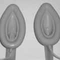
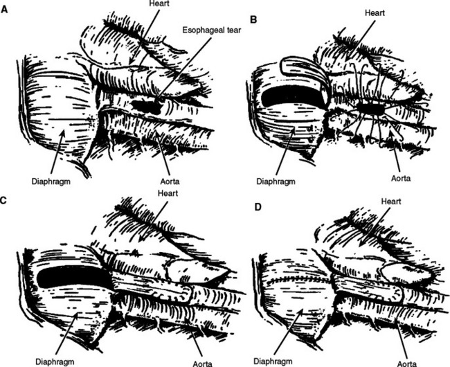
We have demonstrated that injuries to the esophagus involving up to two-thirds of the circumference can be adequately closed with muscle flaps without increased incidence of stricture. The rhomboid muscle may be mobilized for repair of upper- and mid-esophageal injuries (Figure 1). The muscle is dissected from its attachment of the scapula through a parascapular incision and transferred as a pedicle flap into the thoracotomy. For distal esophageal lesions that are too proximal to be buttressed with the gastric fundus, we advocate the use of a diaphragm muscle flap (Figure 2). The central portion of the diaphragm is excised as a pedicle flap and sutured over the defect or buttressed to the repair. The flap should be posteriorly based and great care should be taken to avoid the phrenic nerve. Diaphragm pedicle flaps may reach to the level of the azygous vein. The edges of the diaphragm are then reapproximated with heavy nonabsorbable sutures. A two layer closure is performed if feasible. This has the additional benefit of plicating the diaphragm.

Delayed diagnosis of thoracic esophageal injuries presents a significant surgical challenge. Prolonged perforations are often difficult to visualize secondary to mediastinitis and subsequent empyema. Successful management of uncontained esophageal leaks requires debridement of necrotic tissue, copious irrigation, and complete mediastinal and pleural drainage. Often, a decortication must also be performed at the time of operation if the diagnosis has been delayed for several days. While primary repair remains the preferred method of management, we have found that this is essentially futile after delayed diagnosis and treatment.

All thoracic esophageal injuries should be widely drained. The mediastinal pleura should be widely incised, and a large bore chest tube should be placed in the mediastinum adjacent to the repair. In those with large esophageal injuries, delayed diagnosis of the injury, or those believed at high risk for failure, we support the routine placement of a gastrostomy tube for gastric decompression. The placement of a jejunostomy tube for distal feeding should also be considered in order to maintain adequate nutrition.

DEVASTATING INJURIES

Occasionally, devastating injuries of the esophagus occur. If repair is not feasible, there are several less than desirable options, including resection, T-tube placement, and cervical esophagostomy with or without esophageal exclusion.

Esophageal resection may be indicated in some unusual circumstances, but the senior author has never been forced to resect the esophagus for trauma in 30 years of experience. Clearly, injuries to a damaged esophagus or caustic injuries may require emergency esophagectomy. If this unusual circumstance arises, delayed reconstruction using stomach or colonic bypass would likely be the treatment of choice.

Occasionally, large defects in the esophagus are encountered that cannot be closed. Although we attempt to cover such injuries with an onlay muscle flap, creation of a controlled fistula with a T-tube has been reported. In this technique, the short limb of the T-tube is placed in the defect and the long limb is brought out through the chest. In our experience, if a perforation is amenable to closure around a tube, it can be closed with a tissue flap even if the treatment is delayed.

Cervical esophagostomy should not be used routinely because it represents extremely difficult problems to reconstruct. In our experience, a diversion should be performed for an uncontrolled leak that results in sepsis or a life-threatening problem. If possible, a loop esophagostomy should be performed. This permits the possibility of a one-stage closure of the esophagostomy if and when the esophageal injury has healed. End esophagostomy invariably requires complex reconstruction.

If diversion is performed, we rarely use esophageal exclusion in which the distal esophagus is ligated. This creates a distal obstruction and permits bacterial overgrowth in an organ whose only egress is through the original injury. For this reason, we do no attempt to exclude the esophagus even if a proximal diversion is performed. An esophagus damaged badly enough to require exclusion is best treated by resection. Using muscle flaps, we have been able to preserve esophageal function in many patients with complex injuries.

MANAGEMENT OF COMPLICATIONS

The morbidity associated with cervical esophageal injuries has been estimated as high as 16%, with most complications related to the duration of diagnostic delay and subsequent contamination from esophageal contents. Gunshot wounds exhibit a greater preponderance for complications, whereas stab wounds often fare better secondary to less tissue destruction.

Increased morbidity from diagnostic delays and subsequent delays in operative treatment has been well established. However, little literature exists addressing long-term complications and esophageal function after traumatic esophageal injuries. Certainly, the most concerning complications associated with esophageal injuries are major vascular or airway injuries producing massive hemorrhage or acute respiratory decompensation. The development of anastomotic leaks with subsequent mediastinitis, sepsis, and pulmonary failure presents significant management challenges. Shock, extensive mobilization, inadequate debridement, and tension are well-known risk factors for suture line breakdown and subsequent esophageal leakage. Approximately 50% of esophageal leaks are asymptomatic being noticed only on post-operative esophagograms. Once a leak is identified, the initiation of adequate drainage, wide-spectrum antibiotics, parenteral or distal enteral nutrition, and limited oral intake are the mainstays of therapy. The development of interventional radiological techniques with the advent of percutaneous drainage has certainly assisted in the management of these complicated injuries. Most leaks will subsequently resolve without the need for further operative intervention. Increasingly, endoluminal stents have been used to manage esophageal leaks. We have no experience using stents as primary treatment, but we have used them on two occasions for the treatment of delayed leaks resulting from trauma.

SUGGESTED READINGS

Andrade-Alegre R. T-tube intubation in the management of late esophageal perforations: case report. J Trauma. 1994;37:131-132.

Asensio JA, Berne J, Demetriades D, et al. Penetrating esophageal injuries: time interval of safety for preoperative evaluation—how long is safe? J Trauma. 1997;43:319-324.

Asensio JA, Chawan S, Forno W, et al. Penetrating esophageal injuries: multi-center study of the American Association for the Surgery of Trauma. J Trauma. 2001;50:289-296.

Back MR, Baumgartner FJ, Klein SR. Detection and evaluation of aerodigestive tract injuries caused by cervical and transmediastinal gunshot wounds. J Trauma. 1997;42:680-686.

Bufkin BL, Miller JI, Mansour KA. Esophageal perforation: emphasis on management. Ann Thorac Surg. 1996;61:1447-1452.

Cheadle W, Richardson JD. Options in the management of trauma to the esophagus. Surg Gynecol Obstet. 1982;155:380-384.

Conn TI E, Hubbard A, Patton G. Management of esophageal injuries. Ann Thorac Surg. 1989;48:309-314.

Glatterer MSJr, Toon RS, Ellestad C, et al. Management of blunt and penetrating external esophageal trauma. J Trauma. 1985;25:784-792.

Grillo HC, Wilkins EWJr. Esophageal repair following late diagnosis of intrathoracic perforation. Ann Thorac Surg. 1975;20:387-399.

Karmy-Jones RC, Wagner JW, Lewis JWJr. Esophageal injury. In: Trunkey DD, Lewis FR, editors. Current Therapy of Trauma. 4th ed. St. Louis, MO: Mosby; 1999:209-216.

Pass LJ, LeNarz LA, Schreiber JT, Estrera AS. Management of esophageal gunshot wounds. Ann Thorac Surg. 1987;44:253-256.

Popovsky J, Lee YC, Berk JL. Gunshot wounds of the esophagus. J Thorac Cardiovasc Surg. 1976;72:609-612.

Port JL, Kent MS, Korst RJ, et al. Thoracic esophageal perforations: a decade of experience. Ann Thorac Surg. 2003;75:1071-1074.

Richardson JD. Management of esophageal perforations: the value of aggressive surgical treatment. Am J Surg. 2005;190:161-165.

Richardson JD, Flint LM, Snow NJ, et al. Management of transmediastinal gunshot wounds. Surgery. 1981;90:671-676.

Richardson JD, Tobin GR. Closure of esophageal defects with muscle flaps. Arch Surg. 1994;129:541-548.

Riley RD, Miller PR, Meredith JW. Injury to the esophagus, trachea, and bronchus. In: Moore EE, Feliciano DV, Mattox KL, editors. Trauma. 5th ed. New York: McGraw-Hill; 2004:539-552.

Triggiani E, Belsey R. Oesophageal trauma: incidence, diagnosis and management. Thorax. 1977;32:241-249.