Traumatic Injuries of the Ear and Temporal Bone

Published on 22/03/2015 by admin

Filed under Pediatrics

Last modified 22/04/2025

Print this page

rate 1 star rate 2 star rate 3 star rate 4 star rate 5 star
Your rating: none, Average: 0 (0 votes)

This article have been viewed 1087 times

Chapter 634 Traumatic Injuries of the Ear and Temporal Bone

Auricle and External Auditory Canal

Auricle trauma is common in certain sports, and quick drainage of a hematoma can prevent irreversible damage. Hematoma, with accumulation of blood between the perichondrium and the cartilage, can follow trauma to the pinna and is especially common in teenagers related to wrestling or boxing. Immediate needle aspiration or, when the hematoma is extensive or recurrent, incision and drainage and a pressure dressing are necessary to prevent perichondritis, which can result in cartilage loss and a cauliflower ear deformity. Sports helmets should be worn when appropriate during activities when head trauma is possible.

Frostbite of the auricle should be managed by rapidly rewarming the exposed pinna with warm irrigation or warm compresses.

Foreign bodies in the external canal are common in childhood. Often these can be removed in the office setting without general anesthesia if the child is mature enough to understand and cooperate and is properly restrained; if an adequate headlight, surgical head otoscope, or otomicroscope is used for visualizing the object; and if appropriate instruments, such as alligator forceps, wire loops or a blunt cerumen curette, or suction are used, depending on the shape of the object. Gentle irrigation of the ear canal with body temperature water or saline may be used to remove very small objects, but only if the tympanic membrane (TM) is intact. Attempts to remove an object from a struggling child or with poor visualization and inadequate tools results in a terrified child with a swollen and bleeding ear canal and can then mandate general anesthesia to remove the object. Difficult foreign bodies, especially those that are large, deeply embedded, or associated with canal swelling, are best removed by an otolaryngologist and/or under general anesthesia. Disk batteries are removed emergently because they leach a basic fluid that can cause severe tissue destruction. Insects in the canal are first killed with mineral oil or lidocaine and are then removed under otomicroscopic examination.

After a foreign body is removed from the external canal, the TM should be inspected carefully for possible traumatic perforation or for a pre-existing middle-ear effusion. If a foreign body has resulted in acute inflammation of the canal, eardrop treatment as described for acute external otitis should be instituted (Chapter 631).

Tympanic Membrane and Middle Ear

Traumatic perforation of the TM usually occurs as a result of a sudden external compression, such as a slap, or penetration by a foreign object such as a stick or cotton-tipped applicator. The perforation may be linear or stellate. It is most commonly in the anterior portion of the pars tensa when it is caused by compression, and it may be in any quadrant of the TM when caused by a foreign object. Systemic antibiotics and topical otic medications are not required unless suppurative otorrhea is present. Traumatic TM perforations often heal spontaneously, but it is important to evaluate and monitor the patient’s hearing to ensure that spontaneous healing occurs. If the TM does not heal within several months, surgical graft repair should be considered. As long as the perforation is present, otorrhea can occur from water entering the middle ear from the ear canal, which can occur during swimming or bathing; appropriate precautions should be taken. Perforations resulting from penetrating foreign bodies are less likely to heal than those caused by compression. Audiometric examination reveals a conductive hearing loss (CHL), with larger air-bone gaps seen in larger perforations. Immediate surgical exploration may be indicated if the injury is accompanied by one or more of the following: vertigo, nystagmus, severe tinnitus, moderate to severe hearing loss, or cerebrospinal fluid (CSF) otorrhea. At the time of exploration, it is necessary to inspect the ossicles, especially the stapes, for possible dislocation or fracture and to clear sharp objects that might have penetrated the oval or round windows. Sensorineural hearing loss (SNHL) results if the stapes subluxates or dislocates into the oval window or if either the oval or round window is penetrated. Children should not be given access to cotton-tipped applicators, because the applicators commonly cause ear trauma. Contact with small objects should be limited to times of parental supervision.

Perilymphatic fistula (PLF) can occur after barotrauma or an increase in CSF pressure. It should be suspected in a child who develops a sudden SNHL or vertigo after physical exertion, deep water diving, air travel, playing a wind instrument, or significant head trauma. The leak characteristically is at the oval or the round window and may be associated with congenital abnormalities of these structures or an anatomic abnormality of the cochlea or semicircular canals. PLFs occasionally close spontaneously, but immediate surgical repair of the fistula is recommended to control vertigo and to stop any progression of the SNHL; even timely surgery does not usually restore the SNHL. No reliable test is known for PLF, so middle-ear exploration is required for diagnosis and treatment.

Temporal Bone Fractures

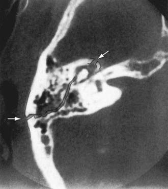
Children are particularly prone to basilar skull fractures, which usually involve the temporal bone. Temporal bone trauma should be considered in head injuries, and the status of the ear and hearing should be evaluated. Temporal bone fractures are divided into longitudinal (70-80%), transverse, and mixed. Longitudinal fractures (Fig. 634-1) are commonly manifested by bleeding from a laceration of the external canal or tympanic membrane; postauricular ecchymosis (Battle sign); hemotympanum (blood behind an intact TM); CHL resulting from TM perforation, hemotympanum, or ossicular injury; delayed onset of facial paralysis (which usually improves spontaneously); and temporary CSF otorrhea or rhinorrhea (from CSF running down the eustachian tube). Transverse fractures of the temporal bone have a graver prognosis than longitudinal fractures and are often associated with immediate facial paralysis. Facial paralysis might improve if caused by edema, but surgical decompression of the nerve is often recommended if there is no evidence of clinical recovery and facial nerve studies are unfavorable. If the facial nerve has been transected, surgical decompression and anastomosis offer the possibility of some functional recovery. Transverse fractures are also associated with severe SNHL, vertigo, nystagmus, tinnitus, nausea, and vomiting associated with loss of cochlear and vestibular function; hemotympanum; rarely, external canal bleeding; and CSF otorrhea, either in the external auditory canal or behind the TM, which can exit the nose via the eustachian tube.

image

Figure 634-1 High-resolution axial CT of uncomplicated longitudinal fracture (arrows). A hematoma is present. The course of the fracture has been touched.

(From Schubiger O, Valavanis A, Stuckman G, et al: Temporal bone fractures and their complications: examination with high resolution CT. Neuroradiology 28:93–99, 1986.)

If temporal bone fracture is suspected or seen on radiographs, gentle examination of the pinna and ear canal is indicated; lacerations or avulsion of soft tissue is common with temporal bone fractures. Vigorous removal of external auditory canal blood clots or tympanocentesis is not indicated, because removing the clot can further dislodge the ossicles or reopen CSF leaks. The effectiveness of prophylactic antibiotics to prevent meningitis in patients with basilar skull fractures and CSF otorrhea or rhinorrhea cannot be determined because studies to date are flawed by biases. If a patient is afebrile and the drainage is not cloudy, watchful waiting without antibiotics is indicated. Surgical intervention is reserved for children who require repair of a nonhealing TM perforation, who have suffered dislocation of the ossicular chain, or who need decompression of the facial nerve. SNHL can also follow a blow to the head without an obvious fracture of the temporal bone (labyrinthine concussion).