Transmissible Spongiform Encephalopathy (Creutzfeldt–Jakob Disease)

Published on 03/03/2015 by admin

Filed under Neurology

Last modified 22/04/2025

Print this page

rate 1 star rate 2 star rate 3 star rate 4 star rate 5 star
Your rating: none, Average: 0 (0 votes)

This article have been viewed 1642 times

19 Transmissible Spongiform Encephalopathy (Creutzfeldt–Jakob Disease)

Clinical Vignette

A 56-year-old man noted increasing clumsiness while writing. A tremor developed in his right hand, and he soon noted slurred speech over 2–3 weeks. Initial neurologic evaluation demonstrated an action tremor in the right hand and some reduction in fine dexterity. Brain magnetic resonance imaging (MRI) and electroencephalography (EEG) results were normal. During the next 2 months, the patient’s wife believed that her husband became increasingly confused, as illustrated by his calling her by his sister’s name. He had trouble initiating and maintaining sleep. His driving became erratic and dangerous. He was pulled over for driving too slowly and his license was revoked. On follow-up examination, 4 months later, he was disoriented. He spoke slowly and gave inappropriate responses. His dysarthria was more pronounced.

The results of repeated MRI and EEG were normal, as were cerebrospinal fluid (CSF) study results, including protein 14-3-3. Over the next 6 months, the patient’s cognitive and motor function declined precipitously. His gait became progressively ataxic, and he fell numerous times. He lost proper use of his hands and legs, requiring assistance with eating, dressing, and bathing. Occasionally, his limbs would jerk suddenly, and he appeared startled and anxious. Speech output became largely unintelligible and palilalic. He would shout occasionally but mostly stayed quiet and passive.

Repeated EEG showed generalized slowing and intermittent periodic sharp waves, approximately 1 per second. Another MRI now demonstrated bright lesions on T2-weighted and flair imaging. Right frontal lobe biopsy showed vacuolar encephalopathy consistent with a transmissible spongiform encephalopathy (TSE). The final stages were characterized by increasing stupor and aspiration pneumonia. The patient died 14 months after onset. Autopsy confirmed the diagnosis.

Epidemiology

Transmissible spongiform encephalopathy includes rare, subacute, universally fatal neurodegenerative diseases affecting humans and animals. Clinical presentation and distribution of pathologic lesions vary widely, complicating classification of these diseases. Classification focuses on pathogenic mechanisms rather than clinical and pathologic features. Currently, human TSE is classified into three categories: sporadic, familial, and infectious. Most animal TSE cases fall into the infectious category, including scrapie (sheep); bovine spongiform encephalopathy (BSE), also known as mad cow disease (cattle); and chronic wasting disease (deer and elk). The sporadic form of human TSE, Creutzfeldt–Jakob disease (CJD), accounts for 85% of cases of this disorder. Sporadic fatal insomnia cases are also described. The inherited form of human TSE, accounting for 15% of cases, includes familial CJD, fatal familial insomnia, Gerstmann–Straussler–Scheinker disease, and other less well defined clinical syndromes.

The human epidemic forms of TSE, comprising less than 1% of cases, include iatrogenic cases of CJD (exposure via dural grafts, infected surgical instruments, and growth hormone injections), kuru (cannibalism), and variant CJD (vCJD; via ingestion of beef contaminated with BSE). In theory, transfusion of any blood product from an affected patient may pose a risk. To date, only a handful of vCJD cases are attributed to transmission from blood transfusion, much fewer than might be expected given the number of blood transfusions worldwide. There seems to be increased risk of CJD among venison eaters, although this is difficult to confirm. The incidence of human TSE is approximately 1.5/1 million people per year. This rate has remained stable over several decades.

The appearance of symptoms after exposure to “infected” tissue varies widely, and incubation periods lasting several decades are described. One hundred sixty-three vCJD cases have been reported in Europe since 1996. These occurred predominantly in the United Kingdom, where most cases of BSE occurred. No instances of vCJD originated in the United States. Given the potentially long incubation period (several years) of vCJD, all cases of TSE are reported and monitored by several European surveillance centers. A similar laboratory, the National Prion Diseases Pathology Surveillance Center, operates in the United States.

Pathogenesis

The histopathologic hallmarks of TSE include severe neuronal loss, spongiform vacuolization, and astrocytosis. There is no associated inflammatory response, and some disease forms have an accumulation of amyloid plaques. However, TSE amyloid differs from β-amyloid typically found in Alzheimer disease. The agent responsible for TSE is a “proteinaceous infectious particle,” termed prion protein (PrPC). This is a normally occurring cell-surface glycoprotein, encoded on chromosome 20, which is highly conserved in mammals. Its normal function is not well defined, but it may have importance in response to oxidative stress.

All three TSE forms are thought to result from conversion of PrPC into PrPSc, also known as scrapie protein. PrPSc is a self-replicating and infectious agent lacking nucleic acid. The mechanisms by which PrPSc leads to neurodegeneration remain largely unknown. It is questionable whether the prion is the sole pathogenic mechanism of TSE. The disease can be experimentally transmitted between various animals, with some variability, by inoculation of infected nervous tissue. However, inoculation with pure PrPSc has not led to transmission of disease. Therefore, PrPSc alone does not account for horizontal transmission.

More than 20 pathogenetic mutations of the prion gene are identified, accounting for 15% of inherited human TSE. Mutations of the PrP gene predispose mutant PrP to transform into the PrPSc tertiary structure. A difference in just 1 amino acid can drastically affect the disease phenotype. With infectious TSE, PrPSc enters the central nervous system (CNS) via ingestion or iatrogenically and precipitates the conversion of PrPC into PrPSc. In sporadic cases, the conversion may occur via rare stochastic (random) changes of the normal protein. Individual susceptibility to conversion of PrPC also may be determined by genetic polymorphisms, some of which are identified and associated with varying phenotypic expression.

For example, patients who are homozygous for methionine at residue 129 tend to present with cognitive loss, aphasia, myoclonus, or insomnia and tend not to have plaque-like lesions at autopsy. Patients who are homozygous for valine at residue 129 present primarily with cognitive loss or insomnia but not aphasia or myoclonus, and all have plaquelike lesions at autopsy. Moreover, individuals who are homozygous for methionine at residue 129 are more susceptible to vCJD than individuals with other polymorphisms at the same residue.

Clinical Presentation

Initial symptoms depend on the brain region involved. Cognitive impairment is often the first sign and may be recognized by attentive patients who note subtle changes in their intellectual capacities that are sometimes difficult for the examining physician to detect. Intellectual impairment may affect any cognitive domain. Behavioral and personality disturbances such as impulsivity, disinhibition, or apathy often occur. Progressive dementia eventually develops in all individuals, often with significant psychiatric and behavioral disturbance. A variety of motor signs and symptoms often develop, typically extrapyramidal features, and sometimes precede onset of cognitive deficits. Cerebellar ataxia develops in some patients and at times may be the presenting feature. Myoclonus develops in others; some also have a very hyperactive startle response. At times, these features do not occur until relatively late in the clinical course. Progressive parkinsonism, weakness with neurogenic muscle atrophy, and bulbar dysfunction also may occur. Motor signs may be unilateral or asymmetric, reflecting focal or multifocal disease, respectively. As symptoms progress, a multifocal pattern emerges with global cognitive impairment, severe loss of motor control, and marked behavioral changes.

Terminal TSE stages are characterized by progressive stupor, leading to coma and aspiration. Sporadic CJD cases usually present between the fifth and eighth decades of life, but vCJD occurs in younger adults and teenagers. The mean survival time for patients with sporadic disease is approximately 6 months; most expire within 12 months. Inherited forms of the disease and vCJD have younger onset ages and more protracted courses than do sporadic cases.

Diagnosis

The results of routine laboratory studies are normal. Identification of pathogenic mutations in hereditary cases is available via rapid screening tests that can be performed on non-CNS tissue, including peripheral blood. Sporadic disease remains a diagnostic challenge. A family of proteins called 14-3-3 is increased in the CSF of affected patients. Although initially reported to have greater than 90% sensitivity and specificity for TSE, it is likely overestimated because 14-3-3 proteins are essentially markers for any acute to subacute brain neuronal damage. Nevertheless, CSF assays for protein 14-3-3 are useful when TSE is suspected. Noting that false positives do occur, positive results must not be considered diagnostic of TSE.

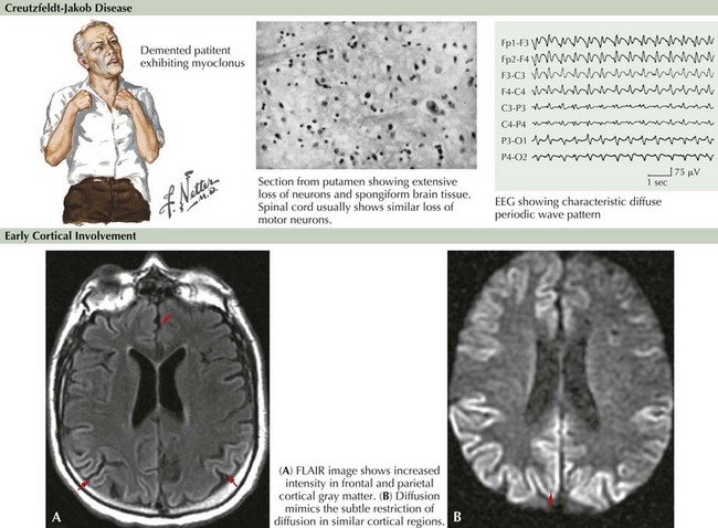
Brain MRI is now very useful in TSE diagnosis. In some cases of vCJD, bright lesions on T2-weighted and fluid-attenuated inversion recovery (FLAIR) imaging occur within the pulvinar, the so-called pulvinar sign. In CJD, focal hyperintense lesions may be found in the basal ganglia on T2-weighted images or multifocally within the cortex on diffusion-weighted images (Fig. 19-1). Diffusion-weighted MRI imaging has shown more than 90% sensitivity and specificity in some series, although the timing of MRI imaging along the course of illness is important. Repeated imaging may be required to detect typical changes. MRI abnormalities are also of supportive diagnostic value, similar to 14-3-3, but are not diagnostic of TSE. EEG may demonstrate 0.5- to 1-Hz periodic sharp waves focally or diffusely at some stage of the disease, particularly when there is clinical cerebral cortical dysfunction but certainly not all CJD individuals such as those presenting with cerebellar or striatal dysfunction. Nonspecific EEG slowing is more common but less specific than periodic sharp waves.

Brain biopsy needs to be considered in every case, particularly when the results of other studies are negative; however, brain biopsy per se is also susceptible to sampling error. This study will often differentiate CJD from other neurodegenerative disease and, more importantly, may help exclude potentially treatable conditions, such as CNS vasculitis. Frontal lobe and cerebellar biopsies can be particularly successful. In suspected vCJD, biopsy of lymphoid tissue such as the tonsils proves very reliable. CSF samples and brain tissue need to be sent to the National Surveillance Center for analysis and monitoring. Autopsy evaluation must be discussed with the family in advance of death because it is essential in confirming the diagnosis. Confirmation of a CJD diagnosis allows epidemiologic surveillance of disease activity as well as providing families a definitive conclusion to their previously unresolved tragic experience with a loved one.

Additional Resources

Appleby BS, Appleby KK, Crain BJ, et al. Characteristics of established and proposed sporadic Creutzfeldt-Jakob disease variants. Arch Neurol. 2009:208-215.

Brandel JP, Heath CA, Head MW, et al. Variant Creutzfeldt-Jakob disease in France and the United Kingdom: Evidence for the same agent strain. Ann Neurol. 2009 Mar;65(3):233-235.

Brown P. Transmissible spongiform encephalopathy in the 21st century: neuroscience for the clinical neurologist. Neurology. 2008 Feb 26;70(9):713-722. Review

Collins SJ, Sanchez-Juan P, Masters CL, et al. Determinants of diagnostic investigation sensitivities across the clinical spectrum of sporadic Creutzfeldt-Jakob disease. Brain. 2006;129:2278-2287.

Glatzel M, Stoeck K, Seeger H, et al. Human prion diseases; molecular and clinical aspects. Arch Neurol. 2005;62:545-552. Review

Goudsmit J. Obituary: Daniel Carleton Gajdusek (1923-2008). Nature. 2009 Jan 22;457(7228):394.

Josephs KA, Ahlskog JE, Parisi JE, et al. Rapidly progressive neurodegenerative dementias. Arch Neurol. 2009;66:201-207.

Kovacs GG, Budka H. Prion diseases: from protein to cell pathology. The American Journal of Pathology. 2008;172(3):555-565.

Shiga Y, Miyazawa K, Sato S, et al. Diffusion weighted MRI abnormalities as an early diagnostic marker for Creutzfeldt-Jakob disease. Neurology. 2004;63:443-449.

Ward HJ, Everington D, Cousens SN, et al. Risk factors for sporadic Creutzfeldt-Jakob disease. Ann Neurol. 2008 Mar;63(3):347-354.

Warren JD, Schott JM, Fox NC, et al. Brain biopsy in dementia. Brain. 2005 Sep;128:2016-2025. Review