Transfusion Medicine and Coagulation Disorders

Published on 07/02/2015 by admin

Filed under Anesthesiology

Last modified 22/04/2025

Print this page

rate 1 star rate 2 star rate 3 star rate 4 star rate 5 star
Your rating: none, Average: 0 (0 votes)

This article have been viewed 2370 times

Chapter 24 Transfusion Medicine and Coagulation Disorders

Coagulation and bleeding assume particular importance when operations are performed on the heart using extracorporeal circulation. This chapter begins with a discussion of the depth and breadth of hemostasis relating to cardiac procedures, beginning with coagulation pathophysiology. The pharmacology of heparin and protamine is described next. This background is then applied to treatment of the bleeding patient.

OVERVIEW OF HEMOSTASIS

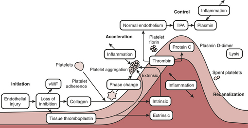
Proper hemostasis requires the participation of innumerable biological elements (Box 24-1). They can be divided into four topics to facilitate understanding: coagulation factors, platelet function, the endothelium, and fibrinolysis. The reader must realize this is for simplicity of learning and that in biology the activation creates many reactions and control mechanisms, all interacting simultaneously. The interaction of the platelets, endothelial cells, and proteins to either activate or deactivate coagulation is a highly buffered and controlled process. It is perhaps easiest to think of coagulation as a wave of biological activity occurring at the site of tissue injury (Fig. 24-1). Although there are subcomponents to coagulation itself the injury/control leading to hemostasis is a four-part event: initiation, acceleration, control, and lysis (recanalization/fibrinolysis). The initiation phase begins with tissue damage, which is really begun with endothelial cell destruction or dysfunction. This initiationphase leads to binding of platelets, as well as protein activations; both happen nearly simultaneously and each has feedbacks into the other. Platelets adhere and create an activation or acceleration phase that gathers many cells to the site of injury and creates a large number of biochemical protein cascade events. As the activation phase ramps up into an explosive set of reactions, counter-reactions are spun off, leading to control proteins damping the reactions. The surrounding normal endothelium exerts control over the reactions. Eventually the control reactions overpower the acceleration reactions and lysis comes into play.

image

Figure 24-1 Coagulation is a sine wave of activity at the site of tissue injury. It goes through four stages: initiation, acceleration, control, and lysis/recanalization.

(Redrawn with permission from Spiess BD: Coagulation function and monitoring. In Lichtor JL [ed]: Atlas of Clinical Anesthesia. Philadelphia, Current Medicine, 1996.)

The other key concept is that hemostasis is part of a larger body system—inflammation. Most of the protein reactions of coagulation control have importance in signaling inflammation and other healing mechanisms. It is no wonder that cardiopulmonary bypass (CPB) has such profound inflammatory effects when it is considered that each of the activated coagulation proteins and cell lines then feeds into upregulation of inflammation.

Protein Coagulation Activations

Coagulation Pathways

The coagulation factors participate in a series of activating and feedback inhibition reactions, ending with the formation of an insoluble clot. A clot is the total of platelet-to-platelet interactions leading to the formation of a platelet plug and then the cross-linking of platelets to each other by way of the final insoluble fibrin that leads to a stable clot. Clotting is not simply the activation of proteins leading to more protein deposition.

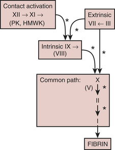
With few exceptions, the coagulation factors are glycoproteins synthesized in the liver, which circulate as inactive molecules termed zymogens. Factor activation proceeds sequentially, with each factor serving as substrate in an enzymatic reaction catalyzed by the previous factor in the sequence. Hence, this has classically been called a “cascade” or “waterfall” sequence. Cleavage of a polypeptide fragment changes an inactive zymogen to an active enzyme. The active form is termed a serine protease because the active site for its protein-splitting activity is a serine amino acid residue. Many reactions require the presence of calcium ion (Ca2+) and a phospholipid surface (platelet phosphatidylserine). The phospholipids occur most often either on the surface of an activated platelet or endothelial cell and occasionally on the surface of white cells. So anchored, their proximity to one another permits reaction rates profoundly accelerated (up to 300,000-fold) from those measured when the enzymes remain in solution. The factors form four interrelated arbitrary groups (Fig. 24-2): the contact activation and the intrinsic, extrinsic, and common pathways.

Modulators of the Coagulation Pathway

Thrombin, the most important coagulation modulator, exerts a pervasive influence throughout the coagulation factor pathways. It activates factors V, VIII, and XIII; cleaves fibrinogen to fibrin; stimulates platelet recruitment and chemotaxis of leukocytes and monocytes; releases tissue plasminogen activator (t-PA), prostacyclin, and nitric oxide from endothelial cells; releases interleukin-1 from macrophages; and with thrombomodulin, activates protein C, a substance that then inactivates factors Va and VIIIa. Note the negative feedback aspect of this last action. The latest thinking on coagulation function centers around the effects of thrombin. The platelets, tissuefactor, and contact activation all are interactive and are activated by a rent in the surface of the endothelium or through the loss of endothelial coagulation control. Platelets adhere to a site of injury and in turn are activated, leading to sequestration of other platelets. It is the interaction of all of those factors together that eventually creates a critical mass. Once enough platelets are interacting together, with their attached surface concomitant serine protease reactions, then a thrombin burst is created. Only when enough thrombin activation has been encountered in a critical time point, then a threshold is exceeded, and the reactions become massive and much larger than the sum of the whole. It is thought that the concentration and ability of platelets to react fully affect the ability to have a critical thrombin burst. CPB may affect the ability to get that full thrombin burst due to its effects on platelet number, platelet-to-platelet interactions, and the decreased amounts of protein substrates.

The many serine proteases that compose the coagulation pathways are balanced by serine protease inhibitors, termed serpins. This biological yin and yang leads to an excellent buffering capacity. It is only when the platelet-driven thrombin burst so overwhelms the body’s localized anticoagulation or inhibitors that clot proceeds forward. Serpins include α1-antitrypsin, α2-macroglobulin, heparin cofactor II,α2-antiplasmin, antithrombin (also termed antithrombin III), and others.

Antithrombin (ATIII) constitutes the most important inhibitor of blood coagulation. It binds to the active site (serine) of thrombin, thus inhibiting thrombin’s action. It also inhibits, to a much lesser extent, the activity of factors XIIa, XIa, IXa, and Xa; kallikrein; and the fibrinolytic molecule plasmin. Thrombin bound to fibrin is protected from the action of antithrombin, thus explaining the poor efficacy of heparin in treating established thrombosis. ATIII is a relatively inactive zymogen. To be most effective antithrombin must bind to a unique pentasaccharide sequence contained on the wall of endothelial cells in the glycosaminoglycan surface known as heparan; the same active sequence is present in the drug heparin. An important note is that activated ATIII is active only against free thrombin. Most thrombin in its active form is either bound to glycoprotein-binding sites of platelets or in fibrin matrices. When blood is put into a test tube and clot begins to form (e.g., in an activated coagulation time [ACT]), 96% of thrombin production is yet to come. The vast majority of thrombin generation is on the surface of platelets and on clot-held fibrinogen. Platelets through their glycoprotein-binding sites and phospholipid folds protect activated thrombin from attack by ATIII. Therefore, the biological role of ATIII is to create an anticoagulant surface on endothelial cells. It is not present biologically to sit and wait for a dose of heparin before CPB.

Another serpin, protein C, degrades factors Va and VIIIa. Like other vitamin K–dependent factors, it requires Ca2+ to bind to phospholipid. Its cofactor, termed protein S, also exhibits vitamin K dependence. Genetic variants of protein C are less active and lead to increased risk for deep vein thrombosis and pulmonary embolism. When endothelial cells release thrombomodulin, thrombin then accelerates by 20,000-fold its activation of protein C. Activated protein C also promotes fibrinolysis.

Regulation of the extrinsic limb of the coagulation pathway occurs via tissue factor pathway inhibitor (TFPI), a glycosylated protein that associates with lipoproteins in plasma.2 TFPI is not a serpin. It impairs the catalytic properties of the factor VIIa/tissue factor complex on factor X activation. Both vascular endothelium and platelets appear to produce TFPI. Heparin releases TFPI from endothelium, increasing TFPI plasma concentrations by as much as sixfold.2

von Willebrand factor (vWF), a massive molecule composed of disulfide-linked glycosylated peptides, associates with factor VIII in plasma, protecting it from proteolytic enzymes. It circulates in the plasma in its coiled inactive form. Disruption of the endothelium either allows for binding of vWF from the plasma or allows for expression of vWF from tissue and from endothelial cells. Once bound, vWF uncoils to its full length and exposes a hitherto cryptic domain in the molecule. This A-1 domain has a very high affinity for platelet glycoproteins. Initially, vWF attaches to the GPIα platelet receptor, which slows platelet shear forces. This is not enough to bind the platelet, but it creates a membrane signal that allows for early shape change and expression of other glycoproteins, GPIb and GPIIb/IIIa. Then, secondary GPIb binding connects to other vWF nearby, binding the platelet and beginning the activation sequence. It bridges normal platelets to damaged subendothelium by attaching to the GPIb platelet receptor. An ensuing platelet shape change then releases thromboxane, β-thromboglobulin, and serotonin and exposes GPIIb/IIIa, which binds fibrinogen.

Platelet Function

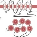
Most clinicians think first of the coagulation proteins when considering hemostasis. Although no one element of the many that participate in hemostasis assumes dominance, platelets may be the most complex. Without platelets, there is no coagulation and no hemostasis. Without the proteins, there is hemostasis, but it lasts only 10 to 15 minutes as the platelet plug is inherently unstable and breaks apart under the shear stress of the vasculature. Platelets provide phospholipid for coagulation factor reactions; contain their own microskeletal system and coagulation factors; secrete active substances affecting themselves, other platelets, the endothelium, and other coagulation factors; and alter shape to expose membrane glycoproteins essential to hemostasis. Platelets have perhaps as many as 30 to 50 different types of cell receptors. The initial response to vascular injury is formation of a platelet plug. Good hemostatic response depends on proper functioning of platelet adhesion, activation, and aggregation (Fig. 24-3).
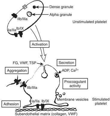
Platelet Adhesion

Capillary blood exhibits laminar flow, which maximizes the likelihood of interaction of platelets with the vessel wall. Red cells and white cells stream near the center of the vessels and marginate platelets. However, turbulence causes reactions in endothelium that leads to the secretion of vWF, adhesive molecules, and tissue factor. Shear stress is high as fast-moving platelets interact with the endothelium. When the vascular endothelium becomes denuded or injured, the platelet has the opportunity to contact vWF, which is bound to the exposed collagen of the subendothelium. A platelet membrane component, glycoprotein (GP)Ib, attaches to vWF, thus anchoring the platelet to the vessel wall. Independently, platelet membrane GPIa and GPIIa and IX may attach directly to exposed collagen, furthering the adhesion stage.

The integrin glycoproteins form diverse types of membrane receptors from combinations of 20 α and 8 β subunits. One such combination is GPIIb/IIIa, a platelet membrane component that initially participates in platelet adhesion. Platelet activation causes a conformational change in GPIIb/IIIa, which results in its aggregator activity.

Platelet adhesion begins rapidly—within 1 minute of endothelial injury—and completely covers exposed subendothelium within 20 minutes. It begins with decreased platelet velocity when GPIb/IX and vWF mediate adhesion, followed by platelet activation, GPIIb/IIIa conformational change, and then vWF binding and platelet arrest on the endothelium at these vWF ligand sites.3

Platelet Activation and Aggregation

Platelet activation results after contact with collagen, when adenosine diphosphate (ADP), thrombin, or thromboxane A2 binds to membrane receptors, or from certain platelet-to-platelet interactions. Platelets then release the contents of their dense (δ) granules and α granules. Dense granules contain serotonin, ADP, and Ca2+; α granules contain platelet factor V (previously termed platelet factor 1), β-thromboglobulin, platelet factor 4, P-selectin, and various integrin proteins (vWF, fibrinogen, vitronectin, and fibronectin). Simultaneously, platelets employ their microskeletal system to change shape from a disk to a sphere, which changes platelet membrane GPIIb/IIIa exposure. Released ADP recruits additional platelets to the site of injury and stimulates platelet G protein, which in turn activates membrane phospholipase. This results in the formation of arachidonate, which platelet cyclooxygenase converts to thromboxane A2. Other platelet agonists besides ADP and collagen include serotonin, a weak agonist, and thrombin and thromboxane A2, both potent agonists. Thrombin is by far the most potent platelet agonist, and it can overcome all other platelet antagonists as well as inhibitors. In total, there are more than 70 agonists that can produce platelet activation and aggregation.

Agonists induce a shape change, increase platelet intracellular Ca2+ concentration, and stimulate platelet G protein. In addition, serotonin and thromboxane A2 are potent vasoconstrictors. The presence of sufficient agonist material results in platelet aggregation. Aggregation occurs when the integrin proteins (mostly fibrinogen) released from α granules form molecular bridges between the GPIIb/IIIa receptors of adjacent platelets (the final common platelet pathway).

Drug-Induced Platelet Abnormalities

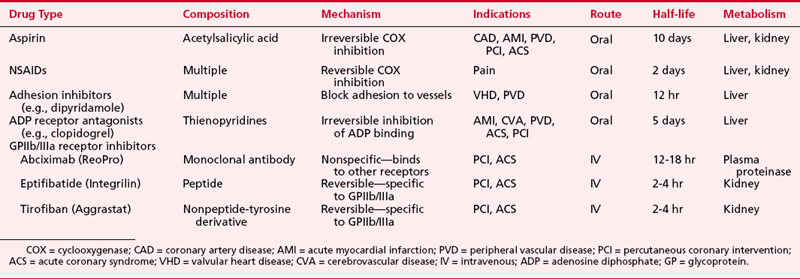
Many other agents inhibit platelet function.4 β-Lactam antibiotics coat the platelet membrane, whereas the cephalosporins are rather profound but short-term platelet inhibitors. Many cardiac surgeons may not realize that their standard drug regimen for antibiotics may be far more of a bleeding risk than aspirin. Hundreds of drugs can inhibit platelet function. Calcium channel blockers, nitrates, and β-blockers are ones commonly utilized in cardiac surgery. Nitrates are effective antiplatelet agents and that may be part of why they are of such benefit in angina, not just for their vaso-relaxing effect on large blood vessels. Nonsteroidal anti-inflammatory drugs (NSAIDs) reversibly inhibit both endothelial cell and platelet cyclooxygenase.

In addition to the partial inhibitory effects of aspirin and the other drugs just mentioned, new therapies have been developed that inhibit platelet function in a more specific manner. These drugs include platelet adhesion inhibitor agents, platelet-ADP-receptor antagonists, and GPIIb/IIIa receptor inhibitors (Table 24-1).

Vascular Endothelium

The cells that form the intima of vessels provide an excellent nonthrombogenic surface. Characteristics of this surface, which may account for its nonthrombogenicity, include negative charge; incorporation of heparan sulfate in the grid substance; the release of prostacyclin, nitric oxide, adenosine, and protease inhibitors by endothelial cells; binding and clearance of activated coagulation factors both directly, as occurs with thrombin, and indirectly, as evidenced by the action of thrombomodulin to inactivate factors Va and VIIIa via protein C; and stimulation of fibrinolysis.

Nitric oxide vasodilates blood vessels and inhibits platelets. Its mechanism involves activation of guanylate cyclase with eventual uptake of calcium into intracellular storage sites. Prostacyclin (PGI2) possesses powerful vasodilator and antiplatelet properties. Endothelium-derived prostacyclin opposes the vasoconstrictor effects of platelet-produced thromboxane A2. Prostacyclin also inhibits platelet aggregation, disaggregates clumped platelets, and, at high concentrations, inhibits platelet adhesion. Prostacyclin increases intracellular concentrations of cAMP, which inhibits aggregation. Thromboxane acts in an opposite manner. The mechanism of prostacyclin action is stimulation of adenylyl cyclase, leading to reduced intracellular calcium concentrations. Some vascular beds (e.g., lung) and atherosclerotic vessels secrete thromboxane, endothelins, and angiotensin, all vasoconstrictors, as well as prostacyclin. Activation of platelets releases endoperoxides and arachidonate. These substances, utilized by nearby damaged endothelial cells, provide substrate for prostacyclin production.

Fibrinolysis

Fibrin breakdown, a normal hematologic activity, is localized to the vicinity of a clot. It remodels formed clot and removes thrombus when endothelium heals. Like clot formation, clot breakdown may occur by intrinsic and extrinsic pathways. As with clot formation, the extrinsic pathway plays the dominant role in clot breakdown. Each pathway activates plasminogen, a serine protease synthesized by the liver, which circulates in zymogen form. Cleavage of plasminogen by the proper serine protease forms plasmin. Plasmin splits fibrinogen or fibrin at specific sites. Plasmin is the principal enzyme of fibrinolysis, just as thrombin is principal to clot formation. Plasma normally contains no circulating plasmin, because a scavenging protein, α2-antiplasmin, quickly consumes any plasmin formed from localized fibrinolysis. Thus, localized fibrinolysis, not systemic fibrinogenolysis, accompanies normal hemostasis.

Clinical Applications

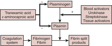
Figure 24-4 illustrates the fibrinolytic pathway, with activators and inhibitors. Streptokinase, ASPAC, and t-PA find application in the lysis of thrombi associated with myocardial infarction. These intravenous agents “dissolve” clots that form on atheromatous plaque. Clinically significant bleeding may result from administration of any of these exogenous activators or streptokinase.

Fibrinolysis also accompanies CPB. This undesirable breakdown of clot after surgery may contribute to postoperative hemorrhage and the need to administer allogeneic blood products. Regardless of how they are formed, the breakdown products of fibrin intercalate into sheets of normally forming fibrin monomers, thus preventing cross-linking. In this way, extensive fibrinolysis exerts an antihemostatic action.

HEPARIN

Pharmacology

Chemical Structure

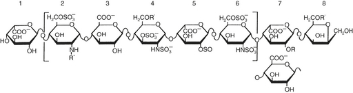
The N-sulfated-D-glucosamine and L-iduronic acid residues of heparin alternate in copolymer fashion to form chains of varying length (Fig. 24-5). As a linear anionic polyelectrolyte, with the negative charges being supplied by sulfate groups, heparin demonstrates a wide spectrum of activity with enzymes, hormones, biogenic amines, and plasma proteins. A pentasaccharide segment binds to antithrombin. Heparin is a heterogeneous compound: the carbohydrates vary in both length and side chain composition, yielding a range of molecular weights from 5 000 to 30,000, with most chains between 12,000 and 19,000. Today, the standard heparin is called unfractionated heparin (UFH).

Pharmacokinetics and Pharmacodynamics

The heterogeneity of UFH molecules produces variability in the relationship of dose administered to plasma level of drug. In addition, the relationship of plasma level to biological effect varies with the test system. A three-compartment model describes heparin kinetics in healthy humans: rapid initial disappearance, saturable clearance observed in the lower dose range, and exponential first-order decay at higher doses. The rapid initial disappearance may arise from endothelial cell uptake. The reticuloendothelial system, with its endoglycosidases and endosulfatases, and uptake into monocytes, may represent the saturable phase of heparin kinetics. Finally, renal clearance via active tubular secretion of heparin, much of it desulfated, explains heparin’s exponential clearance.

Male gender and cigarette smoking are associated with more rapid heparin clearance. The resistance of patients with deep vein thrombosis or pulmonary embolism to heparin therapy may be due to the release from thrombi of platelet factor 4 (PF4), a known heparin antagonist. Chronic renal failure prolongs elimination of high, but not low, heparin doses. Chronic liver disease does not change elimination.

Loading doses for CPB (200 to 400 units/kg) are substantially higher than those used to treat venous thrombosis (70 to 150 units/kg). Plasma heparin levels, determined fluorometrically, vary widely (2 to 4 units/mL) after doses of heparin administered to patients about to undergo CPB. The ACT response to these doses of heparin displays even greater dispersion. However, the clinical response to heparin administered to various patients is more consistent than suggested by in vitro measurements.

Actions and Interactions

Heparin exerts its anticoagulant activity via ATIII, one of the many circulating serine protein inhibitors (serpins), which counter the effects of circulating proteases. The major inhibitor of thrombin and factors IXa and Xa is ATIII; that of the contact activation factors XIIa and XIa is α1-proteinase inhibitor; kallikrein inhibition arises mostly from C1 inhibitor. Antithrombin activity is greatly decreased at a site of vascular damage, underscoring its primary role as a scavenger for clotting enzymes that escape into the general circulation.

Antithrombin inhibits serine proteases even without heparin. The extent to which heparin accelerates antithrombin inhibition depends on the substrate enzyme: UFH accelerates the formation of the thrombin-antithrombin complex by 2000-fold but accelerates formation of the factor Xa-antithrombin complex by only 1200-fold. In contrast, LMWH fragments preferentially inhibit factor Xa. Enzyme inhibition proceeds by formation of a ternary complex consisting of heparin, antithrombin, and the proteinase to be inhibited (e.g., thrombin, factor Xa). For UFH, inhibition of thrombin occurs only on simultaneous binding to both antithrombin and thrombin. This condition requires a heparin fragment of at least 18 residues. A pentasaccharide sequence binds to antithrombin. LMWHs, consisting of chains 8 to 16 units long, preferentially inhibit factor Xa. In this case, the heparin fragment activates antithrombin, which then sequentially inactivates factor Xa; heparin and factor Xa do not directly interact (Fig. 24-6).

Several investigators have demonstrated continued formation of fibrinopeptides A and B, and prothrombin fragment F1.2 and thrombin-antithrombin complexes, despite clearly acceptable anticoagulation for CPB by many criteria. These substances indicate thrombin activity. The clinical significance of this ongoing thrombin activity has had limited study. The ACT must be more prolonged to prevent fibrin formation during cardiac surgery compared with during extracorporeal circulation without surgery, because surgery itself incites coagulation. UFH in conjunction with antithrombin appears to work in plasma only on free thrombin. When considering what is known today about thrombin burst and thrombin activity, heparin appears to be relatively inefficient, because there is not much free thrombin. Thrombin is held on the surface of activated platelets at various glycoprotein binding sites including the GPIIb/IIIa site. Most thrombin is fibrin bound, and heparin-antithrombin complexes do not bind at all to this thrombin unless the level of heparin is pushed far above what is used routinely for CPB. The idea behind using heparin for CPB is that by creating a large circulating concentration of activated antithrombin, whenever a thrombin molecule is produced, an available antithrombin molecule will be there to immediately bind to it before it can have any further activating effect. Clearly, that is unrealistic with the knowledge that thrombin exerts its main activity by binding to the surface of platelets.

Heparin Resistance

Patients receiving UFH infusions exhibit a much diminished ACT response to full anticoagulating doses of UFH for CPB (200 to 400 units/kg). With widespread use of heparin infusions to treat myocardial ischemia and infarction, heparin resistance or, more appropriately, “altered heparin responsiveness” has become more problematic during cardiac surgery (Box 24-2).5

Heparin Rebound

Several hours after protamine neutralization for cardiac surgery, some patients develop clinical bleeding associated with prolongation of coagulation times. This phenomenon is often attributed to reappearance of circulating heparin. Theories accounting for “heparin rebound” include late release of heparin sequestered in tissues, delayed return of heparin to the circulation from the extracellular space via lymphatics, clearance of an unrecognized endogenous heparin antagonist, and more rapid clearance of protamine in relation to heparin. Studies demonstrating uptake of heparin into endothelial cells suggest that these cells may slowly release the drug into the circulation once plasma levels decline with protamine neutralization. It is doubtful how much heparin rebound contributes to actual bleeding. This phenomenon may be caused by TFPI release from the surface of endothelial cells or other causes of bleeding.

Heparin Effects other Than Anticoagulation

Unfractionated heparin was never biologically intended to circulate freely in plasma.7 As such it has a number of underappreciated and untoward effects. All too often the effects of CPB have been asserted as causing a coagulopathy; however, the effect of heparin contributing to this has not been widely studied. That is because there has not been an alternative anticoagulant to compare with heparin until now. In the future, there may be better anticoagulants to use during cardiac surgery.

Heparin exerts its anticoagulant activity by activating a binding site on ATIII; and without antithrombin, heparin has no intrinsic anticoagulation effect. Antithrombin does have anticoagulant effects of its own, but its ability to bind to thrombin is increased 100- to 2000-fold by the presence of the pentasaccharide sequence of heparin. Less than one third of all mucopolysaccharides present in a dose of heparin contain the active pentasaccharide sequence. The other molecules may have a number of adverse properties.

UFH chelates calcium. When a large bolus dose of heparin is given, there is a slow and steady decline in blood pressure, probably due to decreased vascular resistance and decreased preload. Both arterial and venous vessels are dilated by the decrease in the calcium level. The heparin is given while patients are being prepared for CPB, and there are numerous mechanical events (i.e., catheters being inserted into the right atrium and vena cava and arrhythmias) that can be blamed for the hypotension, rather than the heparin itself.

Heparin is important for a number of angiogenesis and repair activities of tissue, and these effects may have something to do with its antineoplastic effect. Heparin also affects lipid, sodium and potassium, and acid-base metabolism. These effects are not usually seen acutely but come into play when patients have been on heparin infusions for days in the intensive care unit.

The immunologic effects of heparin are profound: 30% to 50% of cardiac surgery patients have heparin antibodies present in their blood by the time of hospital discharge. The clinical implications of these prevalent antibodies remain unknown.

Heparin-Induced Thrombocytopenia

Heparin normally binds to platelet membranes at GPIb and other sites and aggregates normal platelets by releasing ADP. A moderate, reversible, heparin-induced thrombocytopenia (HIT), now termed type I, has been known for half a century. The fact that heparin actually triggers an acute drop in platelet count should be considered a biological event, because heparin, even in trace amounts, triggers the expression of many different platelet glycoproteins. Some have termed this activation of platelets, but it is not total activation. Heparin’s prolongation of the bleeding time is probably related to both activation of the platelets as well as heparin binding to the GPIb surface.

In contrast to these predictable effects of heparin, occasional patients develop progressive and severe thrombocytopenia (<100,000/mm3), sometimes accompanied by a debilitating or fatal thrombosis. This syndrome is termed type II heparin-induced thrombocytopenia (HIT II). A platelet count in excess of 100,000/mm3 does not mean that HIT II is not present. A drop in platelet count in excess of 30% to 50% over several days in a patient who is receiving or who has just finished receiving heparin is probably HIT II.

Mechanism

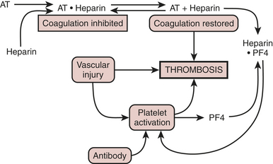
These patients demonstrate a heparin-dependent antibody, usually IgG, although others are described, which aggregates platelets in the presence of heparin.8 During heparin therapy, measured antibody titers remain low, owing to antibody binding to platelets. Titers rise after heparin therapy ceases; but paradoxically, antibody may be undetectable a few months later. Two other features are unexpected: first, the antibody does not aggregate platelets in the presence of excess heparin; and, second, not all reexposed patients develop thrombocytopenia.

The platelet surface contains complexes of heparin and PF4. Affected patients have antibody to this complex. Antibody binding activates platelets via their FcγII receptors and activates endothelium (Fig. 24-7). The activation of the platelet surface triggers a secondary thrombin release. Platelets can attach to each other creating what is known as a white clot syndrome. But if secondary thrombin generation is created through antibody activation of the platelets then a fibrin clot can be the result. In the absence of heparin, the heparin-PF4 antigen cannot form.

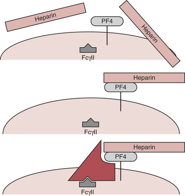
In the absence of an endothelial defect, the only responses to the antibody-antigen interaction are platelet consumption and thrombocytopenia. Atheroma rupture, endovascular interventions such as balloon angioplasty, vascular surgery, and other procedures that disrupt endothelium can provide a nidus for platelet adhesion and subsequent activation. PF4, released with platelet activation, binds to heparin locally, thus not only removing the inhibition of coagulation but also generating additional antigenic material (Fig. 24-8). Clumps of aggregated platelets thrombose vessels, resulting in organ and limb infarction. Amputation and/or death often occurs with established HIT with thrombosis (HITT). The presence of heparin-PF4 antibodies has recently been associated with other adverse effects. It appears that if a patient undergoes cardiac surgery with positive antibodies, the risk of mortality and/or myocardial infarction may at least double.

Incidence and Diagnosis

Estimates of the true incidence of this syndrome are confounded by different diagnostic thresholds for platelet count, varying efforts to detect other causes, and incomplete reports. After 7 days of therapy with UFH, probably 1% of patients develop HIT; after 14 days of therapy the prevalence is 3%. Using a platelet count of 100,000/mm3, multiple reports comprising more than 1200 patients revealed an overall incidence of HIT of 5.5% with bovine heparin and 1.0% with porcine heparin. Other recent research has found the preoperative incidence of ELISA-positive patients to be between 6.5% to 10%. This means that antibodies are present and that may not mean that thrombocytopenia is occurring. Of great interest is that many more patients develop positive tests for ELISA antibodies by days 7 to 30 after cardiac surgery. Somewhere between 25% and 50% of patients develop these antibodies.

Heparin-induced thrombocytopenia can occur not only during therapeutic heparin administration but also with low prophylactic doses, although the incidence is dose related. Even heparin flush solution or heparin-bonded intravascular catheters can incite HIT. Cases of platelet-to-platelet adhesion creating a “white clot” in otherwise normal patients have been observed in the oxygenator and the reservoir of CPB machines. The fact that such events have been reported even when all other tests appeared normal signals the unpredictable nature of the heparin-PF4 antibody as well as the biological activity of UFH.

Although HIT usually begins 3 to 15 days (median, 10 days) after heparin infusions commence, it can occur within hours in a patient previously exposed to heparin. Platelet count steadily decreases to a nadir between 20,000 and 150,000/mm3. Absolute thrombocytopenia is not necessary; only a significant decrease in platelet count matters, as witnessed by patients with thrombocytosis who develop thrombosis with normal platelet counts after prolonged exposure to heparin. Occasionally, thrombocytopenia resolves spontaneously despite continuation of heparin infusion.

Clinical diagnosis of HIT requires a new decrease in platelet count during heparin infusion. Laboratory confirmation is obtained from several available tests. In the serotonin release assay, patient plasma, donor platelets, and heparin are combined. The donor platelets contain radiolabeled serotonin, which is released when donor platelets are activated by the antigen-antibody complex. Measurement of serotonin release during platelet aggregation at both low and high heparin concentrations provides excellent sensitivity and specificity.

A second assay measures more traditional markers of platelet degranulation in a mixture of heparin, patient plasma, and donor platelets. The most specific test is an enzyme-linked immunosorbent assay for antibodies to the heparin-PF4 complex.

Measurement of platelet-associated IgG is poorly specific for HIT, because of numerous other causes of antiplatelet IgG. This test should not be used in the diagnosis of HIT.

Heparin-Induced Thrombocytopenia with Thrombosis

The incidence of HITT is 1.7% with bovine heparin and 0.3% with porcine heparin; thus, thrombosis accompanies more than one in five cases of HIT. It is clear that the longer patients are on heparin the more likely it is that they will develop antibody; and with the knowledge that today close to 50% of cardiac patients develop antibodies, it is possible that a significant number of long-term or early mortalities might be due to undiagnosed HITT. In several studies in the catheterization laboratory, it has been shown that if HITT antibodies are present before the performance of angioplasty, the mortality and combined morbidity are greatly increased, perhaps double or more. One study has been carried out in almost 500 patients undergoing CABG surgery looking for the presence of antibodies and outcome. The incidence of antibody-positive patients was approximately 15%, and their length of stay in the hospital and mortality were more than doubled. Occasional rare situations in which the CPB circuit suddenly clots or when there is early graft thrombosis or whole-body clotting may all be variants of HITT, but none of these cases can be readily studied because they are so rare. If such an occurrence does happen, HITT should be in the differential diagnosis. The occurrence of thrombosis at first seems paradoxical. However, HITT has as its hallmark a huge thrombin burst that can occur all over the body. With such massive thrombin generation the triggering of thrombosis is natural. Thrombosis may then activate the fibrinolytic system to produce a picture of consumptive coagulopathy.

From 15% to 30% of patients who develop HIT with thrombosis will develop severe neurologic complications, require amputation of a limb, or die. Lower limb ischemia constitutes the most frequent presentation. Venous clots occur probably as frequently as arterial ones but are not detected as often. Unfortunately, no test predicts the thrombosis component of HIT; thrombosis should be anticipated in the presence of vascular injury, such as puncture sites for catheterization.

Treatment and Prevention

In the absence of surgery, bleeding from thrombocytopenia with HIT is rare. In contrast to other drug-induced thrombocytopenias, in which severe thrombocytopenia commonly occurs, more moderate platelet count nadirs characterize HIT. Platelet transfusions are not indicated and may incite or worsen thrombosis. Heparin infusions must be discontinued, and an alternative anticoagulant should be instituted. LMWHs can be tested in the laboratory using serotonin release before patient administration. Although thrombosis may be treated with fibrinolytic therapy, surgery is often indicated. No heparin should be given for vascular surgery. Monitoring catheters should be purged of heparin flush, and heparin-bonded catheters should not be placed. Antiplatelet agents, such as aspirin, ticlopidine, or dipyridamole, which block adhesion and activation and, thus, PF4 release, provide ancillary help.

The patient presenting for cardiac surgery who has sustained HIT in the past presents a therapeutic dilemma. Antibodies may have regressed; if so, a negative serotonin release assay using the heparin planned for surgery will predict that transient exposure during surgery will be harmless. However, no heparin should be given at catheterization or in flush solutions after surgery.

Patients with HIT who require urgent surgery may receive heparin once platelet activation has been blocked with aspirin and dipyridamole or, in the past, the prostacyclin analog iloprost. Unfortunately, iloprost is no longer available. The problem with this strategy is obtaining sufficient blockade of platelet activity.

Another alternative, delaying surgery to wait for antibodies to regress, may fail because of the variable offset of antibody presence and the unpredictable nature of platelet response to heparin rechallenge. Plasmapheresis may successfully eliminate antibodies and allow benign heparin administration. Finally, methods of instituting anticoagulation without heparin may be chosen.

LMWH heparin, as an alternative to UFH, has been used for urgent surgery. While LMWHs can also induce thrombocytopenia, by displaying different antigenic determinants, they may prove acceptable alternatives for patients who develop HIT from UFH. Table 24-2 summarizes the therapeutic options available for urgent cardiac surgery in patients with HIT.

Table 24-2 Therapeutic Options for Anticoagulation for Bypass in Patients with Heparin-Induced Thrombocytopenia*

In all cases

* No agent is indicated for anticoagulation in CPB at this time.

New Modes of Anticoagulation

The hemostatic goal during CPB is complete inhibition of the coagulation system. Unfortunately, even large doses of heparin do not provide this, as evidenced by formation of fibrinopeptides during surgery. Despite being far from the ideal anticoagulant, heparin still performs better than its alternatives. Current substitutes for heparin include ancrod, a proteinase obtained from snake venom that destroys fibrinogen; heparin fragments, which provide less thrombin inhibition than the parent, unfractionated molecule; direct factor Xa inhibitors; and direct thrombin inhibitors (Box 24-4).

Direct Thrombin Inhibitors

Hirudin, a single-chain polypeptide containing 65 amino acids with a molecular weight of 7000 and produced by the medicinal leech Hirudo medicinalis, binds directly to thrombin without need of a cofactor or enzyme, inhibiting all the proteolytic functions of thrombin. This inhibition includes actions on fibrinogen; factors V, VIII, and XIII; and platelets.

Modifications of hirudin include hirugen, a synthetic peptide containing residues 53-64 of the native hirudin, and hirulog, formed by attaching the amino acid sequence d-phe-pro-arg-pro-(gly) to the amino-terminal end of hirugen. Hirugen inhibits thrombin’s action on fibrinogen, but not on factor V. Hirulog has full inhibitory properties but is slowly cleaved by thrombin itself to a hirugen-like molecule.

Hirudin depends on renal excretion; renal failure prolongs its elimination half-life of 0.6 to 2.0 hours. Although there are no known direct neutralizing agents for these drugs, administration of prothrombin complex may partially restore coagulation by enhancing thrombin generation. Clinical trials of hirudin compounds have yielded mixed results. It has been used for patients with HITT but the longer half-life of approximately 90 minutes means that many of these patients bleed after cardiac surgery. Hirudin is highly antigenic and will lead to immune complexes being created to itself in about 40% of patients. If it is used a second time, the overall incidence of anaphylaxis may be as high as 10% of all patients who have received it before.

New direct thrombin inhibitors are now available (Fig. 24-9).9 These include argatroban and bivalirudin. Argatroban is a derivative of arginine and is a relatively small molecule. It binds at the active cleavage site of thrombin and stops thrombin’s action on serine proteases. It is completely hepatically cleared and has a half-life of 45 to 55 minutes with prolongation when liver function is depressed or liver blood flow is decreased. There is no reversal agent for argatroban. It has been approved by the U.S. Food and Drug Administration (FDA) for anticoagulation in the face of HITT, but there has not been, to date, a large-scale prospective randomized trial for cardiac surgery or any type of comparison to heparin/protamine. Some case reports do exist of successful usage of argatroban in HITT patients with acceptable amounts of postoperative bleeding. It has been more commonly utilized in the ICU for patients with hypercoagulable syndromes and HITT.

Bivalirudin is a synthetic peptide based on the structure of hirudin (previously called hirulog). Pharmacologists have taken the active amino acids at either end of the hirudin molecule and biosynthesized them. One active site competitively binds to the fibrinogen-binding site of thrombin, and the other 20-amino-acid sequence binds to the active serine cleavage site of thrombin. The two sequences of amino acids are connected together by a tetraglycine spacer. This fully manufactured molecule is highly specific for thrombin and has the unique property that it binds to both clot-bound and free thrombin. Heparin binds only to free plasma thrombin. Bivalirudin has a shorter half-life than argatroban and hirudin; the half-life is approximately 20 to 25 minutes. Like the other direct thrombin inhibitors, it also has no reversal agent analogous to protamine. So when it is used, it must wear off. Bivalirudin undergoes destruction by the molecule to which it binds and deactivates, thrombin; it spontaneously is destroyed by thrombin (proteolytic cleavage). The more thrombin activation that is present (i.e., the less bivalirudin that is present), the shorter is the half-life. Only about 20% of the molecular activity is eliminated by renal clearance. In mild to moderate renal failure, the effect on bivalirudin clearance is fairly small.

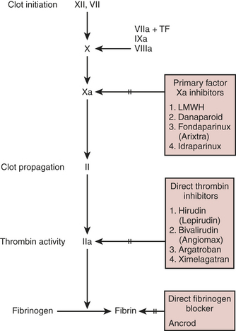
Several clinical trials of bivalirudin for cardiology procedures or cardiac surgery have been completed and published. Other ongoing and pivotal trials aiming for FDA approval are under way comparing bivalirudin to heparin/protamine for both on- and off-pump CABG cases as well as for patients with HITT. Bivalirudin has been FDA approved as a primary anticoagulant for angioplasty. In trials comparing bivalirudin to either heparin/protamine alone or heparin plus the use of a IIb/IIIa inhibitor, bivalirudin was found to have at least equal or better safety and less bleeding than either of the other therapies. When compared with heparin/protamine alone in PCI, bivalirudin was found to be superior, not just in bleeding, but also in terms of morbidity and mortality (as a combined endpoint). In a trial of 100 off-pump CABG patients randomized to receive either bivalirudin or heparin/protamine, bleeding and outcome were equal between the groups.10 These patients underwent recatheterization at 3 months, and it was found that the bivalirudin patients had overall better flow down their grafts than did the patients who had received heparin/protamine. A phase I/II safety trial of bivalirudin in 30 on-pump CABG patients has also shown good safety, but no comparison was carried out to look at advantages against heparin/protamine. At this time, considerable research is ongoing and whether bivalirudin will prove superior to heparin/protamine for routine CABG is yet to be seen.11 When used, the doses for CPB have been a 0.75-mg/kg bolus followed by an infusion at 1.75 mg/kg/hr titrated to the ACT (target, 2.5 times baseline). The CPB system is also primed with 50 mg, and no stasis can be allowed in the CPB circuit due to metabolism of bivalirudin during CPB. The infusion is stopped 15 to 30 minutes before CPB is discontinued, and patients bleed for up to 4 to 6 hours. In off-pump coronary artery bypass grafting similar doses to ACT targets of 350 to 450 seconds have been used.

In the face of HITT syndrome, case reports continue to show effectiveness and utility of bivalirudin. This is an off-label use of the drug because it has not been FDA approved.

PROTAMINE

Pharmacology

Protamine neutralizes heparin-induced anticoagulation. It is a nitrogenous alkaline substance from sperm heads of salmon. Composed of nearly two-thirds arginine, protamines contain many positive charges. Their biological role is to associate with the negatively charged phosphate groups of nucleic acids.

Neutralization of heparin-induced anticoagulation is the primary use of protamine. Formation of complexes with the sulfate groups of heparin forms the basis for this “antidote” effect. Protamine neutralizes the antithrombin effect of heparin far better than its anti–factor Xa effect. This distinction may arise from the need for thrombin, but not factor Xa, to remain complexed to heparin for antithrombin to exert its inhibitory effect. Because porcine mucosal heparin has more potent anti–factor Xa activity than bovine lung heparin, today’s available heparin may prove to be more difficult to neutralize with protamine. Protamine’s poor efficacy in neutralizing anti–factor Xa activity limits the utility of LMWH compounds as anticoagulants for CPB.

In the presence of circulating heparin, protamine forms large complexes with heparin. Excess protamine creates larger complexes. The reticuloendothelial system may then dispose of these particles by endocytosis. Although this action has not been proved, macrophages in the lung may constitute the site for elimination of these complexes, because intravenous administration of protamine permits formation of heparin/protamine complexes in the pulmonary circulation first. Proteolytic degradation of the protamine complexed to heparin conceivably results in free heparin. Protamine degradation in vivo proceeds by the action of circulating proteases, among them carboxypeptidase N, an enzyme that also clears anaphylatoxins and kinin pathway products. The time course of protamine disappearance from plasma in patients remains poorly investigated.

The recommended dose of protamine to neutralize heparin varies widely. This variability has been accounted for by differences in timing, temperature, and other environmental factors; choices for coagulation tests and outcome variables; and speculation and unproven assumptions. Protamine titration tests at the conclusion of CPB can determine the amount of heparin remaining in the patient. With automated versions of this test and simple assumptions regarding the volume of distribution of heparin, the amount needed to neutralize the heparin detected in the patient’s vasculature can easily be calculated.

An alternative scheme splits a calculated dose of protamine of 1 mg/100 units into two separate doses: an initial dose (75% of the total) after CPB, with the remainder after reinfusion of blood from the bypass circuit. This scheme prevented increased plasma heparin levels and prolongation of the activated partial thromboplastin time (aPTT), compared with a control group. The ACT remained unchanged, perhaps a reflection of its insensitivity to small amounts of circulating heparin.

Adverse Reactions

The potential for a deleterious response to protamine administration raises serious questions and difficult choices in clinical care before, during, and after cardiac operations.

Anaphylactoid Reaction

Pulmonary Vasoconstriction

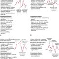
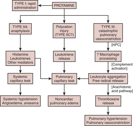
Several years after pulmonary artery catheters achieved common usage and case reports sensitized clinicians to adverse responses to protamine, Lowenstein and associates reported a series of cases in which protamine caused systemic hypotension, decreased left atrial pressure, elevated rather than decreased pulmonary artery pressure, and right ventricular distention and failure.14 Unlike in anaphylaxis, plasma histamine levels do not change during this idiosyncratic, catastrophic pulmonary vasoconstriction, thus justifying a separate classification for this unusual response. The duration of pulmonary hypertension may vary substantially from brief episodes to those requiring reinstitution of CPB.

Animal models of type III protamine responses demonstrate that heparin must precede the protamine, that heparin/protamine complexes activate the complement pathway, and that blockade of complement activation attenuates pulmonary damage. Furthermore, leukocytes respond to complement activation by forming free radicals, which stimulate the arachidonate pathway. Blockade of this pathway mitigates the pulmonary response, whereas antihistamines do not. Figure 24-10 summarizes the speculative mechanisms of various adverse responses to protamine.

image

Figure 24-10 Speculative mechanisms of some protamine reactions. HPC = heparin-protamine complex.

(From Horrow JC: Heparin reversal of protamine toxicity: Have we come full circle? J Cardiothorac Vasc Anesth 4:539, 1990.)

Theoretically, slow administration should limit type III reactions, because large heparin/protamine complexes would less likely form. Slow dilute infusion (see later) has decreased this adverse response to protamine.

On detection of sudden pulmonary hypertension and systemic hypotension, protamine infusion should cease, as should administration of any cardiovascular depressant. Administration of a heparin bolus should be considered in an attempt to reduce heparin/protamine complex size. Excess heparin would theoretically attract protamine away from large complexes to yield a larger number of smaller size particles. If hemodynamics have not deteriorated sufficiently to warrant immediate reinstitution of CPB, 70 units/kg of heparin should be tried first, then 300 units/kg if that fails. Inotropic support should be selected so as not to worsen the pulmonary hypertension; isoproterenol (0.1 to 0.2 μg/kg bolus followed by 0.1 to 0.3 μg/kg/min)or milrinone appear best suited for this purpose. Milder cases may revert without intervention, merely by halting protamine administration, a highly desirable outcome insofar as the treatments outlined above all extract a price, whether it be arrhythmias from inotropes or bleeding from heparin. Rechallenge with protamine should be avoided.

Alternatives to Protamine

This section discusses techniques for neutralizing heparin other than administration of protamine.

BLEEDING PATIENT

After cardiac surgery, some patients bleed excessively. Chest tube drainage of more than 10 mL/kg in the first hour after operation or a total of more than 20 mL/kg over the first 3 hours after operation for patients weighing more than 10 kg is considered significant. Also, any sudden increase of 300 mL/hr or more after minimal initial drainage in an adult usually indicates anatomic disruption warranting surgical intervention.

The TEG has been extensively tested both alone and in conjunction with a number of other tests including prothrombin time (PT), platelet count, and fibrinogen. The TEG has been shown to have the best predictive accuracy for postoperative bleeding.16 Using an algorithm based on the TEG and other tests, blood product utilization was cut considerably. Chest tube bleeding was not different, but the TEG did predict which patients might bleed abnormally. Work with TEG monitoring has shown that it can detect both hypocoagulable states as well as hypercoagulable states. New additives to the testing make it sensitive to the ADP receptor platelet antagonists as well as the GPIIb/IIIa inhibitors.

Insult of Cardiopulmonary Bypass

More so than patient factors, CPB itself acts to impair hemostasis. Bypass activates fibrinolysis, impairs platelets, and affects coagulation factors. Hypothermia, employed in most centers during CPB, adversely affects hemostasis as well.

Prevention of Bleeding

The possible transmission of serious viral illness during transfusion of blood products and impairment of immune function generate concern among clinicians and patients. Many techniques attempt to limit viral exposure, including donation of autologous blood or directed blood, blood scavenging during and after surgery, and efforts to limit perioperative hemorrhage.

Desmopressin

This analog of vasopressin provides more potent and longer lasting antidiuretic activity than vasopressin, with little vasoconstriction (Box 24-5). Like the parent compound and like epinephrine and insulin, desmopressin releases coagulation system mediators from vascular endothelium. Factor VIII coagulant activity increases 2- to 20-fold and is maximal 30 to 90 minutes after injection. Factor XII levels also increase. In response to desmopressin, endothelium releases the larger multimers of vWF, as well as t-PA and prostacyclin. Nevertheless, the overall effect of desmopressin is procoagulant, perhaps because of the impact of factor VIII and vWF.

The optimal dose of desmopressin is 0.3 μg/kg. Intravenous, subcutaneous, and intranasal routes are all acceptable. After plasma redistribution with an 8-minute half-life, metabolism in liver and kidney and urinary excretion yield a plasma half-life of 2.5 to 4 hours. Levels of factor VIII persist in plasma long after desmopressin excretion due to the release of vWF. Rapid intravenous administration decreases systemic blood pressure and SVR, possibly by prostacyclin release or stimulation of extrarenal vasopressin V2 receptors. The drug’s antidiuretic action poses no problem in the absence of excessive free water administration.

aprotinin

Bovine lung provides the source of this 58-residue polypeptide serine protease inhibitor. Aprotinin inhibits a host of proteases, including trypsin, plasmin, kallikrein, and factor XIIa activation of complement (Fig. 24-11). The adult intravenous dose for surgical hemostasis is 2 million kallikrein inhibitor units (KIU) for both patient and bypass pump, followed by 600,000 KIU/hr. The elimination half-life of aprotinin, 7 hours, is considerably longer than that of the synthetic antifibrinolytics; after 6 days, aprotinin continues to be excreted in the urine.

image

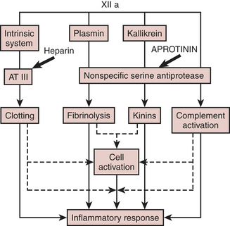
Figure 24-11 Actions of aprotinin on the contact coagulation system, fibrinolytic pathway, and complement activation.

(From Royston D: The serine antiprotease aprotinin [Trasylol]: A novel approach to reducing postoperative bleeding. Blood Coag Fibrin 1:55, 1990.)

Royston and coworkers documented more than a fourfold reduction in blood loss during repeat cardiac surgery. Subsequent studies using high-dose aprotinin confirmed conservation of blood products and a reduction in bleeding, ranging from 29% to 50%. Studies clearly demonstrate decreased fibrinolysis in aprotinin-treated patient groups; preservation of platelet GPIb, or blockade of a plasmin-mediated platelet defect may also explain the hemostatic mechanism of aprotinin.

High-dose aprotinin alone prolongs the celite ACT. Most investigators simply avoid the celite ACT and use kaolin ACT. The kaolin ACT adsorbs about 98% of aprotinin and any intrinsic antithrombin effect that aprotinin has is therefore mitigated. It is recommended to use the kaolin ACT and keep the length of ACT time the same as if aprotinin was not being used. An animal protein, aprotinin can cause anaphylaxis, although this is uncommon (<1 in 1000). Aprotinin costs significantly more than equivalent doses of synthetic antifibrinolytic drugs. The discussion and argument about cost effectiveness of aprotinin versus other antifibrinolytics has continued for more than 10 years. The major concerns today with aprotinin are its adverse effects on renal function and overall cardiovascular outcome; These concerns led to its removal from the market in 2007.18
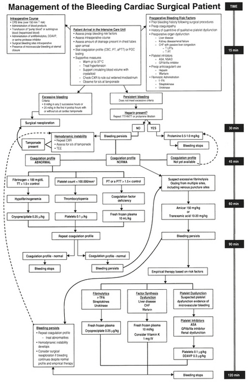
Determine the Cause

The complexity of human hemostasis, augmented by unexpected behavior of coagulation tests, can lead to confusion in the diagnosis of bleeding after cardiac surgery. Anatomic sources of bleeding frequently present once systemic blood pressure achieves sufficient magnitude. Some clinicians prefer to identify these sources before chest closure with a provocative test (i.e., allowing brief periods of hypertension). Generous chest tube drainage early after operation suggests an anatomic source. Retained mediastinal clot may engender a consumptive coagulopathy. A widened mediastinum on chest radiograph suggests the need for surgical drainage.

Nonsurgical causes of bleeding (platelets, coagulation factors, and fibrinolysis) usually manifest as a generalized ooze. Inspection of vascular access puncture sites aids in this diagnosis. Bleeding from other areas not manipulated during surgery (stomach, bladder) may also occur.

Coagulation tests aid diagnosis. Because the PT and aPTT are usually prolonged by several seconds after CPB, only values more than 1.5 times control suggest factor deficiency. Elevation of the ACT should first suggest unneutralized heparin, then factor deficiency.

A decreased platelet count, usually denoting hemodilution or consumption, requires correction with exogenous platelets in any bleeding patient. However, bleeding patients with insufficient functional platelets may demonstrate normal platelet counts early after operation. For this reason, clinicians have sought rapid diagnostic tests of platelet function and attempted correlation with bleeding after CPB.

Low plasma fibrinogen occurs from excessive hemodilution or factor consumption and is corrected with cryoprecipitate or FFP. The thrombin time is useful here. Most clinical laboratories can perform this test with rapid turnaround. A prolonged thrombin time denotes unneutralized heparin, insufficient fibrinogen, or high concentrations of fibrin degradation products. Finally, direct measurement of fibrin degradation products denotes fibrinolytic activity. In the absence of a cause for a consumptive coagulopathy, antifibrinolytic therapy may be useful.

Table 24-3 lists a treatment plan for excessive bleeding after cardiac surgery. Interventions appear not in order of likelihood but rather by priority of consideration. Thus, surgical causes should be ruled out before seizing on the diagnosis of a consumptive coagulopathy. The priority will also vary among institutions, depending on the availability and cost of resources. This table provides a simple algorithm for treating postoperative bleeding. More complete schemes present a daunting level of complexity that deter implementation (Fig. 24-12).

Table 24-3 A Treatment Plan for Excessive Bleeding after Cardiac Surgery

Action Amount Indication
Rule out surgical cause No oozing at puncture sites; chest radiograph
More protamine 0.5 to 1 mg/kg ACT > 150 s or aPTT > 1.5 times control
Warm the patient “Core” temperature < 35°C
Apply PEEP* 5 to 10 cm H2O
Desmopressin 0.3 μg/kg IV Prolonged bleeding time
Aminocaproic acid 50 mg/kg, then 25 mg/kg/hr Elevated D‑dimer or teardrop-shaped TEG tracing
Tranexamic acid 10 mg/kg, then 1 mg/kg/hr Elevated D-dimer or teardrop-shaped TEG tracing
Platelet transfusion 1 U/10 kg Platelet count < 100,000/mm3
Fresh frozen plasma 15 mL/kg PT or aPTT > 1.5 times control
Cryoprecipitate 1 U/4 kg Fibrinogen < 1 g/L or 100 mg/dL

ACT = activated coagulation time; PT = prothrombin time; aPTT = activated partial thromboplastin time; TEG = thromboelastographic.

* Positive end-expiratory pressure (PEEP) is contraindicated in hypovolemia.

image

Figure 24-12 Algorithm for treating excessive bleeding.

(From Milas B, Johes D, Gorman R: Management of bleeding and coagulopathy after heart surgery. Semin Thorac Cardiovasc Surg 12:326, 2000.)

Blood Products

The bleeding patient becomes subject to additional hemostatic derangements. The need to maintain intravascular blood volume arises in nearly all cases before identification of the cause of bleeding. Clear fluid or colloid will replenish intravascular volume. However, red blood cells, platelets, and coagulation factors become diluted when continued bleeding is treated with such replacement. Also, packed red blood cells and banked whole blood do not provide platelets or sufficient factor V or factor VIII to maintain hemostasis. Although routine prophylactic administration of FFP or platelets plays no role in modern cardiac surgical care, demonstration of a platelet count below 100,000/mm3 or prolongation of the PT or aPTT despite adequate heparin neutralization in a patient actively bleeding is an indication for platelet or plasma replacement.

Banked blood should be infused to maintain a hemoglobin concentration that allows appropriate oxygen delivery to tissues. Because patients undergoing cardiac surgery also experience the hemodiluting and antihemostatic effects of CPB, the prudent clinician will commence platelet and factor replenishment earlier in the course of hemorrhage while awaiting laboratory confirmation. Each unit of platelet concentrate supplies about 1011 platelets, which increases the platelet count by about 20,000/mm3 in the adult. Enough plasma accompanies platelet concentrates to supply the equivalent of 1.0 to 1.5 units of plasma for each 6 units of platelets.

Shed mediastinal blood can be collected and given back to patients using a closed drainage system. The drainage fluid, often collected in citrate, contains little fibrinogen. Presence of tissue and other debris in this fluid suggests the need for filtration before reinfusion. Shed mediastinal fluid supplies red blood cells without risk of viral transmission via allogeneic blood products.

SUMMARY

REFERENCES

1. Edmunds L.H.Jr. Blood-surface interactions during cardiopulmonary bypass. J Card Surg. 1993;8:404.

2. Fischer R., Kuppe H., Koster A. Impact of heparin management on release of tissue factor pathway inhibitor during cardiopulmonary bypass. Anesthesiology. 2004;100:1040.

3. Savage B., Saldivar E., Ruggeri Z.M. Initiation of platelet adhesion by arrest onto fibrinogen or translocation on von Willebrand factor. Cell. 1996;84:289.

4. Lange R.A., Hillis L.D. Antiplatelet therapy for ischemic heart disease. N Engl J Med. 2004;350:277.

5. Koster A., Fischer T., Grunendel M., et al. Management of heparin: Resistance and cardiopulmonary bypass: The effect of 5 different anticoagulation strategies on hemostatic activation. J Cardiothorac Vasc Anesth. 2004;18:131.

6. Avidan M., Levy J., Scholz J., et al. A phase III, double blind, placebo-controlled, multicenter study on the efficacy of recombinant human antithrombin in heparin-resistant patients scheduled to undergo cardiac surgery necessitating cardiopulmonary bypass. Anesthesiology. 2005;102:276.

7. Day J.R.S., Landis R.C., Taylor K.M. Heparin is more than just an anticoagulant. J Cardiothorac Vasc Anesth. 2004;18:93.

8. Godal H.C. Heparin-induced thrombocytopenia. In: Lane D.A., Lindahl U., editors. Heparin. Boca Raton, FL: CRC Press; 1989:533.

9. Dinisio M., Middeldorp S., Buller H. Direct thrombin inhibitors. N Engl J Med. 2005;353:1028.

10. Merry A.F., Raudkivi P., White H.D., et al. Anticoagulation with bivalirudin (a direct thrombin inhibitor) vs heparin: A randomized trial in OPCAB graft surgery. Ann Thorac Surg. 2004;77:925.

11. Koster A., Spiess B.D., Chew D.P., et al. Effectiveness of bivalirudin as a replacement for heparin during cardiopulmonary bypass in patients undergoing coronary artery bypass grafting. Am J Cardiology. 2004;93:356.

12. Welsby I., Newman M., Phillips-Bute B., et al. Hemodynamic changes after protamine administration. Anesthesiology. 2005;102:308.

13. Horrow J.C. Protamine allergy. J Cardiothorac Vasc Anesth. 1988;2:225.

14. Kirklin J.K. Prospects for understanding and eliminating deleterious effects of cardiopulmonary bypass. Ann Thorac Surg. 1991;51:529.

15. Michelsen L.G., Kikura M., Levy J.H., et al. Heparinase I (Neutralase) reversal of systemic anticoagulation. Anesthesiology. 1996;85:339.

16. Cammerer U., Dietrich W., Rampf T., et al. The predictive value of modified computerized thromboelastography and platelet function analysis for postoperative blood loss in routine cardiac surgery. Anesth Analg. 2003;96:51.

17. Shore-Lesserson L., Reich D.L., Vela-Cantos F., et al. Tranexamic acid reduces transfusions and mediastinal drainage in repeat cardiac surgery. Anesth Analg. 1996;83:18.

18. Smith P.K., Shah A.S. The role of aprotinin in a blood-conservation program. J Cardiothoracic Vasc Anesth. 2004;18(suppl):S24.

19. Roberts H., Monroe D., Hoffman M. Safety profile of recombinant factor VIIa. Semin Hematol. 2004;41(suppl 1):101.

20. Tanaka K., Waly A., Cooper W., Levy J. Treatment of excessive bleeding in Jehovah’s Witness patients after cardiac surgery with recombinant factor VIIa. Anesthesiology. 2003;98:1513.