Similar lesion may represent a pulsion diverticulum, due to esophageal dysmotility
• Chest x-ray PA view: Calcified perihilar lymph nodes
• Videofluoroscopic esophagogram (barium studies)
Mid esophagus: Traction diverticulum
Tented or triangular in shape with pointed tip, wide mouth
Diverticulum tends to empty when esophagus is collapsed (because it contains all layers)
TOP DIFFERENTIAL DIAGNOSES
• Zenker diverticulum
Posterior hypopharyngeal diverticulum
• Pulsion diverticulum
Mid and distal esophageal pulsion diverticula tend to remain filled after most of barium is emptied (lack of muscle)
Associated with motility disorders
• Esophageal perforation
Sealed-off leak seen as self-contained extraluminal collection of contrast medium that communicates with adjacent esophagus
May be indistinguishable from traction diverticulum without history
PATHOLOGY
• Pathogenesis: Acutely inflamed, enlarged subcarinal nodes indent and adhere to esophageal walls
As inflammation subsides, nodes shrink and retract adherent esophagus
CLINICAL ISSUES
• Small diverticula: Usually asymptomatic
• Large diverticula: ± dysphagia or regurgitation
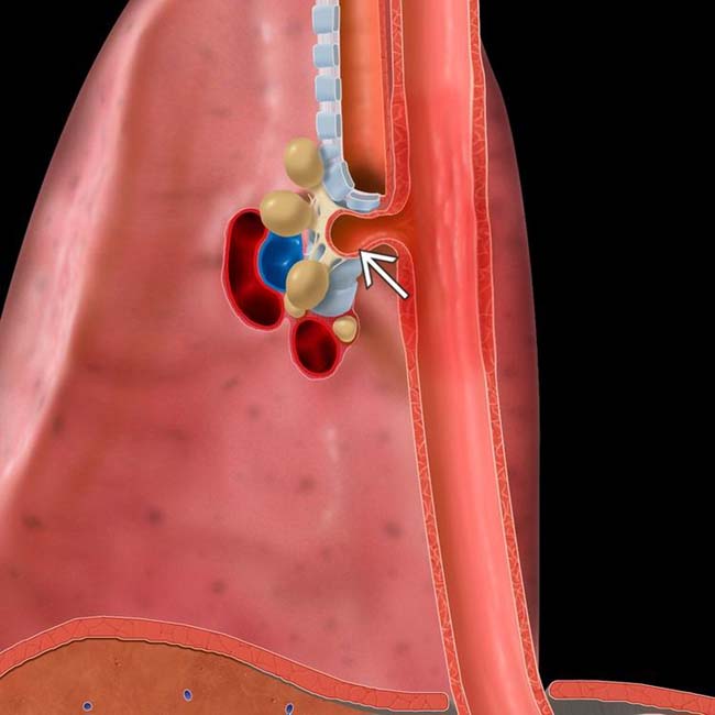
(Left) Graphic shows subcarinal lymph nodes that are adherent to the esophageal wall, resulting in a traction diverticulum .
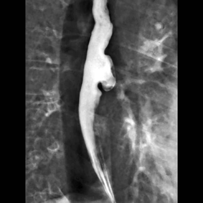
(Right) Spot film from an esophagram shows a barium-filled tented outpouching from the mid esophagus. Calcified subcarinal lymph nodes were more evident on chest radiograph (not shown).
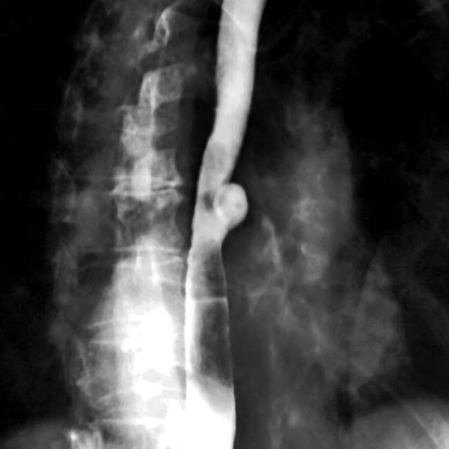
(Left) Oblique view from a barium esophagram demonstrates a saccular outpouching from the mid esophagus just below the tracheal carina, a typical appearance for a traction diverticulum.
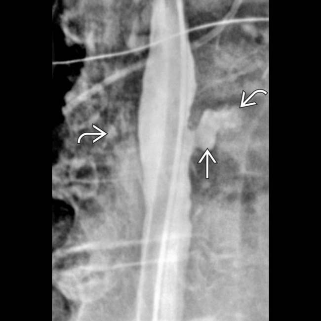
(Right) Spot film from a barium esophagram shows a focal outpouching from the left mid esophagus. Although this may represent a traction diverticulum, it may rather be a normal outpouching of the esophageal wall between the extrinsic indentations by the aortic arch and the left mainstem bronchus.
Esophagram shows a mid esophageal diverticulum.
Esophagram shows outpouching at the subcarinal level.


 .
.
 from the mid esophagus. Calcified subcarinal lymph nodes
  were more evident on chest radiograph (not shown).
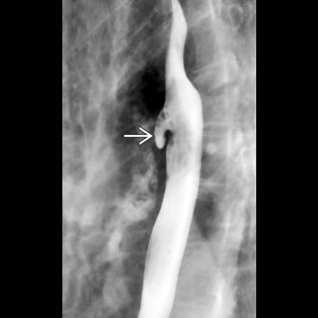
 from the mid esophagus just below the tracheal carina, a typical appearance for a traction diverticulum.
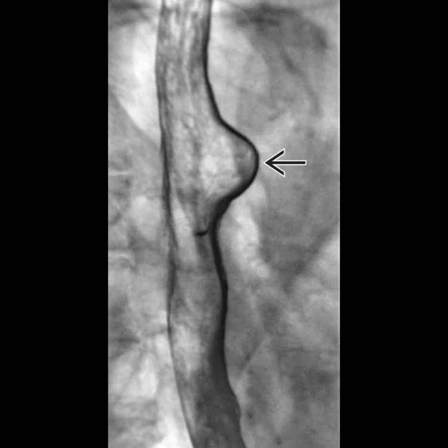
 from the left mid esophagus. Although this may represent a traction diverticulum, it may rather be a normal outpouching of the esophageal wall between the extrinsic indentations by the aortic arch and the left mainstem bronchus.