Tracheotomy and Cricothyrotomy

Published on 16/04/2015 by admin

Filed under Surgery

Last modified 22/04/2025

Print this page

rate 1 star rate 2 star rate 3 star rate 4 star rate 5 star
Your rating: none, Average: 2 (1 votes)

This article have been viewed 10774 times

Chapter 2

Tracheotomy and Cricothyrotomy

image

Indications and Principles of Tracheotomy

Indications for tracheotomy are multiple and include the need to bypass an airway obstruction caused by congenital anomaly, vocal cord paralysis, inflammatory disease, benign or malignant laryngeal pathology, laryngotracheal trauma, facial trauma, or severe sleep apnea refractory to other interventions. Additional indications for tracheotomy include the need to provide an airway for patients receiving mechanical ventilation for respiratory failure and for those with chronic aspiration secondary to inadequate cough. Tracheotomy may also allow for a more secure and comfortable airway for home ventilation in patients with neuromuscular or other chronic diseases.

Preoperative Considerations

Once a tracheotomy is planned, certain factors influence whether patients should have an open tracheotomy or a percutaneous dilatational tracheotomy, as first described by Ciaglia in 1985. Regardless of the tracheotomy method chosen, a patient’s overall medical condition must be optimized, body habitus assessed, and coagulation profile addressed. Other preoperative factors to consider include the urgency of the procedure (emergency vs. elective); need for general or local anesthesia, adult or pediatric patient, current status of the airway (intubated vs. nonintubated patient), availability of proper equipment, patient portability, surgeon’s experience (open vs. percutaneous technique), and capability of the institution to perform bedside procedures. This will determine which team performs the procedure and whether it will be done in the operating room or at the bedside in the intensive care unit.

Surgical Anatomy and Tracheotomy Procedure

External Anatomy

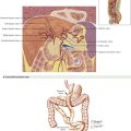
The patient is placed in the supine position. The surgeon might consider placing the neck into slight extension, but this should not be too far past the neutral position, so that the skin incision remains in line with the tracheal incision. A shoulder roll may be used in some patients to assist with positioning (Fig. 2-1).

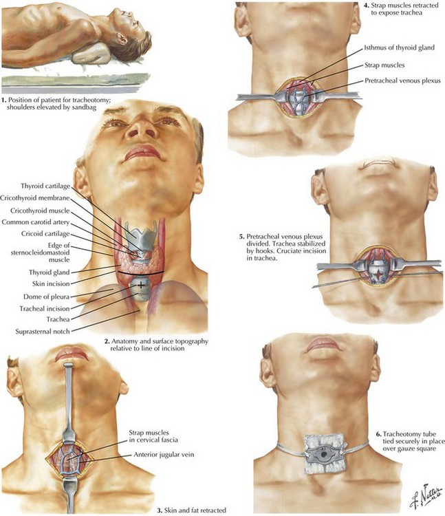
The thyroid notch superiorly, cricoid cartilage, and suprasternal notch inferiorly can usually be palpated and should be marked (Fig. 2-1). If an awake tracheotomy is being performed, the skin is injected with 1% lidocaine with 1 : 100,000 epinephrine solution for hemostasis and anesthesia. According to surgeon preference, this injection may also be done for general anesthesia patients. A vertical or horizontal incision is made in the midline of the neck, about 2 cm above the sternal notch, and is carried down until the strap muscles are visible.

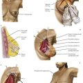
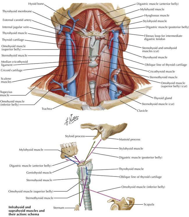
Strap Muscles and Midline Raphe

The anterior jugular veins are typically located on the strap musculature and may require ligation if encountered in the midline (Fig. 2-2). Small cricothyroid arteries traverse the superior aspect of the cricothyroid space, forming an anastomosis near the midline. This may cause problematic bleeding in the setting of emergent airway access or if dissection is carried out above the cricoid cartilage. In the lower neck, the surgeon must be aware that the innominate artery crosses over anterior to the trachea at the level of the thoracic inlet and is higher on the right side. Before dissection of the strap muscles, the surgeon should palpate for innominate pulsations in the suprasternal notch and should be cognizant of the pathway of the surgical dissection in the setting of a high-riding vessel.

Midline dissection is essential for hemostasis and avoidance of paratracheal structures, including the great vessels of the neck. The midline raphe between the paired sternohyoid and sternothyroid muscles can be easily identified. Lateral retraction of the strap muscles along the midline raphe will expose the underlying thyroid gland. Palpation of the trachea can help maintain a midline course of dissection in those individuals with thick subcutaneous tissues.

Thyroid Isthmus

The strap muscles are separated using blunt dissection and retracted to either side until the thyroid isthmus is visible. The isthmus of the thyroid gland generally lies across the first to fourth tracheal rings. It must be divided when overlying the tracheotomy site, because this will make reinsertion safer and easier in the setting of accidental dislodgement. The isthmus can be addressed in one of several ways. First, the fascial attachments of the thyroid to the anterior trachea may be dissected free, thus allowing the gland to be retracted above or below the planned entry site into the trachea. If the thyroid is enlarged and cannot be retracted out of the way, it will need to be divided by further dissecting it from the anterior tracheal wall in the immediate pretracheal plane to establish a bloodless plane of dissection. By identifying the bright-white layer of the tracheal cartilage, the surgeon will minimize bleeding from trauma to the posterior aspect of the gland.

Once the thyroid isthmus is elevated from the trachea, the surgeon may use two clamps on either side, then cutting in the midline with a Bovie cautery device. Once divided, the two ends of isthmus are then suture-ligated using a running or figure-of-eight 2-0 silk stitch. If available, other methods of dividing the gland include using the Harmonic scalpel or other device, based on surgeon preference. Use of cautery alone to divide the thyroid is discouraged, to minimize risk of postoperative hemorrhage.

Anatomy with Trachea Visualized

A cricoid hook may be used to help stabilize the position of the trachea before entering the airway. A hook may also be used to elevate the trachea out of the chest in the patient with kyphosis or a low-lying laryngotracheal complex. Once the anterior wall of the trachea is visualized, the space between the second and third tracheal rings is identified by palpation using a hemostat. A horizontal incision is made between the rings with a scalpel and can be extended laterally in each direction using scissors. Care is taken not to rupture the cuff of the endotracheal tube (ETT) by either deflating it before entering the airway or advancing it distally. It is preferable to maintain the ETT inflated during the procedure. It allows ventilation and minimizes the spray of blood and secretions into the surgical field.

Surgeon preference and age of the patient may influence the type of tracheal incision used. In children, a vertical incision may be used, but in adults the most common technique is to create an anterior tracheal window by removing a section of a single ring. Another technique creates an inferiorly based “trapdoor” flap (Björk flap) composed of an anterior portion of a single tracheal ring and interspace tissue below. After an intercartilaginous incision is made, scissors are used to cut downward on either side to create an inferiorly based flap of tracheal tissue. The superior edge of this flap is then stitched to the skin edge to exteriorize and secure the trachea. Although some consider this to be the safest method because the airway is secured to the skin, this may lead to future complications.