Thyroiditis

Published on 25/03/2015 by admin

Filed under Pediatrics

Last modified 22/04/2025

Print this page

rate 1 star rate 2 star rate 3 star rate 4 star rate 5 star
Your rating: none, Average: 0 (0 votes)

This article have been viewed 1192 times

Chapter 560 Thyroiditis

Lymphocytic Thyroiditis (Hashimoto Thyroiditis, Autoimmune Thyroiditis)

Lymphocytic thyroiditis is the most common cause of thyroid disease in children and adolescents and accounts for many of the enlarged thyroids formerly designated “adolescent” or “simple” goiter. It is also the most common cause of acquired hypothyroidism, with or without goiter.

One to 2% of younger school-aged children and 4-6% of adolescents have positive antithyroid antibodies as evidence of autoimmune thyroid disease.

Etiology

This typical organ-specific autoimmune disease is characterized histologically by lymphocytic infiltration of the thyroid. Early in the course of the disease, there may be hyperplasia only; this is followed by infiltration of lymphocytes and plasma cells between the follicles and by atrophy of the follicles. Lymphoid follicle formation with germinal centers is almost always present; the degree of atrophy and fibrosis of the follicles varies from mild to moderate.

Intrathyroidal lymphocyte subsets differ from those in blood. About 60% of infiltrating lymphoid cells are T cells, and about 30% express B-cell markers; the T-cell population is represented by helper (CD4+) and cytotoxic (CD8+) cells. The participation of cellular events in the pathogenesis is clear. Certain HLA haplotypes (HLA-DR4, HLA-DR5) are associated with an increased risk of goiter and thyroiditis, and others (HLA-DR3) are associated with the atrophic variant of thyroiditis.

A variety of different thyroid antigen autoantibodies are also involved. Thyroid antiperoxidase antibodies (TPOAbs; formerly called antimicrosomal antibodies) and antithyroglobulin antibodies are demonstrable in the sera of 90% of children with lymphocytic thyroiditis and in many patients with Graves disease. TPOAbs inhibit enzyme activity and stimulate natural killer cell cytotoxicity. Antithyroglobulin antibodies do not appear to play a role in the autoimmune destruction of the gland. Thyrotropin receptor–blocking antibodies are often present, especially in patients with hypothyroidism, and it is now believed that they are related to the development of hypothyroidism and thyroid atrophy in patients with autoimmune thyroiditis. Antibodies to pendrin, an apical protein on thyroid follicular cells, have been demonstrated in 80% of children with autoimmune thyroiditis.

Clinical Manifestations

The disorder is 2-4 times more common in girls than in boys. It can occur during the first 3 yr of life but becomes sharply more common after 6 yr of age and reaches a peak incidence during adolescence. The most common clinical manifestations are goiter and growth retardation. The goiter can appear insidiously and may be small or large. In most patients, the thyroid is diffusely enlarged, firm, and nontender. In about 30% of patients, the gland is lobular and can seem to be nodular. Most of the affected children are clinically euthyroid and asymptomatic; some may have symptoms of pressure in the neck, including difficulty swallowing and shortness of breath. Some children have clinical signs of hypothyroidism, but others who appear clinically euthyroid have laboratory evidence of hypothyroidism. A few children have manifestations suggesting hyperthyroidism, such as nervousness, irritability, increased sweating, and hyperactivity, but results of laboratory studies are not necessarily those of hyperthyroidism. Occasionally, the disorder coexists with Graves disease. Ophthalmopathy can occur in lymphocytic thyroiditis in the absence of Graves disease.

The clinical course is variable. The goiter might become smaller or might disappear spontaneously, or it might persist unchanged for years while the patient remains euthyroid. Most children who are euthyroid at presentation remain euthyroid, although a percentage of patients acquire hypothyroidism gradually within months or years. In children who initially have mild or subclinical hypothyroidism (elevated serum TSH, normal free T4 level), over several years about 50% revert to euthyroidism, about 50% continue to have subclinical hypothyroidism, and a few develop overt hypothyroidism. Thyroiditis is the cause of most cases of nongoitrous (atrophic) hypothyroidism.

Familial clusters of lymphocytic thyroiditis are common; the incidence in siblings or parents of affected children may be as high as 25%. Autoantibodies to thyroglobulin and thyroid peroxidase in these families appear to be inherited in an autosomal dominant fashion, with reduced penetrance in males. The concurrence within families of patients with lymphocytic thyroiditis, “idiopathic” hypothyroidism, and Graves disease provides cogent evidence for a basic relationship among these 3 conditions.

The disorder has been associated with many other autoimmune disorders. Autoimmune thyroiditis occurs in 10% of patients with type I autoimmune polyglandular syndrome (APS-1), characterized by autoimmune polyendocrinopathy, candidiasis, and ectodermal dysplasia (APCED). APS-1 consists of 2 of the triad of hypoparathyroidism, Addison disease, and mucocutaneous candidiasis (HAM syndrome). This relatively rare autosomal recessive disorder occurs in childhood and is caused by mutations in the autoimmune regulatory (AIRE) gene on chromosome 21q22.3.

Autoimmune thyroiditis occurs in 70% of patients with APS-2 (Schmidt syndrome). APS-2 consists of the association of Addison disease with type 1 diabetes mellitus (T1DM) or autoimmune thyroid disease. The etiology is unknown, and it typically occurs in early adulthood. Autoimmune thyroid disease also tends to be associated with pernicious anemia, vitiligo, or alopecia. TPOAbs are found in approximately 20% of white and 4% of black children with T1DM. Autoimmune thyroid disease has an increased incidence in children with congenital rubella.

Lymphocytic thyroiditis is also associated with certain chromosomal disorders, particularly Turner syndrome and Down syndrome. In children with Down syndrome, one study reported that 28% had antithyroid antibodies (predominantly anti-TPOs), 7% had subclinical hypothyroidism, 7% had overt hypothyroidism, and 5% had hyperthyroidism. In a study of girls with Turner syndrome, 41% had antithyroid antibodies (again, predominantly anti-TPOs), 18% had goiter, and 8% had subclinical or overt hypothyroidism. Another study of 75 girls with Turner syndrome found that autoimmune thyroid disease increased from the 1st (15%) to the 3rd (30%) decade of life. Boys with Klinefelter syndrome are also at risk for autoimmune thyroid disease. The differential diagnosis is noted in Table 560-1.

Laboratory Findings

Thyroid function tests (free T4 and TSH) are often normal, although the level of TSH may be slightly or even moderately elevated in some patients, termed subclinical hypothyroidism. The fact that many children with lymphocytic thyroiditis do not have elevated levels of TSH indicates that the goiter may be caused by the lymphocytic infiltrations or by thyroid growth-stimulating immunoglobulins. Young children with lymphocytic thyroiditis have serum antibody titers to TPO, but the antithyroglobulin test for thyroid antibodies is positive in <50%. Antibodies to TPO and thyroglobulin are found equally in adolescents with lymphocytic thyroiditis. When both tests are used, approximately 95% of patients with thyroid autoimmunity are detected. Levels in children and adolescents are lower than those in adults with lymphocytic thyroiditis, and repeated measurements are indicated in questionable instances because titers might increase later in the course of the disease.

Thyroid scans and ultrasonography usually are not needed. If they are done, thyroid scans reveal irregular and patchy distribution of the radioisotope, and in about 60% or more, the administration of perchlorate results in a >10% discharge of iodide from the thyroid gland. Thyroid ultrasonography shows scattered hypoechogenicity in most patients. The definitive diagnosis can be established by biopsy of the thyroid; this procedure is rarely clinically indicated.

Antithyroid antibodies may also be found in almost 50% of the siblings of affected patients and in a significant percentage of the mothers of children with Down syndrome or Turner syndrome without demonstrable thyroid disease. They are also found in 20% of children with DM and in 23% of children with the congenital rubella syndrome.

Other Causes of Thyroiditis

Acute suppurative thyroiditis is uncommon; it is usually preceded by a respiratory infection. The left lower lobe is affected predominantly. Abscess formation can occur. Anaerobic organisms, with or without aerobes, are the typical infectious agent. The most common organism is viridans streptococcus, followed by Staphylococcus aureus and pneumococcus. Recurrent episodes or detection of a mixed bacterial flora suggests that the infection arises from a piriform sinus fistula or, less commonly, from a thyroglossal duct remnant. Exquisite tenderness of the gland, swelling, erythema, dysphagia, and limitation of head motion are characteristic findings. Fever, chills, and sore throat are not uncommon, and leukocytosis is present. Scintigrams of the thyroid often reveal decreased uptake in the affected areas, and ultrasonography might show a complex echogenic mass. Thyroid function is usually normal, but thyrotoxicosis due to escape of thyroid hormone has been encountered in a child with suppurative thyroiditis resulting from Aspergillus. When abscesses form, incision and drainage and administration of parenteral antibiotics are indicated. After the infection subsides, a barium esophagram or CT scan with contrast is indicated to search for a fistulous tract; if one is found, surgical excision is indicated.

Subacute granulomatous thyroiditis (de Quervain disease) is rare in children. It is thought to have a viral cause and remits spontaneously. The disorder becomes manifested by an upper respiratory infection with vague tenderness over the thyroid and low-grade fever, followed by severe pain in the region of the thyroid gland. Inflammation results in leakage of preformed thyroid hormone from the gland into the circulation. Serum levels of T4 and T3 are elevated while TSH is suppressed, and mild symptoms of hyperthyroidism may be present, but radioiodine uptake is depressed. The erythrocyte sedimentation rate is increased. The course is variable but usually characterized by four phases: hyperthyroidism, usually followed by a euthyroid phase and then a hypothyroid phase, and remission usually occurring in several months, with recovery to euthyroidism.

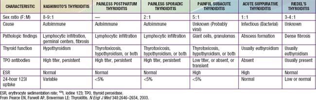
Specific conditions such as tuberculosis, sarcoidosis, mumps, and cat-scratch disease are rare causes of thyroiditis in children. Other forms of thyroiditis seen in adults, such as painless sporadic thyroiditis and Riedel thyroiditis, are rare in children (see Table 560-1).

Bibliography

Corrias A, Cassio A, Weber G, et al. Thyroid nodules and cancer in children and adolescents affected by autoimmune thyroiditis. Arch Pediatr Adolesc Med. 2008;162:526-531.

Demirbilek H, Kandemir N, Gonc EN, et al. Hashimoto’s thyroiditis in children and adolescents: a retrospective study on clinical, epidemiological and laboratory properties of the disease. J Pediatr Endocrinol Metab. 2007;20:1199-1205.

de Vries L, Bulvik S, Phillip M. Chronic autoimmune thyroiditis in children and adolescents: at presentation and during long-term follow-up. Arch Dis Child. 2009;94:33-37.

Dittmar M, Kahaly GJ. Immunoregulatory and susceptibility genes in thyroid and polyglandular autoimmunity. Thyroid. 2005;15:239-250.

Gopalakrishnan S, Chugh PK, Chhillar M, et al. Goitrous autoimmune thyroiditis in a pediatric population: a longitudinal study. Pediatrics. 2008;122:e670-e674.

Hollowell JG, Staehling NW, Flanders D, et al. Serum TSH, T4, and thyroid antibodies in the United States population (1988 to 1994): National Health and Nutrition Examination Survey (NHANES III). J Clin Endocrinol Metab. 2002;87:489-499.

Radetti G, Gottardi E, Bona G, et al. The natural history of euthyroid Hashimoto’s thyroiditis in children. J Pediatr. 2006;149:827-832.

Svensson J, Ericsson UB, Nilsson P, et al. Levothyroxine treatment reduces thyroid size in children and adolescents with chronic autoimmune thyroiditis. J Clin Endocrinol Metab. 2006;91:1729-1734.

Wang SY, Tung YC, Tsai WY, et al. Long-term outcome of hormonal status in Taiwanese children with Hashimoto’s thyroiditis. Eur J Pediatr. 2006;165:481-483.

Yoshida A, Hisatome I, Taniguchi S, et al. Pendrin is a novel autoantigen recognized by patients with autoimmune thyroid diseases. J Clin Endocrinol Metab. 2009;94:442-448.