The Yeasts

Published on 08/02/2015 by admin

Filed under Basic Science

Last modified 22/04/2025

Print this page

rate 1 star rate 2 star rate 3 star rate 4 star rate 5 star
Your rating: none, Average: 0 (0 votes)

This article have been viewed 3230 times

The Yeasts

General Characteristics

Yeasts are eukaryotic, unicellular organisms that are round to oval and range in size from 2 to 60 µm. The microscopic morphologic features have limited usefulness in helping to differentiate or identify these organisms. The microscopic morphology on cornmeal agar is most useful when considered in conjunction with the biophysical profile (i.e., a combination of the biochemical and physical characteristics used in the identification of a microorganism) obtained using a commercial system. Differentiation of yeasts in direct microscopic and histopathologic examination of clinical specimens is often impossible, but sometimes particular characteristics are seen that suggest the identification or are pathognomonic (i.e., unique) for a particular organism. Important morphologic characteristics that are useful in differentiating yeasts include the size of the yeasts, the presence or absence of a capsule, and broad-based or narrow-necked budding. For example, variability in size with evidence of a capsule and narrow-necked budding are features that can be helpful for distinguishing Cryptococcus spp. from Candida spp. The medically important yeasts and yeastlike organisms belong to different taxonomic groups, including the Ascomycota, Basidiomycota, and Deuteromycota.

In general, the yeasts reproduce asexually by blastoconidia formation (budding) (Figure 63-1) and sexually by the production of ascospores or basidiospores. The process of budding begins with a weakening and subsequent outpouching of the yeast cell wall. This process continues until the bud, or daughter cell, is completely formed. The cytoplasm of the bud is contiguous with the cytoplasm for the original cell. Finally, a cell wall septum is created between mother and daughter yeast cells. The daughter cell often eventually detaches from the mother cell, and a residual defect occurs at the budding site (i.e., a bud scar).

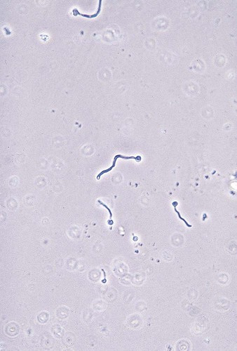
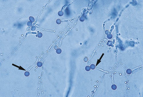
With certain environmental stimuli, yeast can produce different morphologies. An outpouching of the cell wall that becomes tubular and does not have a constriction at its base is called a germ tube; it represents the initial stage of true hyphae formation (Figure 63-2). Alternatively, if buds elongate, fail to dissociate, and form subsequent buds, pseudohyphae are formed; to some, these resemble links of sausage (Figure 63-3). Pseudohyphae have cell wall constrictions rather than true intracellular septations delineating the fungal cell borders.

The number of fungal infections caused by yeasts and yeastlike fungi has increased significantly during recent years. Most of these infections have been caused by various Candida spp. However, other yeasts also cause significant disease, particularly in immunocompromised hosts, as yeasts are the cause of many opportunistic infections. In addition to causing disease in immunocompromised patients, infections also are common in postsurgical patients, trauma patients, and patients with long-term indwelling venous catheters. Some of these yeasts are resistant to commonly used antifungal agents, which emphasizes the need for prompt, appropriate identification and, in some cases, antifungal susceptibility testing.

The extent to which the laboratory should identify all yeast species is the subject of debate. Each laboratory director must decide how much time, effort, and expense is to be spent on the identification of yeasts in the laboratory.

The development of commercially available yeast identification systems has provided laboratories of all sizes with accurate, standardized methods. However, these methods should be used in conjunction with cornmeal agar morphology to prevent misidentifications. Some commercial systems have extensive computer databases that include biochemical profiles of a large number of yeasts. Variations in the reactions of carbohydrates and other substrates utilized are considered in the identification of yeasts provided by these systems.

Commercially available systems are recommended for all laboratories. They may be used in conjunction with some less expensive and rapid screening tests that provide presumptive identification of C. neoformans and definitive identification of Candida albicans.

More recent diagnostic tools that have been introduced for quicker characterization of yeasts include CHROMagar Candida (BD Diagnostics, Sparks, Maryland) and C. albicans PNA FISH (AdvanDX, Woburn, Massachusetts). Because some laboratories might prefer to use conventional systems, the information presented in this section discusses rapid screening methods for presumptive identification of yeasts, commercially available systems, and a conventional schema for identifying commonly encountered yeast species.

Epidemiology

Candida spp.

Candida spp. are responsible for the most frequently encountered opportunistic fungal infections. Currently Candida spp. are the fourth most common cause of hospital-acquired bloodstream infections (BSIs) in the United States, and a mortality rate as high as 49% has been seen with these infections. Candida infections are caused by a variety of species. C. albicans is the most commonly isolated yeast, but other, emerging species include Candida glabrata, Candida parapsilosis, Candida tropicalis, Candida dubliniensis, and Candida krusei. The frequency with which these organisms are isolated varies by institution. Until recently C. albicans was the most common yeast isolated from infections, accounting for at least 60% to 70% of yeast infections. Epidemiologic data for the past decade reveal a paradigm shift in candidal infections. Studies from intensive care units (ICUs) confirm an increase in C. glabrata and C. krusei (isolated in approximately 25% of infections) and C. albicans (about 50% of infections). C. albicans and other Candida spp. are part of the human body’s microbiota (i.e., normal flora), but they also have become endemic in most hospitals. Infections may be caused by endogenous yeasts or may be acquired in the hospital. Differentiating among the Candida spp. in the clinical laboratory is very important because of the differences in the virulence of the species and in their susceptibility to antifungal drugs.

Cryptococcus spp.

Cryptococcus neoformans-Cryptococcus gatti complex has been divided into the two species and five serotypes. Serotype A has been referred to as C. neoformans var. grubii with some groups recognizing the variant as a distinct species. Serotype D is now classified as C. neoformans var. neoformans. Both C. neoformans serotypes produce the teleomorphic state Flobasidiella neoformans. A C. grubii/C. neoformans hybrid exists that is a variant of serotypes A and D. Serotypes B and C are recognized as the independent species, C. gattii. Both serotypes B and C produce the teleomorph referred to as Filobasidiella bacillispora. Standard laboratory tests do not differentiate among the serotypes of C. neoformans and C. gattii.Cryptococcus exists as a saprobe in nature. It is most associated with avian excreta, particularly pigeons. C. neoformans is thought to be widely distributed in nature, and aerosolization is a prerequisite to most infections.

Pathogenesis and Spectrum of Disease

Candida Albicans

Candidiasis is an infection caused by a Candida spp. It may include oroesophageal candidiasis, intertriginous candidiasis (in which skin folds are involved), paronychia, onychomycosis, perlèche respiratory infections, vulvovaginitis, thrush, pulmonary infection, eye infection, endocarditis, meningitis, fungemia or candidemia, or disseminated infection. Paronychia is an infection of the tissues surrounding the nails, and onychomycosis is an infection of the nail and nail bed. Thrush, an infection of the mucous membranes in the mouth, is considered a localized infection. Thrush can be seen in newborns, patients with human immunodeficiency virus (HIV) infection, individuals with diabetes, and patients undergoing chemotherapy. Creamy patches or colonies appear on the tongue and mucous membranes. Candida organisms may be recovered from the oropharynx, gastrointestinal (GI) tract, genitourinary tract, and skin.

The clinical significance of candidal organisms recovered from respiratory tract secretions is difficult to determine, because Candida spp. are considered part of the normal oropharyngeal flora of humans. A study at the Mayo Clinic evaluated the clinical significance of yeasts other than C. neoformans that are recovered from respiratory secretions. These researchers concluded that such yeasts are part of the normal flora and do not need to be routinely identified. Similarly, Barenfanger et al. demonstrated that routine identification of yeasts from respiratory specimens results in unnecessary antifungal therapy, an extended hospital stay, and increased health care costs without demonstrable benefit. Simultaneous recovery of the same species of yeast from several body sites, including urine, is a good indicator of disseminated infection and fungemia.

The pathogenesis of candidal infections is extremely complex and probably varies with each species. Adhesion of Candida organisms to the epithelium of the gastrointestinal or urinary tract is a crucial factor. Candida spp. commonly colonize mucosal surfaces. Their ability to invade and cause infection depends on adherence to the surface before infection. Three distinct aspartyl proteases have been described in C. albicans, and strains with high levels of proteases have been shown to have an increased ability to cause disease in experimental animal models. Hydrophobic molecules on the surface of Candida spp. also appear to be important in pathogenesis, and a strong correlation exists between adhesion and surface hydrophobicity. In addition, high levels of phospholipase, found in strains of C. albicans, have correlated with a higher mortality rate in experimental animals compared with experimental infections caused by strains that produce a lower level of phospholipase. Phenotypic switching (i.e., the ability to produce pseudohyphae and hyphae), seen in C. albicans, also may play a role in pathogenesis.

Non-Albicans Candida

The other Candida spp. (also called non-albicans Candida), once thought not to cause disease, are emerging as agents of infection in certain patient populations. The incidence of infection with C. glabrata is higher in older adults than in young adults and children. Recent studies have demonstrated the ability of C. glabrata to become resistant to common antifungal drugs. C. tropicalis has been shown to be prevalent in patients with hematologic malignancies, especially those who are neutropenic. Mouse models of infection and human studies have shown C. tropicalis in the tissues surrounded by necrotic tissues, which may indicate that the organism can invade the GI tract efficiently, particularly in oncology patients. This phenomenon most likely is due to the expression and secretion of aspartyl proteases and tropiase (acid proteinase), a virulence factor found in Candida organisms. Because C. krusei is inherently resistant to the azole class of antifungal drugs, identification of this species is essential to proper clinical management of the patient. C. parapsilosis is the primary cause of fungemia in the neonatal intensive care unit (NICU). C. parapsilosis is also the second most frequently isolated Candida spp. in positive blood cultures; this could be due partly to its known selective growth in hyperalimentation solutions and also to its ability to grow on intravascular catheters. Historically C. parapsilosis was categorized into three groups (I to III); however, it now has been typed by molecular methods into three different Candida species: C. parapsilosis, C. orthopsilosis, and C. metapsilosis.

Cryptococcus Neoformans

Genus Cryptococcus

Cryptocococcosis is an acute, subacute, or chronic fungal infection that has several manifestations. Crytococcus neoformans var. grubii, C. neoformans var. neofromans, and C. gatti are considered the major human pathogens. Differences in the infections by Cryptococcus neoformans appear to be dependent on the host immune status and not the variant. C. neoformans infections can present initially as a chronic or subacute pulmonary infection. C. neoformans eventually makes its way to the central nervous system, where the yeast can cause cryptococcal meningitis. Disseminated disease with meningitis is commonly seen in immunocompromised patients. Patients with a moderately compromised immune system or who are early in the disease process of cryptococcal fungemia may present without concomitant meningitis. Disseminated cryptococcosis and cryptococcal meningitis became well recognized in patients with acquired immunodeficiency syndrome (AIDS), and they remain an important cause of morbidity and mortality in these patients in resource-poor countries that do not have access to highly active antiretroviral therapy.

Patients with disseminated infection may have painless papular skin lesions that may ulcerate. Other, less common manifestations of cryptococcosis include endocarditis, hepatitis, renal infection, and pleural effusion. Interestingly, a review of patient records at the Mayo Clinic revealed that more than 100 immunocompetent patients with C. neoformans colonization of the respiratory tract did not develop subsequent infection. Follow-up on these patients was as long as 6 years, and none in this group were considered to be immunocompromised. This makes the clinical significance of C. neoformans somewhat difficult to assess. However, given the severity of disease it can cause, its presence in clinical specimens should be considered significant. In many instances, the clinical symptoms are suppressed by corticosteroid therapy, which is a risk factor for disease, and culture or serologic evidence (detection of cryptococcal antigens) provides the earliest proof of infection. Cryptococcal infection is strongly associated with such debilitating diseases as leukemia and lymphoma and the immunosuppressive therapy that may be required for these and other underlying diseases. In some cases the presence of C. neoformans in clinical specimens precedes the symptoms of an underlying disease.

C. neoformans can exhibit a very characteristic polysaccharide capsule. The capsule collapses and protects the yeast from desiccation under drying conditions. The capsule of C. neoformans is thought to help the organism survive through the pigeon gut before it is excreted. The reduction in the yeast’s cell size caused by capsular collapse places the organism in the ideal size range for alveolar deposition in the human host. In addition, a virulent property of the polysaccharide capsule is that it contains compounds that phagocytes do not recognize. The so-called acapsular strains of C. neoformans, which actually just have a very reduced capsule, are more easily phagocytosed. In some instances, C. neoformans elicits minimal tissue response in infected individuals, particularly severely immunocompromised patients.

Phenoloxidase, an enzyme found in C. neoformans, is responsible for melanin production. Some have speculated that melanin might act as a virulence factor by making the organism resistant to leukocyte attack. Evidence also has been presented that increased melanin production can decrease immune system functions, such as lymphocyte proliferation and tumor necrosis factor production. Whether phenoloxidase is truly a virulence factor has yet to be determined (Li SS, et al, 2010. (Cryptococcus. 2010 Proc Am Thorac Soc Vol 7 pp 186-196, 2010). An interesting question is whether the interactions of substances in the brain that are known to react with phenoloxidase may play a role in the affinity of C. neoformans and certain neurotropic dematiaceous fungi for invading the central nervous system (Li et al, 2010).

Trichosporon spp.

Trichosporonosis is caused by a variety of Trichosporon spp., which have undergone changes in nomenclature based on DNA sequence comparisons. The yeastlike fungus causes disease almost exclusively in immunocompromised patients, particularly those with leukemia. Disseminated trichosporonosis is the most common clinical manifestation. Skin lesions accompanied by fungemia are frequently seen. Endocarditis, endophthalmitis, and brain abscess have been reported. Trichosporon organisms occasionally are recovered from respiratory tract secretions, skin, the oropharynx, and the stool of patients with no evidence of infection and may represent transient fungal colonization of those individuals.

White piedra, an uncommon fungal infection of immunocompetent patients, is found in both tropical and temperate regions of the world. It is characterized by the development of soft, yellow or pale brown aggregations around hair shafts in the axillary, facial, genital, and scalp regions of the body. The Trichosporon spp. that cause this disease frequently invade the cortex of the hair, causing damage.

Laboratory Diagnosis

Specimen Collection, Transport, and Processing

See Chapter 59.

Stains

Candida spp.

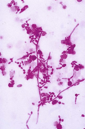
Direct microscopic examination of clinical specimens containing Candida organisms reveals budding yeast cells (blastoconidia) 2 to 4 μm in diameter and/or pseudohyphae (Figure 63-4) showing regular points of constriction, resembling links of sausage. True septate hyphae (filamentation) may also be produced by C. albicans and C. dubliniensis. The blastoconidia, hyphae, and pseudohyphae are strongly gram positive. The approximate number of such forms should be reported, because the presence of large numbers in a fresh clinical specimen may have diagnostic significance. Microscopically C. glabrata blastoconidia are notably smaller (at 1 to 4 µm) than those of other medically significant Candida spp.

Cryptococcus spp.

Traditionally, the India ink preparation has been the most widely used method for the rapid detection of C. neoformans in clinical specimens. This method is still used as a rapid and inexpensive assessment tool in many institutions and has considerable diagnostic value in resource-poor settings. This method delineates the large capsule of C. neoformans, because the ink particles cannot penetrate the capsular polysaccharide material. Although this test is useful, many laboratories have replaced it with the more sensitive cryptococcal latex agglutination test that detects cryptococcal antigen. (The cryptococcal antigen detection [CAD)]test is described later in the chapter.) The India ink preparation is commonly positive in specimens from patients with AIDS and has been shown to have a sensitivity of 50% in patients who do not have HIV or AIDS. Laboratories that examine many specimens from these patients may want to continue using this procedure in combination with the CAD test and culturing.

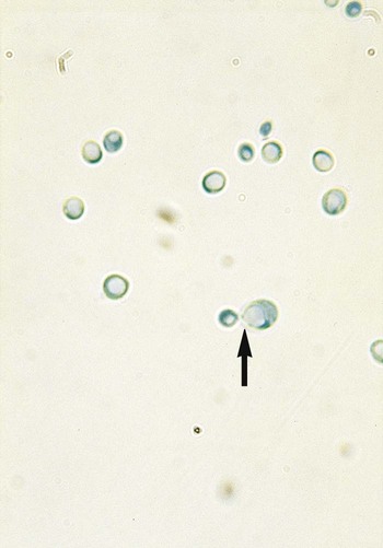
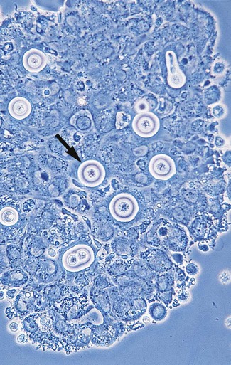
Microscopic examination of other clinical specimens, including respiratory secretions, can be valuable for making a diagnosis of cryptococcosis. C. neoformans appears as a spherical, single or multiple budding, thick-walled yeast 2 to 15 μm in diameter. It usually is surrounded by a wide, refractile polysaccharide capsule (Figure 63-5). Perhaps the most important characteristic of C. neoformans is the extreme variation in the size of the yeast cells; this is unrelated to the amount of polysaccharide capsule present. It is important to remember that not all isolates of C. neoformans exhibit a discernible capsule.

Trichosporon spp.

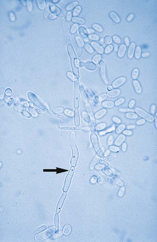
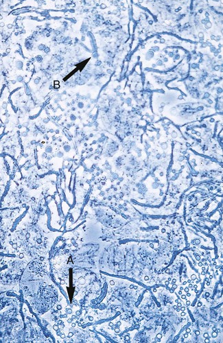
Microscopic examination of clinical specimens that contain Trichosporon spp. reveals hyaline hyphae, numerous round to rectangular arthroconidia, and occasionally a few blastoconidia. Usually hyphae and arthroconidia predominate. In white piedra, white nodules are removed and observed using the potassium hydroxide (KOH) preparation after light pressure is applied to the coverslip to crush the nodule. Hyaline hyphae 2 to 4 μm wide and arthroconidia are found in the preparation of the cementlike material that binds the hyphae together. The organism may be identified in culture by the presence of true hyphae, blastoconidia, and arthroconidia in conjunction with a positive urease (see Figure 60-40). Although Trichosporon asahii may be distinguished from other Trichosporon species by its biophysical profile (carbohydrate and substrate utilization), these organisms are likely best distinguished at the species level with molecular tools, such as DNA sequencing.

Molecular Assays

Nucleic acid amplification tests (NAATs) have been developed for a variety of yeast species. However, these are usually performed in research settings. Most are labeled as “laboratory-developed tests” or “home-brewed tests,” and few have been approved by the U.S. Food and Drug Administration (FDA). Real-time polymerase chain reaction (PCR) methods are now commercially available in the TaqMan system (Applied Biosystems, USA) and LightCycler (Roche Molecular Systems, Indianapolis, Indiana). These methods are very expensive but have proven to be much more specific than conventional yeast identification methods. Ligouri et al. recently demonstrated the Multiplex PCR method for identifying Candida spp. that has high concordance with commercially available phenotypic identification systems, such as the API 20C AUX (bioMérieux, Durham, North Carolina) and Vitek 2 YST card (bioMérieux, Durham, North Carolina). The Multiplex PCR method is much faster and more sensitive than the current phenotypic tests. A newer molecular test, the PNA FISH kit, uses in situ hybridization to detect Candida organisms in blood culture bottles by targeting specific rRNA sequences. Subculturing should follow this method, because whether more than one species is present cannot be determined. Colonies may also display protrusions from the colony, resembling a star or foot-like projections on blood agar.

Cultivation

Cryptococcus spp.

C. neoformans is easily cultured on routine fungal culture media without cycloheximide. The organism is inhibited by the presence of cycloheximide at 25° to 30°C. For optimal recovery of C. neoformans from cerebrospinal fluid, a 0.45-mm membrane filter should be used with a sterile syringe. The filter is placed on the surface of the culture medium and is removed at daily intervals so that growth under the filter can be visualized. An alternative to the membrane filter technique is culture after centrifugation.

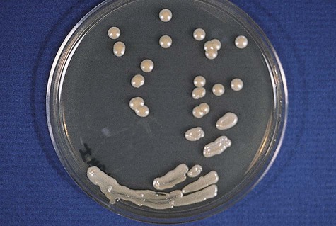
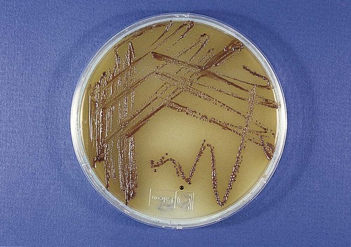
Colonies of C. neoformans usually appear on culture media within 1 to 5 days. The growth begins as a smooth, white to tan colony that may be mucoid to creamy (Figure 63-7). It is important to recognize the colonial morphology on different culture media, because variation does occur; for example, on inhibitory mold agar, C. neoformans appears as a golden yellow, nonmucoid colony. Textbooks typically characterize the colonial morphology as being Klebsiella-like because of the large amount of polysaccharide capsule material present. In reality, most isolates of C. neoformans do not have large capsules and may not have the typical mucoid appearance.

Approach to Identification

The general approach to yeast identification consists of evaluating the carbohydrate and substrate utilization profile with a commercial system and observing the morphology in a cornmeal preparation. This latter aspect is particularly important for discovering any errors in identification that may have been made by the commercial system and prevents the release of an erroneous identification to the clinician. For example, if a commercial system designates an isolate as C. glabrata but pseudohyphae are seen in the cornmeal preparation, additional testing is needed to identify the isolate correctly, because C. glabrata does not produce pseudohyphae. This traditional approach could be modified to use newer methods of confirmation, such as CHROMagar Candida.

Candida spp.

C. albicans may be identified by the production of germ tubes or chlamydoconidia (Figure 63-8; also see Figure 63-2). Other Candida spp. are most commonly identified by the utilization of specific substrates and the fermentation or assimilation of particular carbohydrates. For instance, C. glabrata ferments and assimilates only glucose and trehalose, whereas C. tropicalis ferments and assimilates sucrose and maltose. Another method of identifying C. albicans and differentiating it from other Candida spp. is based on the presence of chlamydoconidia (see Figure 63-8) on cornmeal agar containing 1% Tween 80 and trypan blue incubated at room temperature for 24 to 48 hours. (Many of the finer points of yeast identification are discussed in The Yeasts: A Taxonomic Study, by Kreger-Van Rij.) The morphologic features of yeasts on cornmeal agar containing Tween 80 often allow for tentative identification of selected species and, an important benefit, may detect misidentifications by commercial systems (Table 63-1).

TABLE 63-1

Characteristic Microscopic Features of Commonly Encountered Yeasts on Cornmeal Tween 80 Agar

Organism Arthroconidia/Blastoconidia Pseudohyphae or Hyphae
Candida albicans Spherical clusters at regular intervals on pseudohyphae Chlamydoconidia present on hyphae
Candida glabrata Small, spherical, and tightly compacted None produced
Candida krusei Elongated; clusters occur at septa of pseudohyphae Branched pseudohyphae
Candida parapsilosis Present but not characteristic Sagebrush or “shaggy star” appearance; large (giant) hyphae present
Candida kefyr (pseudotropicalis) Elongated, lie parallel to pseudohyphae Pseudohyphae present but not characteristic
Candida tropicalis Produced randomly along hyphae or pseudohyphae Pseudohyphae present but not characteristic
Cryptococcus spp. Round to oval, vary in size, separated by a capsule Rare; usually not seen
Saccharomyces spp. Large and spherical Rudimentary hyphae sometimes present
Trichosporon spp. Numerous, resemble Geotrichum spp.; septate hyphae present May be present but difficult to find

Colonies that appear star-like or possess feet-like projections on agar, as previously described on blood agar, may be identified as C. albicans, according to the Clinical Laboratory Standards Institute document M35-A2. However, this method is not as sensitive as traditional methods when colonies are examined within 18 to 24 hours versus 24- to 48-hour incubation times. In addition, species such as Trichosporon spp. may give false-positive results for possessing the pseudohyphal fringe that appears as starting on blood agar. A Gram stain of the isolate would provide a means of differentiation of the isolate as Trichosporon spp. by the characteristic presence of arthroconidia. The susceptibility profiles of C. albicans and Trichosporon spp. would also be significantly different.

Germ Tube Test

The germ tube test (see Procedure 63-1 on the Evolve site) is the most generally accepted and economical method used in the clinical laboratory to identify yeasts. Approximately 75% of the yeasts recovered from clinical specimens are C. albicans, and the germ tube test usually provides sufficient identification of this organism within 3 hours.

Germ tubes appear as early hyphal-like extensions of yeast cells that are produced without a constriction at the point of origin from the yeast cell (see Figure 63-2). Another Candida species, C. dubliniensis, has been shown to also produce true germ tubes. Although C. dubliniensis is infrequently encountered, supplemental biochemical or morphologic testing may be needed to differentiate it from C. albicans. C. tropicalis produces what has been called “pseudo-germ tubes,” which are constricted at the base or point of germ tube origin from the yeast cell. Unless this is recognized and the laboratorian has developed the skills to distinguish between true germ tubes and pseudo-germ tubes, C. tropicalis isolates will be misidentified as C. albicans.

The search for a more rapid, less subjective method of identifying C. albicans and other Candida spp. continues. C. albicans produces beta-galactose aminidase and L-proline aminopeptidase. Other Candida spp. may produce one enzyme but not both. Assays such as BactiCard Candida (Remel Laboratories, Lenexa, Kansas), were designed to detect these enzymes.

Heelan et al. compared the germ tube test to BactiCard, Murex C. albicans-50, Albicans-sure, and the API 20C AUX yeast identification systems. All rapid enzymatic screening methods were sensitive and specific for rapid identification of C. albicans. Compared with the germ tube test, all required less time (5 to 30 minutes), were more expensive, and required some additional equipment. Overall, all methods provided rapid and objective alternatives to the germ tube test.

CHROMagar Candida is another product that uses enzymatic reactions to differentiate C. albicans and several other yeast species. More recently C. albicans PNA FISH was released for detection and differentiation of C. albicans from non-albicans Candida spp. directly in positive blood cultures that contain yeast. The use of any new or additional testing for identification of yeasts should be submitted to a financial impact and outcomes analysis and should be compared with the traditional identification methods. Thereafter, the medical director, in conjunction with the medical staff, may determine the optimal approach for the laboratory.

Cryptococcus neoformans

Microscopic examination of colonies of C. neoformans may be helpful for providing a tentative identification of C. neoformans, because the cells are spherical and vary considerably in size. A presumptive identification of C. neoformans may be based on rapid urease production and failure to utilize an inorganic nitrate substrate. Final identification of C. neoformans usually is based on typical substrate utilization patterns and, in some laboratories, pigment production on niger seed (thistle or birdseed) agar (Figure 63-9). Immunocompromised patients suffering from infection die each year, with an estimated 500,000 in Africa alone. Diagnosis is typically made by identifying the encapsulated yeast in the spinal fluid using India ink. The use of serological techniques for the detection of the cryptococcal polysaccharide capsule glucuronoxylamannan (GXM) may be completed using latex agglutination or enzyme-linked immunosorbent assay (ELISA). A recent study demonstrated that plasma and urine can be used to identify cryptococcal antigen, thereby eliminating the need for the invasive spinal tap. The use of alternate specimens has the potential to improve screening of patients infected with HIV and reduced CD4 lymphocyte counts before the exacerbation of symptoms, which may not be apparent early enough for successful treatment of the infection. Additional tests useful for identifying C. neoformans and other species of cryptococci are discussed later in the chapter.

Rapid Urease Test

The rapid urease test (see Procedure 63-2 on the Evolve site) is a most useful tool for screening for urease-producing yeasts recovered from respiratory secretions and other clinical specimens. Alternatives to this method include use of a heavy inoculum of the tip of a slant of Christensen’s urea agar and subsequent incubation at 35° to 37°C. In many instances, a positive reaction occurs within several hours; however, 1 to 2 days of incubation may be required. Interestingly, strains of Rhodotorula spp., some Candida spp., and Trichosporon spp. hydrolyze urea with time, so a distinction should be made between a traditional urease test, which takes hours, and the rapid urease test.

The microscopic morphologic features of the yeast in question are helpful for interpreting the usefulness of the traditional urease test. An alternative to traditional urease testing is the rapid selective urease test (see Procedure 63-3 on the Evolve site). This method appears to be useful for rapidly detecting C. neoformans. These screening tests are helpful for making a presumptive identification of C. neoformans. When inoculum is limited, the laboratorian must use tests that can be performed and then prepare a subculture so that additional tests can be performed later. Often, inoculating the organism onto the surface of a plate of niger seed agar is just as fast, and results may be obtained during the same day of incubation at 25°C (this is discussed later in this section).

All the tests mentioned provide a tentative identification of C. neoformans; however, they must be supplemented with additional tests (usually the results of a commercial system in conjunction with cornmeal agar morphology) before a final identification can be reported. Additional tests useful in the identification of potential cryptococci are the nitrate reduction test and the detection of phenoloxidase production.

Commercially Available Yeast Identification Systems

Commercially available yeast identification systems have provided laboratories of all sizes with standardized identification methods. For the most part, the methods are rapid, providing results within 24 to 72 hours. The major advantage is that the systems provide an identification based on a database of thousands of yeast biotypes that considers a number of variations and substrate utilization patterns. Another advantage is that manufacturers of these products provide computer consultation services to help the laboratorian identify isolates that produce an atypical result. Although these systems are powerful tools, they should not be used as the sole method of identification; traditionally, they are most effectively used in conjunction with yeast morphology on blood, chocolate, or cornmeal agar.

API-20C AUX Yeast System

The API-20C AUX yeast identification system has perhaps the most extensive computer-based data set of all commercial systems available. The system consists of a strip that contains 20 microcupules, 19 of which contain dehydrated substrates for determining the utilization profiles of yeasts. Reactions are compared with growth (turbidity) in the first cupule, which lacks a carbohydrate substrate. Reactions are read and results are converted to a seven-digit biotype profile number. Most of the yeasts are identified within 48 hours; however, some Cryptococcus and Trichosporon spp. may require up to 72 hours. The API-20C AUX yeast identification system, as well as all other commercially available products, requires that the microscopic morphologic features of yeast grown on cornmeal agar containing 1% Tween 80 and trypan blue be used in conjunction with the substrate utilization patterns. This is particularly helpful when more than one possibility for an identification is provided; the microscopic morphologic features can be used to distinguish between the possibilities given by the profile register.

Several evaluations of the API-20C AUX yeast identification system have been performed, and the results have all been favorable. This system is limited in that it cannot identify unusual species; however, most of those seen in the clinical laboratory are accurately identified to the species level, especially the non-germ tube forming Candida spp. When this system is used to differentiate C. albicans from C. dubliniensis, assimilation results for xylose and alpha-methyl-d-glucoside may help distinguish between the two species. The results are negative for C. dubliniensis in 100% and 95% of the strains and positive for C. albicans in 100% and 95% of the strains.

Microscan Yeast Identification Panel

The MicroScan Yeast Identification Panel (Siemens, Deerfield, Illinois) is a 96-well, microtiter plate containing 27 dehydrated substrates. It was introduced as an alternative to the API-20C AUX yeast identification system. It uses chromogenic substrates to assess specific enzyme activity, which can be detected within 4 hours. Specific enzyme profiles have been generated for many of the yeasts commonly encountered in the clinical microbiology laboratory. The most recent evaluation of the method showed that it was moderately accurate within 4 hours using no supplementary tests. When supplementary tests were used, the sensitivity was excellent compared with that of the API-20C AUX yeast identification system. Accuracy for identification of common yeasts was high, and uncommon yeasts were identified in most instances.

Vitek Biochemical Cards

The Yeast Biochemical Card (bioMérieux, Durham, North Carolina) is a 30-well, disposable plastic card that contains conventional biochemical tests and negative controls. It is used with the automated Vitek II system (BioMérieux, Durham, North Carolina), which is used for bacterial identification and susceptibility testing in many laboratories. The most recent evaluation of this system showed an overall accuracy of identification near 100% compared with API-20C AUX. Fewer than one fourth of the yeasts required supplemental biochemical or morphologic features to confirm their identification. Of all correctly identified yeasts, more than half were reported after 24 hours of incubation. The accuracy of identification of common and uncommon species was satisfactory. Identifying germ tube–positive yeasts is not necessary with this system. For laboratories already using this system, accurate and reliable identification of most commonly encountered yeasts can be accomplished.

Interest in commercially available yeast identification systems has taken precedence over the more cumbersome, labor-intensive conventional yeast identification methods. Currently the rapid identification methods are financially feasible and provide laboratories of all sizes with the capability to identify yeasts. Commercially available systems, which provide accurate and rapid identification of yeasts and yeastlike organisms, are recommended for all laboratories. In general, they are easy to use, easy to interpret, and relatively inexpensive compared with conventional methods. In most cases they are faster than conventional systems, provide more standardized results, and require less technical skill to perform. As with any system, uncommon identifications should be scrutinized to prevent misidentifications.

Chromagar Candida

CHROMagar is a differential medium useful for the recovery of Candida organisms in clinical specimens, differentiation of Candida spp., and isolation of colonies. Distinct enzymes of different Candida spp. react with chromogenic substrates to yield a characteristic colony color. When used with colonial morphologic features, this system can provide a presumptive identification. Sand-Millan et al. reported an evaluation of 1537 isolates of yeast, which after 48 hours of incubation at 37°C showed that CHROMagar had a sensitivity and specificity near 100% for C. albicans, C. tropicalis, and C. krusei. Another evaluation by Pfaller et al. showed that more than 95% of stock and clinical isolates of C. albicans, C. tropicalis, and C. krusei were correctly identified. A similar sensitivity was observed for C. glabrata. CHROMagar also was evaluated as a recovery medium and was found to detect mixed cultures of Candida spp. Considering that the previously mentioned species account for approximately 90% of the yeast recovered in the clinical laboratory, CHROMagar appears to be a suitable alternative to other yeast identification systems.

Matrix-Assisted Laser Desorption Ionization Time-of-Flight (MALDI-TOF)

Matrix-assisted laser desorption ionization time-of-flight mass spectrometry (MALDI-TOF) is emerging as a potential rapid technique for the identification of yeast and fungal isolates within the microbiology laboratory. See Chapter 7 for more information. MALDI-TOF requires that isolates be cultured overnight and then processed using a standardized extraction procedure. Several studies report the correct identification of yeast to the species level up to approximately 98% in comparison to traditional culture methods. In addition, some laboratories have required the direct isolation of organisms from blood culture systems without subculturing to another medium to facilitate identification. Evidence suggests that MALDI-TOF is able to resolve species discrepancies more accurately than traditional culture methods and will reduce the time needed to identify an organism significantly.

Conventional Yeast Identification Methods

A few laboratories still prefer to use conventional methods to identify yeasts. Regardless of the type of identification system used, the germ tube test often is the first step in screening a large number of isolates, unless another screening test for C. albicans has been used (e.g., PNA FISH). As previously mentioned, approximately 75% of yeasts recovered in the clinical laboratory can be identified using the germ tube test.

Cornmeal Agar Morphology

The second major step in this practical identification schema is to use cornmeal agar morphology as a means to determine whether the yeast produces blastoconidia, arthroconidia, pseudohyphae, true hyphae, and/or chlamydoconidia (see Procedure 63-4 on the Evolve site). Cornmeal agar morphology can be used to detect characteristic chlamydoconidia produced by C. albicans. This method currently is satisfactory for definitive identification of C. albicans when the germ tube test is negative. In other instances, microscopic morphologic features on cornmeal agar help differentiate the genera Cryptococcus, Saccharomyces, Candida, Geotrichum, and Trichosporon. Previously, it was believed that the morphologic features of the common Candida spp. were distinct enough to provide a presumptive identification. This can be accomplished for C. albicans, C. glabrata, C. krusei, C. parapsilosis, C. tropicalis, and C. kefyr, keeping in mind that numerous other species, uncommonly recovered in the clinical laboratory, might resemble microscopically any of the previously mentioned species. In general, this method performs well, because the previously mentioned genera and species are more commonly seen in clinical laboratories.

Cornmeal agar morphology has less value for uncommonly encountered isolates. It should be used as an adjunct test with most commercially available yeast identification systems (e.g., to differentiate C. albicans from C. dubliniensis). It aids the differentiation of yeasts that yield similar biochemical profiles and helps prevent misidentifications, particularly of less commonly encountered species that may not be well represented in the commercial database.

Carbohydrate Utilization

Carbohydrate utilization patterns are the most commonly used conventional methods for definitive identification of yeasts recovered in a clinical laboratory. Various methods have been advocated for use in determining carbohydrate utilization patterns by clinically important yeasts, and all work equally well. Procedure 63-5, which can be found on the Evolve site, outlines the method previously found to be most useful by the Mayo Clinic Mycology Laboratory; however, this method is not commonly used in clinical laboratories. Most use commercially available methods.

Once the carbohydrate utilization profile has been obtained, reactions may be compared with those listed in tables in most mycology laboratory manuals. In most instances, carbohydrate utilization tests provide definitive identification of an organism, and additional tests are unnecessary. Some laboratories prefer carbohydrate fermentation tests, which are simply performed using purple broth containing different carbohydrate substrates. In general, carbohydrate fermentation tests are unnecessary and are not recommended for routine use.

Phenoloxidase Detection Using Niger Seed Agar

Use of a simplified Guizotia abyssinica (niger seed) medium is a definitive method for detecting phenoloxidase production by yeasts (see Procedure 63-6 on the Evolve site). Most isolates of C. neoformans readily produce phenoloxidase; however, some do not. In addition, in some instances, cultures of C. neoformans have been shown to contain both phenoloxidase-producing and phenoloxidase-deficient colonies in the same culture.

If conventional methods are used, all criteria, including urease production, carbohydrate utilization, and the phenoloxidase test, must be met before a final identification of C. neoformans is made.

Chapter Review

1. Which of the following is not a virulence factor of Candida spp.?

2. Which method has improved the isolation of Candida spp. from positive blood cultures?

3. The specimen needed to isolate Trichosporon spp. is:

4. Germ tube formation is seen with which two yeasts?

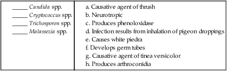
5. Matching: Match each description in the right-hand column with the correct organism in the left-hand column. More than one answer may be correct.

image

6. Short Answer