Chapter 99 The Umbilicus
Umbilical Cord
When the cord sloughs after birth, portions of these structures remain in the base. The blood vessels are functionally closed but anatomically patent for 10-20 days. The arteries become the lateral umbilical ligaments; the vein, the ligamentum teres; and the ductus venosus, the ligamentum venosum. During this interval, the umbilical vessels are potential portals of entry for infection. The umbilical cord usually sloughs within 2 wk. Delayed separation of the cord, after more than 1 mo, has been associated with neutrophil chemotactic defects and overwhelming bacterial infection (Chapter 124).
Patency of the omphalomesenteric (vitelline) duct may be responsible for intestinal obstruction, intestinal fistula with fecal or bilious draining, prolapse of the bowel, a polyp (cyst), or a Meckel diverticulum (Chapter 323.2). Therapy is surgical excision of the anomaly.
Congenital Omphalocele
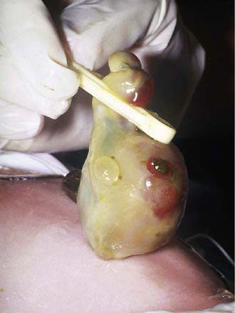
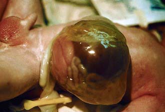
An omphalocele is a herniation or protrusion of the abdominal contents into the base of the umbilical cord (Figs. 99-1 and 99-2). In contrast to the more common umbilical hernia, the sac is covered with peritoneum without overlying skin. The size of the sac that lies outside the abdominal cavity depends on its contents. Herniation of intestines into the cord occurs in about 1/5,000 births, and herniation of liver and intestines in 1/10,000 births. The abdominal cavity is proportionately small because the impetus to grow and develop is deficient. Immediate surgical repair, before infection has taken place and before the tissues have been damaged by drying (saline-soaked sterile dressings should be applied immediately) or by rupture of the sac, is essential for survival. Mersilene mesh or similar synthetic material may be used to cover the viscera if the sac has ruptured or if excessive mobilization of the skin would be necessary to cover the mass and its intact sac. The majority (≈75%) of infants with omphalocele have associated congenital anomalies/syndromes, including Beckwith-Wiedemann syndrome (omphalocele, macrosomia, hypoglycemia), and other chromosomal (29%, including trisomies 13 and 18) and nonchromosomal (45%) multiple and isolated congenital anomalies (musculoskeletal, 24%; urogenital, 20%; cardiovascular, 15%; and central nervous system, 9%). The survival rate is about 80% overall, but in infants with isolated omphalocele, the survival rate is > 90%.
Ameh EA, Nmadu PT. Major complications of omphalitis in neonates and infants. Pediatr Surg Int. 2002;18:413-416.
Deshpande SA, Jog S, Watson H, et al. Do babies with isolated single umbilical artery need routine postnatal renal ultrasonography? Arch Dis Child Fetal Neonatal Ed. 2009;94:F265-F267.
Hwang P, Kousseff B. Omphalocele and gastroschisis: an 18-year review study. Genetics Med. 2004;6:232-236.
Krakowiak P, Smith EN, de Bruyn G, et al. Risk factors and outcomes associated with a short umbilical cord. Obstet Gynecol. 2004;103:119-127.
Mullany LC, Darmstadt GL, Khatry SK. Topical applications of chlorhexidine to the umbilical cord for prevention of omphalitis and neonatal mortality in southern Nepal: a community-based, cluster-randomized trial. Lancet. 2006;367:910-918.
Pomeranz A. Anomalies, abnormalities, and care of the umbilicus. Pediatr Clin North Am. 2004;51:819-827.
Salihu HM, Emusu D, Aliyu ZY, et al. Omphalocele and gastroschisis: black-white disparity in infant survival. Am J Med Genet A. 2005;1(135):161-165.
Stoll C, Alembik Y, Dott B, et al. Omphalocele and gastroschisis and associated malformations. Am J Med Genet A. 2008;146A:1280-1285.
Weber DM, Freeman NV, Elhag KM. Periumbilical necrotizing fasciitis in the newborn. Eur J Pediatr Surg. 2001;11:86-91.
Zupan J, Garner P, Omari AA: Topical umbilical cord care at birth, Cochrane Database Syst Rev (3):CD001057, 2004.


