The Role of Spinal Fusion and the Aging Spine: Stenosis without Deformity

Published on 11/04/2015 by admin

Filed under Orthopaedics

Last modified 22/04/2025

Print this page

rate 1 star rate 2 star rate 3 star rate 4 star rate 5 star
Your rating: none, Average: 0 (0 votes)

This article have been viewed 1351 times

50 The Role of Spinal Fusion and the Aging Spine

Stenosis without Deformity

Introduction

Spinal stenosis is defined as any condition that results in a narrowing of the spinal canal, nerve root canal, or intervertebral foramina. This narrowing can occur at several locations at the same spinal level, or it can affect multiple levels at similar locations at each level. Spinal stenosis may be due to soft tissue impingement from a herniated intervertebral disc or an infolded ligamentum flavum, from bony processes such as osteophytes or hypertrophied facet joints, or from a combination of these.

Spinal stenosis usually involves the lumbar spine and tends to affect patients in the sixth or seventh decade of life. With an increase in the average age of the patient population, the incidence of patients seeking medical care for symptomatic spinal stenosis is also increasing. Not only are patients living longer, but also they are remaining more active in their older years, thereby making symptoms of neurogenic claudication more apparent. Additionally, surgeries such as total hip and knee arthroplasties are helping patients remain very active further into their lives. Patients with lumbar spinal stenosis often complain of symptoms consistent with neurogenic claudication, including pain, numbness, and paresthesias in the posterolateral portions of the legs and thighs. These symptoms are improved by activities in which the lumbar spine is held in flexion, such as leaning over a walker or shopping cart or riding a bicycle. Spinal stenosis also occurs in the cervical and thoracic spine. The clinical presentations are different, as is the decision-making process for treatment options.

The initial treatment for symptomatic spinal stenosis is nonoperative and consists of activity modification, oral medications, epidural steroid injections, and physical therapy. For those cases recalcitrant to nonoperative treatment, surgery is considered. Surgical options include decompression alone, decompression with a noninstrumented fusion, decompression with an instrumented fusion, and, recently, decompression followed by posterior pedicle-based dynamic stabilization. Newer technologies involving minimally-invasive approaches to the spine have been developed that indirectly improve the dimensions of the spinal canal and neuroforamina in select cases of moderate spinal canal stenosis (interspinous process spacers). Minimally invasive surgical (MIS) techniques have also been developed for spinal decompression as well as fusion.

Fusion in the aging spine can be problematic, due to poor bone quality. Spinal arthrodesis should be incorporated in the surgical treatment of spinal stenosis, either when potentially significant instability is present preoperatively, or if postoperative instability is expected secondary to the extent of decompression performed. The indications for spinal arthrodesis for spinal stenosis will be discussed in this chapter, as well as the arguments both for and against fusion. Outcomes and potential complications of spinal arthrodesis for stenosis will also be discussed.

Basic Science

As with other segments of the spine, the etiology and specific location of the narrowing is important when treating cervical spinal stenosis. Cervical spinal stenosis can occur from a herniated cervical disc, an ossified posterior longitudinal ligament, a redundant ligamentum flavum, an ossified ligamentum flavum, a hypertrophied facet joint, or from any combination of these potentially compressive pathologies.

Stenosis in the cervical spine is of particular importance, due to the relatively small canal diameter as compared to the caliber of the spinal cord. Some surgeons now recommend surgical treatment for severe asymptomatic cervical spinal stenosis for prophylaxis against paralysis, while others recommend observation.

The clinical presentation of stenosis in the cervical spine can be that of myelopathy, radiculopathy, or both. Patients with compressive pathology secondary to a herniated disc pressing on the spinal cord may report symptoms of myelopathy, including loss of hand dexterity and gait abnormalities. Physical exam will show signs of upper motor neuron pathology, which can be manifested as hyperactive deep tendon reflexes, a positive Hoffmann sign, or as a Babinski sign. If, instead, this herniated disc places pressure on an exiting nerve root, or if the loss of intervertebral disc height causes a loss of cross-sectional area of the neuroforamen, then the patient may exhibit signs and symptoms of a particular nerve root radiculopathy. This will result in motor weakness, paresthesias, and loss of deep tendon reflexes (if applicable) for that particular cervical nerve root.

Radiographically, cervical stenosis is often diagnosed using a radiographic measurement called the Pavlov ratio. This ratio is defined as the ratio between the sagittal diameter of the spinal canal and the sagittal diameter of the vertebral body, as measured on a lateral radiograph. A ratio of greater than 1 is considered normal, while a ratio of less than 0.8 is considered to be diagnostic for spinal stenosis. A cervical MRI can help determine the etiology of the compression if the source is a herniated disc or a redundant ligamentum flavum. Additionally, an MRI can show any evidence of spinal cord compression, such as a lack of cerebrospinal fluid around the spinal cord and/or myelomalacia within the cord itself. A CT scan can be used to diagnose bony abnormalities such as osteophytes or hypertrophied facet joints.

Stenosis in the Cervical Spine

Case 1

A 63-year-old female presented to clinic with an 8-year history of worsening neck pain radiating to her bilateral shoulders and scapulae. The pain radiated primarily down her bilateral biceps and radial forearms into the thumb and index fingers of both hands. Additionally, she had noted a gradually worsening weakness in her legs, with loss of balance, worsening handwriting, and difficulty buttoning buttons and manipulating small objects with her hands. She had no bowel or bladder dysfunction. Despite nonoperative management that included activity modification, physical therapy, and nerve root and trigger point injections, her symptoms persisted and seemed to be worsening.

Physical examination revealed tenderness to palpation in the cervical paraspinal musculature as well as in the midline. She had an unsteady gait with a positive Romberg sign. Range of motion was limited by pain, and neck extension reproduced the pain, numbness, and tingling in her arms. She had weakness in her right greater than left biceps, wrist extensors, and hand intrinsics, but intact sensation throughout all dermatomes. Reflex testing was significant for global hyperreflexia and a positive Hoffmann sign bilaterally.

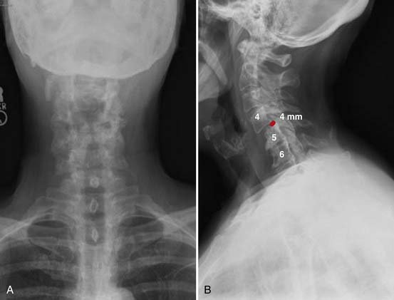
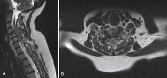
Radiographs, seen in Figure 50-1A and B, demonstrate loss of normal cervical lordosis, severe spondylosis, and a spondylolisthesis of C4 on C5. Sagittal and coronal MRI cuts, seen in Figure 50-2A and B, demonstrate significant spinal stenosis, loss of normal disc height, and severe spinal cord compression with myelomalacia.

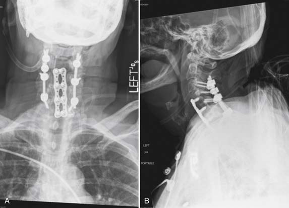
The patient was taken to the operating room for a combined anterior and posterior cervical decompression and fusion. Discectomies and interbody fusion were performed with allograft spacers and an anterior plate at C4-5, C5-6, and C6-7. This portion of the procedure restored normal lordosis and addressed the anterior pathology, including reduction of the spondylolisthesis at C4-5. The posterior procedure included laminectomy from C3 to C7 with screw and rod fixation from C3 to C7 as well (Figure 50-3A and B).

Postoperatively, the patient did well, with complete resolution of her arm pain. At last visit, her gait and balance were steadily improving, with overall improved function compared to preoperatively.

Clinical Practice Guidlines

Cervical radiculopathy can be successfully treated nonoperatively with activity modification, oral medications, and selective nerve root block injections. Operative indications include symptoms that are recalcitrant to nonoperative treatment as well as development of myelopathy. The natural history of cervical myelopathy is that of a stepwise progression of symptoms alternating with periods of nonprogressive neurological symptoms.

The location of the compressive pathology in cervical stenosis is important, as it dictates the operative approach. Cervical stenosis due to a central or moderate-sized posterolateral cervical herniated nucleus pulposus is best treated with an anterior approach, in order to adequately remove the compressive pathology. This anterior cervical discectomy is typically combined with a fusion. Fusion is achieved with or without instrumentation, consisting of an anterior plate and screws. Anterior cervical discectomy and fusion (ACDF) has been described with good results without the use of instrumentation. An anterior cervical discectomy (ACD) without fusion is rarely performed today, and is almost never performed for multilevel disease. Discectomy without fusion has been reported in a prospective, randomized trial to be equivalent to ACDF for the treatment of cervical radiculopathy.1 For the treatment of myelopathy, ACD without fusion has been reported to result in good relief of neck and arm pain as well as a 76% rate of return to work.2 However, ACD without fusion has been shown in other case series to be associated with worsening of preexisting cervical myelopathy in 3.3% of cases.3 Worsening of symptoms after ACD without fusion was also reported by Nandoe Tewarie et al in a retrospective review of 102 patients evaluated up to 18 years after surgery.4 While ACD alone has been shown to be successful in the treatment of cervical myeloradiculopathy, the possibility of worsening of symptoms, combined with the difficulty of revision of anterior cervical surgery, makes this a possible yet unattractive surgical option.

If the compressive pathology is secondary to redundant ligamentum flavum, hypertrophied facet joints, or other posterior pathology, then a posterior approach allows the surgeon to directly decompress the offending agent. A posterior-only approach is only indicated if neutral or lordotic alignment of the cervical spine is maintained. A kyphotic deformity in the cervical spine often mandates an anterior approach to restore the normal cervical sagittal alignment. Decompression from a posterior approach consists of laminotomy, laminectomy, or laminoplasty. Removal of significant portions of the facet joints should be avoided in order to avoid causing iatrogenic postlaminectomy cervical kyphosis. Raynor et al reported a cadaveric study comparing the potential degrees of instability on biomechanical testing of intact specimens, and following 50% facetectomy, and 70% facetectomy. The conclusion of this study was the recommendation that a facetectomy should involve less than 50% of the facet joint in the absence of fusion, in order to avoid spinal instability.5 Postlaminectomy kyphosis after posterior cervical decompression alone is common when there is evidence of hypermobility on preoperative flexion-extension radiographs. A cervical fusion should be considered after posterior decompression if:

For multiple-level posterior cervical spinal cord compression, laminoplasty is another option. In this procedure, the space available for the spinal cord is increased by cutting the affected lamina on one side and scoring the contralateral lamina. The posterior elements are then “booked” open, utilizing the scored side as a hinge, and held open with suture, structural grafts/spacers, or plate and screws. With the newly increased space available for the spinal cord, the spinal cord can float away from the vertebral body. This treatment plan only works in cases in which a neutral or lordotic curve of the cervical spine is maintained. In a kyphotic cervical spine, the spinal cord will remain draped over the vertebral bodies regardless of the increased space posterior to the cord.

A combined anterior and posterior approach may be needed for kyphotic deformities with spinal stenosis and for multiple level disease. Posterior instrumentation and fusion is usually warranted when performing three or more cervical corpectomies or when doing four or more cervical discectomies.

Clinical Practice Guidelines

Operative indications for stenosis in the thoracic spine are similar to those in the cervical spine: symptoms recalcitrant to nonoperative measures and cases in which myelopathy develops.

Stenosis in the Thoracic Spine

Case 2

A 58-year-old woman with no significant past medical history presented to an outside hospital with progressive bilateral lower extremity weakness, right greater than left, and decreased sensation below the nipple line. In addition, she reported 5 out of 10 pain in the midthoracic area, urinary retention, mild constipation, and an inability to bear weight. Emergency department records indicated that she had twisted her back one week prior to admission and complained of subsequent onset of these progressive symptoms. Of note, several months prior to presentation, the patient noted irritation and a possible mass in the upper outer quadrant of her right breast. Radiographs obtained at an outside hospital demonstrated a pathologic fracture of her sixth thoracic vertebrae. She was transferred to our facility for evaluation and for further radiologic and immunopathic workup for presumed metastatic breast cancer to the thoracic spine.

Physical exam at the time of admission was notable for a 2 × 2 cm purple nodule on her right breast with induration and no discharge. Tenderness to palpation over the cervical spine was noted. Neurological exam demonstrated weakness in bilateral lower extremities with 2/5 hip flexors, 4/5 quadriceps, 1/5 tibialis anterior, 1/5 extensor hallucis longus, and 2.5 gastrocnemius; upper extremity strength was 5/5. Sensation was decreased bilaterally; clonus was present, particularly on the right; Babinski sign was absent; and rectal tone was decreased. The patient was afebrile with stable vital signs, and admission labs were within normal limits.

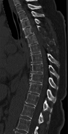
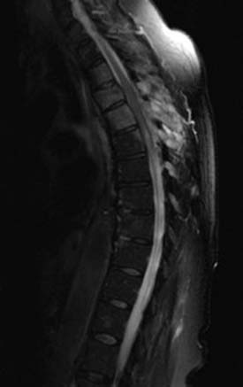
The patient had a CT scan of her spine that demonstrated pathologic fractures of T5 and T6 and lytic lesions at multiple spinal levels with bony destruction, predominantly from T3 to T6, with encroachment upon the adjacent central canal and neural foramen (Figure 50-4). These findings were most suggestive of metastatic lesions. MRI of her spine demonstrated metastatic disease with multilevel involvement and posterior epidural extension in T5 and T6 resulting in spinal cord compression (Figure 50-5).

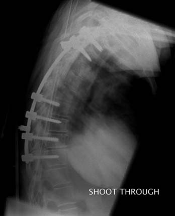
The patient underwent a T3 to T6 laminectomy for extradural tumor, posterolateral fusion of T2 to T10, and open biopsy of the presumed metastatic lesion. Pathologic examination was consistent with metaplastic breast cancer (Figure 50-6). The patient tolerated the procedure well without complication. At the time of discharge from inpatient rehabilitation, lower extremity motor strength had improved to 3/5, and sensation had improved as well. Bladder function did not return and a Foley catheter was maintained. The neurogenic bowel responded well to enema, Dulcolax, Colace, and senna.

The patient was transferred to the oncology service for further evaluation and treatment of her metastatic breast cancer.

Treatment for thoracic stenosis is usually limited to decompression alone. The decision on whether to use an anterior, posterior, or transthoracic approach depends on the source of the compressive pathology. Fusion, historically, has rarely been indicated due to the greater inherent stability of the thoracic spine afforded by the rib cage and sternum. Palumbo et al described a retrospective review of 12 patients treated for thoracic stenosis. All were treated with a decompression alone, without fusion. Although the majority of their patients improved with regard to pain, ambulation, and neurological function, five patients exhibited early deterioration of symptoms due to recurrent stenosis, deformity/instability, or both. The authors imply that a decompression at the thoracolumbar junction was more prone to instability.6

When surgically treating spinal stenosis in the thoracic spine, fusion should be considered in cases where:

Basic Science

Spinal stenosis is most frequently encountered in the lumbar spine. It typically affects patients in their sixth or seventh decade of life. As a normal age-related change of the intervertebral disc, the nucleus pulposus itself loses water content, which in turn leads to a loss of disc height. This loss of water may result in increased motion at the vertebral body–disc interface, leading to increased motion at the level of the facet joint. This excess motion at the facet joint may lead to facet joint arthrosis and hypertrophy. Finally, the loss of intervertebral disc height translates to decreased cross-sectional area of the neural foramina as well as redundancy of the ligamentum flavum. All of these age-related changes contribute to lumbar spinal stenosis.

The clinical syndrome of lumbar spinal stenosis is that of neurogenic claudication. Patients often report lower extremity pain that is worsened with activity, but lessened when the patient is allowed to assume a position in which the lumbar spine is flexed. Activities such as riding a bicycle or leaning over a shopping cart are better tolerated by patients with lumbar stenosis, because they are performed with the lumbar spine in a flexed position.

The radiographic workup of lumbar stenosis begins with plain radiographs of the lumbar spine. Radiographs should be scrutinized for osteophytes, malalignment, facet hypertrophy or any other abornormalities that could potentially decrease the space available for the neural elements. Flexion-extension radiographs are needed to determine the presence of instability. The presence of any preoperative instability necessitates a spinal fusion. A MRI can be useful in determining the specific location (paracentral, lateral recess, or foraminal) of the stenosis. A CT myelogram is useful in localizing the specific area of the compressive lesion, but this has been largely supplanted by MRI.

Clinical Practice Guidelines

Controversy exists in the literature with regard to whether a spinal fusion should be added to decompression. Decompression alone without fusion has been reported to treat lumbar spinal stenosis with good results. However, other reports in the literature, primarily those including patients with instability, report better clinical outcomes with addition of spinal fusion. Yone et al reported on a group of 34 patients with lumbar spinal stenosis.7 Of this group, 17 patients had radiographic spinal instability as described by Posner. Ten patients underwent decompression and fusion, and the remaining seven underwent decompression alone. The decompression-alone patients had significantly worse Japanese Orthopaedic Association back scores. Still others report no difference in clinical outcomes for patients treated with decompression alone and decompression plus fusion.8,9

Expansive laminoplasty has been reported as an alternative to lumbar decompression and fusion, with good results.10 This procedure shares the technical demands present in cervical laminoplasty.

Stenosis in the Lumbar Spine

Case 3

An active 90-year-old man was referred to the spine surgery clinic with a long history of worsening bilateral buttock pain and decreased walking tolerance. He was otherwise in excellent health, but noted a burning pain in his buttocks with radiation down the posterior and lateral thighs after ambulating more than about 50 yards. The pain quickly improved with rest, and would not occur if he had a shopping cart to lean on while ambulating. Descending stairs or inclines would aggravate symptoms but ascending stairs would not. He had been evaluated by the vascular surgery service and was not found to have vascular insufficiency. Despite an intensive program of physical therapy focusing on core strengthening, flexibility, and cardiovascular fitness his symptoms persisted. Interventions by the pain management service, including epidural steroid injections, had been unsuccessful.

Physical examination was relatively unremarkable, with normal strength throughout all muscle groups and intact sensation in all dermatomes. Gait was steady and neurological testing was unremarkable.

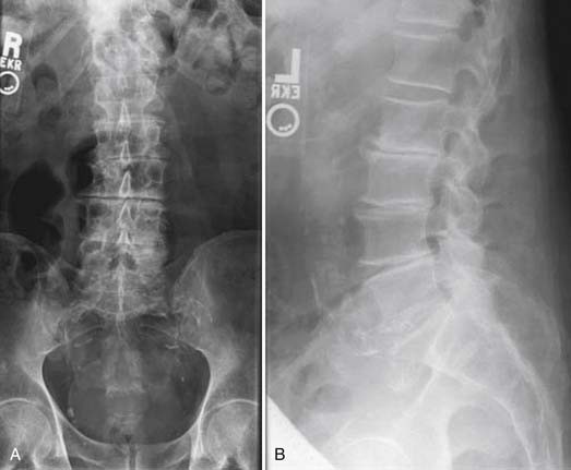
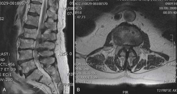
Radiographs, seen in Figures 50-7A and B, demonstrate advanced degenerative changes, with significant disc height loss, large disc osteophyte complexes, and facet hypertrophy from L2 to L5. Sagittal and coronal MR images seen in Figure 50-8A and B show severe central, lateral recess, and foraminal stenosis, with increased fluid in the zygapophyseal joints and buckling of the ligamentum flavum.

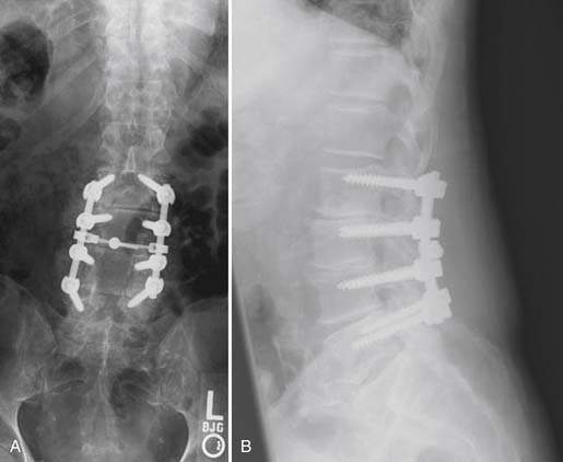
The patient was taken to the operating room for a L2 to L5 decompression and posterolateral instrumented fusion. Postoperative images are shown in Figure 50-9A and B. At last follow-up, the patient had returned to his premorbid level of functioning, with complete resolution of his claudication symptoms and no postoperative pain.

As with stenosis found elsewhere, successful surgical management of lumbar stenosis depends on adequate decompression of the neural elements. Fusion should be considered in cases in which:

The addition of a lumbar interbody fusion to the spinal decompression can restore the disc space back to its normal height. This helps treat the stenosis by reducing any buckling of the ligamentum flavum as well as restoring the native cross-sectional area of the neural foramina. This interbody lumbar fusion can be done from a variety of approaches: anterior (anterior lumbar interbody fusion or ALIF), retroperitoneal (extreme or direct lateral lumbar interbody fusion or XLIF/DLIF), or posterior (transforaminal lumbar interbody fusion or TLIF). ALIF and XLIF/DLIF can be used as a standalone procedure for a spinal fusion, while TLIF must be augmented with posterior pedicle screw instrumentation. Each of these approaches has its pros and cons. An ALIF allows for excellent visualization of the intervertebral disc, but it often requires an access surgeon to mobilize the great vessels off the spinal column. Additionally, since this is done in a supine patient, the patient must be turned prone if posterior decompression and/or instrumentation is needed. An XLIF/DLIF also requires repositioning for a posterior approach. The TLIF allows for 360-degree fusion from a posterior-alone approach. No access surgeon is needed, and no repositioning is required. This approach may require some manipulation of the neural elements, thereby putting those structures at risk.