The acute respiratory distress syndrome

Published on 07/02/2015 by admin

Filed under Anesthesiology

Last modified 22/04/2025

Print this page

rate 1 star rate 2 star rate 3 star rate 4 star rate 5 star
Your rating: none, Average: 0 (0 votes)

This article have been viewed 2336 times

The acute respiratory distress syndrome

Mark T. Keegan, MB, MRCPI, MSc

Background and definitions

The acute respiratory distress syndrome (ARDS) is an inflammatory lung condition with associated noncardiogenic pulmonary edema and impairment of gas exchange. ARDS is a major cause of respiratory failure in patients in the intensive care unit (ICU). In the perioperative period, patients who are undergoing major surgical procedures, who are seriously ill, or who aspirate are susceptible to developing ARDS. In 2012, ARDS was redefined according to the Berlin definition (Table 227-1) in an effort to overcome some of the inadequacies of the previously used American-European Consensus definition, which dated from 1994 (Table 227-2). Compared with the prior definition, the Berlin definition defines “acute,” clarifies the methods to exclude hydrostatic edema, adds minimal ventilator-setting requirements, drops the term “acute lung injury,” and classifies ARDS into three categories of severity.

Table 227-1

Berlin Definition of Acute Respiratory Distress Syndrome

Feature Description
Timing Onset within 1 week of a known clinical insult or new or worsening respiratory symptoms
Chest imaging* Bilateral opacities—not fully explained by effusions, lobar/lung collapse, or nodules
Origin of edema Respiratory failure not fully explained by cardiac failure or fluid overload; need objective assessment (e.g., echocardiography) to exclude hydrostatic edema if no risk factor is present
Oxygenation  
Mild 200 mm Hg < PaO2/FIO2 ≤ 300 mm Hg with PEEP or CPAP ≥ 5 cm H2O
Moderate 100 mm Hg < PaO2/FIO2 ≤ 200 mm Hg with PEEP ≥ 5 cm H2O
Severe PaO2/FIO2 ≤ 100 mm Hg with PEEP ≥ 5 cm H2O

ARDS, Acute respiratory distress syndrome; CPAP, continuous positive airway pressure; FIO2, fraction of inspired O2; PaO2, partial pressure of arterial O2; PEEP, positive end-expiratory pressure.

*Chest radiograph or computed tomography scan.

If altitude is greater than 1000 m, the correction factor should be calculated as follows: (PaO2/FIO2) × (Barometric pressure/760).

This may be delivered noninvasively in the mild ARDS group.

Modified from ARDS Definition Task Force. Acute respiratory distress syndrome: The Berlin definition. JAMA. 2012;307:2526-2533.

ARDS may result from a direct insult to the lung, such as from pneumonia or from aspiration of gastric contents. Direct injury may also be related to pulmonary contusion, fat embolus, inhalation or drowning injury, or transfusion of blood products. Secondary ARDS occurs as part of a systemic illness or may be related to trauma and may be thought of as the lung manifestation of the systemic inflammatory response, just as oliguria, mental status changes, and hypotension are manifestations of sepsis in the kidney, brain, and cardiovascular systems, respectively.

ARDS manifests as noncardiogenic pulmonary edema with hypoxemia. An alteration in the relationship between the alveolar epithelium and the capillary endothelium allows influx of protein-rich edema fluid into the alveoli. Injury to type II alveolar cells results in disruption of epithelial fluid transport, impairs removal of alveolar fluid, and alters surfactant production. These changes lead to abnormalities in gas exchange. Pulmonary neutrophils play a key role in the generation of an inflammatory response, and this inflammatory process may be augmented by inappropriate mechanical ventilation (see later discussion).

A prospective study in 1999 to 2000 estimated an incidence of ARDS in the United States of almost 200,000 adult patients per year. The incidence appears to have decreased over the subsequent decade, probably because of the use of lung-protective ventilation, more conservative use of blood products, and a reduction in nosocomial infections. ARDS remains, however, a major ICU disease entity. Approximately 10% to 15% of patients admitted to the ICU and up to 20% of those requiring mechanical ventilation have ARDS.

ARDS has been divided into a number of pathologic stages. Diffuse alveolar damage seen in the initial “exudative” stage gives way over the first week to a “proliferative” stage, during which type II alveolar cells predominate and interstitial inflammation develops. A later “fibrotic” stage occurs in some patients, during which normal lung architecture is disrupted by deposition of collagen.

Clinical manifestations of ARDS include rapidly worsening dyspnea, tachypnea, and hypoxemia with diffuse rales on lung auscultation. Arterial blood gas analysis shows an elevated alveolar-arterial O2 gradient with severe hypoxemia, consistent with right-to-left shunt physiology. Pulmonary hypertension may develop. Although a respiratory alkalosis may be present in early ARDS, respiratory acidosis usually develops later in the course of the condition. Diffuse “fluffy” bilateral infiltrates are apparent on chest radiography. A computed tomography scan will show areas of alveolar filling, consolidation, and atelectasis, especially in the dependent lung zones. Despite the heterogeneity of the computed tomographic findings, the whole lung is involved in the inflammatory process, and bronchoalveolar lavage of even the relatively spared areas will show inflammatory changes.

Patients with ARDS almost invariably need mechanical ventilation, which is the mainstay of supportive therapy. Noninvasive ventilation, such as continuous positive airway pressure or biphasic positive airway pressure, may be sufficient in some patients, but most patients with moderate or severe ARDS require tracheal intubation. Although oxygenation tends to improve over the course of the first few days as pulmonary edema resolves, the presence of continued hypoxemia, high minute ventilation requirements, and poor lung compliance necessitate prolonged ventilation in a significant number of patients. Large doses of sedative agents and, on occasion, the use of infusions of neuromuscular blocking agents may be required to enable appropriate ventilation of the patient with ARDS. Current guidelines (2012) from the Society of Critical Care Medicine, based on the results of one prospective study, advocate a short course (≤48 h) of a neuromuscular blocking agent for septic patients with ARDS.

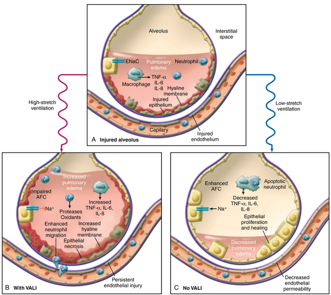
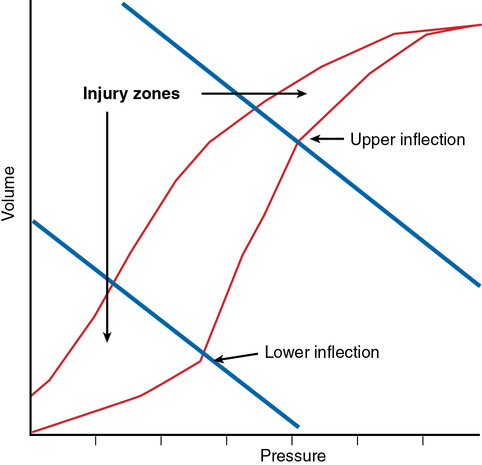
Ventilator-associated lung injury is a major concern (Figure 227-1). Inspired gas flows preferentially to relatively uninvolved alveoli, potentially causing overdistention and lung injury due to volutrauma and barotrauma. Constant opening and closing of derecruited lung units can lead to shear stress (atelectrauma). These physical forces can lead to an increase in the injurious inflammatory response (biotrauma). Laboratory investigations have suggested that a “safe zone” exists on the pulmonary pressure-volume curve defined by lower and upper “inflection points” in which ventilation should occur. At the lower end of the pressure-volume curve, lung units are susceptible to derecruitment and atelectasis, and, at the upper end of the pressure-volume curve, overdistention leads to lung injury (Figure 227-2).

image
Figure 227-1 Mechanisms of ventilator-associated lung injury (VALI). A, Acute respiratory distress syndrome (ARDS) leads to lung endothelial and epithelial injury, increased permeability of the alveolar-capillary barrier, flooding of the airspace with protein-rich pulmonary edema fluid, activation of alveolar macrophages with release of proinflammatory chemokines and cytokines, enhanced neutrophil migration and activation, and fibrin deposition (hyaline membranes). B, If the injured lung is ventilated with high tidal volumes and high inflation pressures (high-stretch ventilation), then lung injury is exacerbated, with increased lung endothelial and epithelial injury and necrosis, enhanced neutrophil margination, release of injurious neutrophil products such as proteases and oxidants, increased release of proinflammatory cytokines from alveolar macrophages and the lung epithelium, increased fibrin deposition, and increased hyaline membrane formation. Injurious mechanical ventilation can also impair alveolar fluid clearance (AFC) mechanisms. C, In contrast, a protective ventilatory strategy (low-stretch ventilation) can limit further lung endothelial and epithelial injury, reduce the release of proinflammatory cytokines, and enhance alveolar fluid clearance through the active transport of sodium and chloride across the alveolar epithelium, thereby reducing the quantity of pulmonary edema and allowing endothelial and epithelial repair to occur. Epithelial repair occurs through migration, proliferation, and differentiation of alveolar epithelial type II cells to repopulate the denuded basement membrane. Acute inflammation resolves through apoptosis of neutrophils, which are phagocytosed by alveolar macrophages. ENaC, Epithelial sodium channels; IL, interleukin; TNF, tumor necrosis factor. (Modified from Matthay MA, Ware LB, Zimmerman GA. The acute respiratory distress syndrome. J Clin Invest. 2012;122:2731-2740.)

The National Heart Lung and Blood Institute ARDSNet investigators have conducted a number of randomized, multicenter, clinical trials evaluating the role of therapies in ARDS. The ARDSNet lower tidal volume trial (ARMA) proved that use of lower tidal volumes (6 mL/kg predicted body weight) decreases fatality caused by lung overdistention and ventilator-associated lung injury. Predicted body weight is based on patient height and sex. Low tidal volume ventilation has become a standard of care in the management of patients with ARDS in the ICU. Although volume-control ventilation was used in the ARDSNet low tidal volume study, pressure-control ventilation—which may provide superior oxygenation because of its flow pattern—can be used instead, providing tidal volumes are limited.

Application of positive end-expiratory pressure (PEEP) has been advocated as a method of improving oxygenation and preventing atelectasis, thus allowing ventilation above the lower inflection point. PEEP levels of up to 22 to 24 cm H2O have been used. The optimal level of PEEP in ARDS has not been determined, despite a number of prospective studies having been conducted.

Treatment

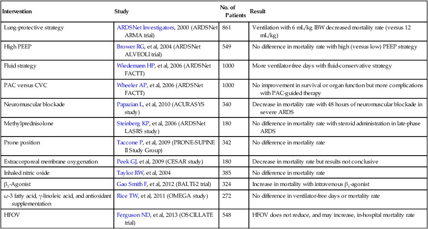
A variety of techniques have been used in patients with ARDS to allow adequate oxygenation and ventilation while attempting to minimize ventilator-induced lung injury. Selected major clinical trials of therapeutic interventions for ARDS are listed in Table 227-3.

Table 227-3

Selected Major Clinical Trials in Acute Respiratory Distress Syndrome

Intervention Study No. of Patients Result
Lung-protective strategy ARDSNet Investigators, 2000 (ARDSNet ARMA trial) 861 Ventilation with 6 mL/kg IBW decreased mortality rate (versus 12 mL/kg)
High PEEP Brower RG, et al, 2004 (ARDSNet ALVEOLI trial) 549 No difference in mortality rate with high (versus low) PEEP strategy
Fluid strategy Wiedemann HP, et al, 2006 (ARDSNet FACTT) 1000 More ventilator-free days with fluid-conservative strategy
PAC versus CVC Wheeler AP, et al, 2006 (ARDSNet FACTT) 1000 No improvement in survival or organ function but more complications with PAC-guided therapy
Neuromuscular blockade Papazian L, et al, 2010 (ACURASYS study) 340 Decrease in mortality rate with 48 hours of neuromuscular blockade in severe ARDS
Methylprednisolone Steinberg KP, et al, 2006 (ARDSNet LASRS study) 180 No difference in mortality rate with steroid administration in late-phase ARDS
Prone position Taccone P, et al, 2009 (PRONE-SUPINE II Study Group) 342 No difference in mortality rate
Extracorporeal membrane oxygenation Peek GJ, et al, 2009 (CESAR study) 180 Decrease in mortality rate but results not conclusive
Inhaled nitric oxide Taylor RW, et al, 2004 385 No difference in mortality rate
β2-Agonist Gao Smith F, et al, 2012 (BALTI-2 trial) 324 Increase in mortality with intravenous β2-agonist
ω-3 fatty acid, γ-linoleic acid, and antioxidant supplementation Rice TW, et al, 2011 (OMEGA study) 272 No difference in ventilator-free days or mortality rate
HFOV Ferguson ND, et al, 2013 (OSCILLATE trial) 548 HFOV does not reduce, and may increase, in-hospital mortality rate

image

ARDSNet, Acute Respiratory Distress Syndrome Network (of the National Heart, Lung, and Blood Institute); CVC, central venous catheter; HFOV, high-frequency oscillatory ventilation; IBW, ideal body weight; PAC, pulmonary artery catheter; PEEP, positive end-expiratory pressure.

Tracheal gas insufflation

In some patients with ARDS, the presence of a large physiologic dead space may require the use of a very high minute ventilation (e.g., 20 L/min). Even such minute ventilation may not maintain normocapnia because increasing respiratory rate to increase minute ventilation without increasing tidal volume does not change the ratio of dead space ventilation to alveolar ventilation. Although the patient may tolerate hypercapnia, the development of cardiac arrhythmias or the presence of intracranial disease may require additional efforts to increase the clearance of CO2. Tracheal gas insufflation involves the continuous flow of fresh O2 (usually ∼6 L/min) through a small tube placed through or alongside the tracheal tube and exiting above the carina. The gas washes out CO2 as an adjunct to the CO2 removal provided by the regular ventilatory circuit. Care must be taken to avoid damaging the carina by means of high pressures or “catheter whip.”

Inhaled nitric oxide and prostaglandin e1

Inhaled nitric oxide (INO), a selective pulmonary vasodilator, may be administered through the breathing circuit by a dedicated delivery system. The gas is delivered to those lung units that are ventilated, and it dilates the local pulmonary arterioles. The pulmonary vasculature of nonventilated lung units is not affected. INO thus improves ventilation and perfusion matching and increases PaO2. INO is rapidly metabolized and has no systemic effects. In addition to improving oxygenation (at doses of 3-20 ppm), when used at higher doses (up to 80 ppm), INO decreases pulmonary arterial pressure and pulmonary vascular resistance, increases right ventricular ejection fraction, and causes mild bronchodilation. Despite improvements in physiologic parameters, the use of INO has not been associated with improvement in outcomes in patients with ARDS.

Inhaled prostaglandin E1 works in a manner similar to INO. The drug is delivered by nebulizer into the breathing circuit and is used at a dose of 10 to 40 ng·kg−1·h−1. As with INO, prostaglandin E1 improves oxygenation but has not been shown to improve outcome in a randomized trial.

High-frequency oscillatory ventilation

The recognition that conventional mechanical ventilation can lead to a worsening of lung injury makes the use of high-frequency oscillatory ventilation (HFOV) attractive. An oscillator is used to deliver very low tidal volume breaths at rates of 3 to 15 Hz. HFOV can provide excellent lung recruitment without causing overdistention while maintaining near-normal arterial blood gases. It permits higher mean airway pressures without exposing the lungs to high pressures during tidal excursions. The technique decouples oxygenation and ventilation: oxygenation is proportional to mean airway pressure, and the pressure amplitude controls ventilation. CO2 removal is decreased by increases in frequency. Gas exchange relies on less obvious mechanisms of gas transport, such as bulk convection, pendelluft, cardiogenic oscillations, augmented dispersion, and molecular diffusion. Although this technique is theoretically attractive, large studies of HFOV in ARDS have failed to show a benefit and have even suggested harm.

Mortality and morbidity

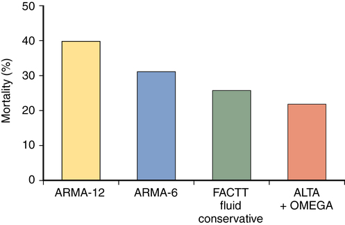
The mortality rate associated with ARDS has decreased over the past decade. Recent clinical trials in ARDS have demonstrated mortality rates of approximately 25% to 45%, with lower mortality rates reported in young patients who developed ARDS following polytrauma and higher mortality rates in older patients admitted to medical ICUs (Figure 227-3). As alluded to previously, although some patients die as a result of hypoxemia, most deaths are due to sepsis, multiple organ failure, and the underlying disease rather than to ARDS itself. Initial indices of oxygenation and ventilation do not predict outcome, although failure to improve after 1 week of therapy and persistently high minute ventilation requirements are signs of poor prognosis. Advanced age and the presence of comorbid conditions also increase the chance of death.

Survivors of ARDS may have significant short-term to medium-term disability. Many patients will require a tracheostomy and a significant period of ventilator weaning. Prolonged immobility may lead to the development of critical illness polymyoneuropathy requiring lengthy periods of rehabilitation. Dysfunction of other organs (e.g., renal failure) may be due to the initiating illness or secondary to nosocomial infection. In the long term, lung function usually returns to near normal, but mild abnormalities on pulmonary function tests may persist.