T Lymphocytes, B Lymphocytes, and Natural Killer Cells

Published on 27/03/2015 by admin

Filed under Pediatrics

Last modified 22/04/2025

Print this page

rate 1 star rate 2 star rate 3 star rate 4 star rate 5 star
Your rating: none, Average: 0 (0 votes)

This article have been viewed 2349 times

Chapter 117 T Lymphocytes, B Lymphocytes, and Natural Killer Cells

Defense against infectious agents is secured through a combination of anatomic physical barriers including the skin, mucous membranes, mucous blanket, and ciliated epithelial cells, as well as the various components of the immune system. The immune system of vertebrates integrates 2 fundamental response mechanisms. Innate (natural) immunity responds to infection regardless of previous exposure to the agent and includes polymorphonuclear leukocytes, dendritic and mononuclear phagocytic cells, various receptors that recognize common pathogen antigens (Toll-like receptors) and the complement system. Acquired (adaptive) immunity is a highly specific response that includes T lymphocytes, B lymphocytes, and natural killer (NK) cells. The immune system also helps protect against malignancy and autoimmunity.

Lymphopoiesis in the Fetus

Origin of the Lymphoid System

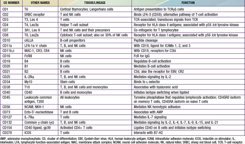
The human immune system arises in the embryo from gut-associated tissue. Pluripotential hematopoietic stem cells 1st appear in the yolk sac at 2.5-3 wk of gestational age, migrate to the fetal liver at 5 wk of gestation, and later reside in the bone marrow, where they remain throughout life (Fig. 117-1). Lymphoid stem cells develop from such precursor cells and differentiate into T, B, or NK cells, depending on the organs or tissues to which the stem cells traffic. Development of the primary lymphoid organs—thymus and bone marrow—begins during the middle of the 1st trimester of gestation and proceeds rapidly. Development of the secondary lymphoid organs—spleen, lymph nodes, tonsils, Peyer patches, and lamina propria—soon follows. These organs serve as sites of differentiation of T, B, and NK lymphocytes from stem cells throughout life. Both the initial organogenesis and the continued cell differentiation occur as a consequence of the interaction of a vast array of lymphocytic and microenvironmental cell surface molecules and proteins secreted by the involved cells. The complexity and number of lymphoid cell surface molecules led to the development of an international nomenclature for clusters of differentiation (CD) (Table 117-1).

image

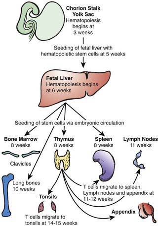
Figure 117-1 Migration patterns of hematopoietic stem cells and mature lymphocytes during human fetal development.

(From Haynes BF, Denning SM: Lymphopoiesis. In Stamatoyannopoulis G, Nienhuis A, Majerus P, editors: Molecular basis of blood diseases, ed 2, Philadelphia, 1994, WB Saunders.)

T and B lymphocytes are the only components of the immune system that have antigen-specific recognition capabilities and are responsible for adaptive immunity. NK cells are lymphocytes that are also derived from hematopoietic stem cells and are thought to have a role in host defense against viral infections, tumor surveillance, and immune regulation, but they do not have antigen receptors. Nonantibody proteins synthesized and secreted by T, B, and NK cells, and by the cells with which they interact, act as intercellular mediators and are referred to as cytokines or interleukins (ILs) (Table 117-2). Cytokines have the ability to act in an autocrine, paracrine, or endocrine manner to promote and facilitate differentiation and proliferation of the cells of the immune system.

Table 117-2 FUNCTIONAL CLASSIFICATION OF CYTOKINES*

2 Lymphocyte regulatory cytokines

GM-CSF, granulocyte macrophage colony stimulating factor; IFN, interferon; IL, interleukin; MHC, major histocompatibility complex; MCP-1, monocyte chemotactic protein; NK, natural killer; RANTES, regulated on activation normal t cell expressed and secreted; SCF, stem cell factor; TGF, transforming growth factor; TNF, tumor necrosis factor.

* This is not an exhaustive list.

Modified from Whiteside TL: Cytokine measurements and interpretation of cytokine assays in human disease, J Clin Immunol 14:327–339, 1994.

T-Cell Development and Differentiation

The primitive thymic rudiment is formed from the ectoderm of the 3rd branchial cleft and endoderm of the 3rd branchial pouch at 4 wk gestation. Beginning at 7-8 wk, the right and left rudiments move caudally and fuse in the midline. Blood-borne T-cell precursors from the fetal liver then begin to colonize the perithymic mesenchyme at 8 wk gestation. These precursor pro–T cells are identified by surface proteins designated as CD7 and CD34. At 8-8.5 wk gestation, CD7 cells are found intrathymically, with some cells that co-express CD4, a protein present on the surfaces of mature T-helper (TH) cells, or CD8, a protein found on both mature cytotoxic T cells and NK cells. In addition, some cells bear single T-cell receptor (Ti) chains (β, δ, or γ), but none bear complete T-cell receptors.

The mature T-cell receptor (TCR) is a heterodimer of 2 chains, either α and β or γ and δ, that is co-expressed on the cell surface with CD3, a complex of 5 polypeptide chains (γ, δ, ε, ζ, η). TCR gene rearrangement occurs by a process in which large, noncontiguous blocks of DNA are spliced together. These segments, known as V (variable), D (diversity), and J (joining), each have a number of variants. VDJ segments are joined to a constant region of the α gene, and VDJ segments are joined to the β gene to complete the receptor polypeptide genes. Random combinations of the segments account for much of the enormous diversity of TCRs that enables humans to recognize millions of different antigens. TCR gene rearrangement requires the presence of recombinase activating genes, RAG-1 and RAG-2, as well as other recombinase components. This process is flawed in mice with severe combined immunodeficiency (SCID) and in some humans with SCID. Rearrangement of TCR genes signifies commitment of pro–T cells to T-lineage development, becoming pre–T cells. TCR gene rearrangement begins shortly after colonization of the thymus with stem cells, and the establishment of the T-cell repertoire begins at 8-10 wk of gestation. By 9.5-10 wk, >95% of thymocytes express CD7, CD2, CD4, CD8, and c (cytoplasmic) CD3, and ≈30% bear the CD1 inner cortical thymocyte antigen. By 10 wk, 25% of thymocytes bear αβ TCRs. Ti αβ T cells gradually increase in number during embryonic life and represent >95% of thymocytes postnatally.

As immature cortical thymocytes begin to express TCRs, the processes of positive and negative selection take place. Positive selection occurs through the interaction of immature thymocytes, which express low levels of TCR, with major histocompatibility complex (MHC) antigens present on cortical thymic epithelial cells. As a result, thymocytes with TCR capable of interacting with foreign antigens presented on self human leukocyte antigen (HLA) molecules are activated and develop to maturity. Mature thymocytes that survive the selection process either express CD4 and are restricted to interacting with self class II HLA antigens or express CD8 and are restricted to interacting with self class I HLA antigens when foreign antigens are presented by these MHC molecules. Negative selection occurs next and is mediated by interaction of the surviving thymocytes, which have much higher levels of TCR expression, with host peptides presented by HLA class I or II antigens present on bone marrow-derived thymic macrophages, dendritic cells, and possibly B cells. This interaction mediates apoptosis, or programmed cell death, of such autoreactive thymocytes. Fetal cortical thymocytes are among the most rapidly dividing cells in the body and increase in number by 100,000-fold within 2 wk after stem cells enter the thymus. As these cells mature and the selection process takes place, 97% of all cortical thymocytes die. The surviving cells are no longer doubly positive for both CD4 and CD8, but are singly positive for either 1 or the other and migrate to the medulla of the thymus.

T-cell functions are acquired concomitantly with the development of single-positive thymocytes, but they are not fully developed until the cells emigrate from the thymus. It has been estimated that 1 stem cell gives rise to approximately 3,000 mature medullary thymocytes, which are resistant to the lytic effects of corticosteroids. T cells begin to emigrate from the thymus to the spleen, lymph nodes, and appendix at 11-12 wk of embryonic life and to the tonsils by 14-15 wk. They leave the thymus via the bloodstream and are distributed throughout the body, with the heaviest concentrations in the paracortical areas of lymph nodes, the periarteriolar areas of the spleen, and the thoracic duct lymph. Recent thymic emigrants co-express the CD45RA isoforms and CD62L (L-selectin). Rearrangement of the TCR locus during intrathymic T-cell development results in the excision of DNA and the excised elements form circular episomes as a by-product. These TCR recombination excision circles (TRECs) can be detected in T cells that are recent thymic emigrants, whereas T cells that develop extrathymically do not contain these episomes. Inability to detect TRECs at birth is one test used for screening for SCID. The homing of lymphocytes to peripheral lymphoid organs is directed by the interaction of a lymphocyte surface adhesion molecule, L-selectin, with carbohydrate moieties on specialized regions of lymphoid organ blood vessels called high endothelial venules. By 12 wk gestation, T cells can proliferate in response to plant lectins, such as phytohemagglutinin (PHA) and concanavalin A (Con A), and to allogeneic cells; antigen-binding T cells have been found by 20 wk gestation. Hassall’s corpuscles (bodies), which are swirls of terminally differentiated medullary epithelial cells, are 1st seen in the thymic medulla at 16-18 wk of embryonic life.

B-Cell Development and Differentiation

In parallel with T-cell differentiation, B-cell development begins in the fetal liver before 7 wk of gestation. Fetal liver CD34 stem cells are seeded to the bone marrow of the clavicles by 8 wk of embryonic life and to that of the long bones by 10 wk (see Fig. 117-1). Antigen-independent stages of B-cell development have been defined according to immunoglobulin gene rearrangement patterns and the surface proteins the cells bear. The pro–B cell is the 1st descendent of the pluripotential stem cell committed to B-lineage development that is detected by the presence of both CD34 and CD10 on its surface, although the immunoglobulin genes remain germ line (Fig. 117-2). The next stage is the pre-pre–B cell, during which immunoglobulin genes are rearranged but there is no cytoplasmic expression of µ heavy chains or surface IgM (sIgM). These cells are further characterized by the co-expression of membrane CD34, CD10, CD19, and CD40, and somewhat later by the additional presence of CD73, CD22, CD24, and CD38. The pre–B cell follows and is distinguished by the expression of cytoplasmic µ heavy chains but no sIgM, because no immunoglobulin light chains are produced yet. These cells also continue to express all CD antigens seen at the pre-pre–B-cell stage except CD34 and CD10, which are lost; in addition, they express CD21. Next is the immature B-cell stage, during which sIgM but not sIgD is expressed. The light-chain genes have been rearranged, and CD38 is lost but all other pre–B-cell CD antigens persist. The last stage of antigen-independent B-cell development is the mature or virgin B cell, which co-expresses both sIgM and sIgD; CD23 is also acquired at this stage, and all of the other CD antigens present on immature B cells persist. Pre–B cells can be found in fetal liver at 7 wk gestation, sIgM+ and sIgG+ B cells at between 7 and 11 wk, and sIgD+ and sIgA+ B cells by 12-13 wk. By 14 wk of embryonic life, the percentage of circulating lymphocytes bearing sIgM and sIgD is the same as in cord blood and slightly higher than in the blood of adults.

image

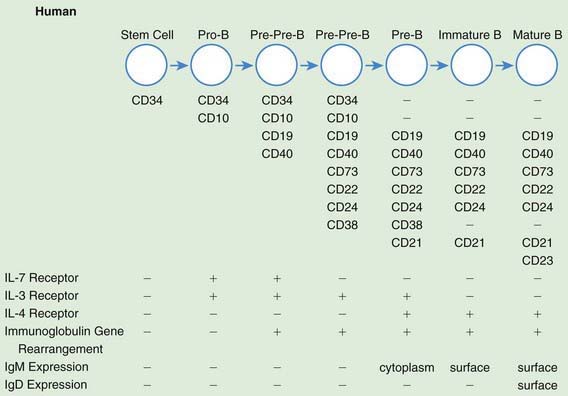
Figure 117-2 Antigen-independent human B-cell development. CD, cluster of differentiation; Ig, immunoglobulin; IL, interleukin.

(From Haynes BF, Denning SM: Lymphopoiesis. In Stamatoyannopoulis G, Nienhuis A, Majerus P, editors: Molecular basis of blood diseases, ed 2, Philadelphia, 1994, WB Saunders.)

Antigen-dependent stages of B-cell development are those that develop after the mature or virgin B cell is stimulated by antigen through its antigen receptor (sIg); the outcome is the differentiation of the cell and its progeny into sIg+ memory (CD27) B cells (for that particular antigen) and plasma cells, which synthesize and secrete antibody, which is antigen-specific immunoglobulin. Deficiency of activation-induced cytidine deaminase (AICDA) or of uracil DNA glycosylase (UNG), as seen in 2 forms of autosomal recessive hyper IgM, can result in a failure of isotype switching so that only IgM antibodies are formed. There are 5 immunoglobulin isotypes, which are defined by unique heavy-chains: IgM, IgG, IgA, IgD, and IgE. IgG and IgM, the only complement-fixing isotypes, are the most important immunoglobulins in the blood and other internal body fluids for protection against infectious agents. IgM is confined primarily to the intravascular compartment because of its large size, whereas IgG is present in all internal body fluids. IgA is the major protective immunoglobulin of external secretions—in the gastrointestinal, respiratory, and urogenital tracts—but it is also present in the circulation. IgE, present in both internal and external body fluids, has a major role in host defense against parasites. Because of high-affinity IgE receptors on basophils and mast cells, however, IgE is the principal mediator of allergic reactions of the immediate type. The significance of IgD is still not clear. There are also immunoglobulin subclasses including four subclasses of IgG (IgG1, IgG2, IgG3, and IgG4) and 2 subclasses of IgA (IgA1 and IgA2). These subclasses each have different biologic roles. For example, antipolysaccharide antibody activity is found predominantly in the IgG2 subclass. Secreted IgM and IgE have been found in abortuses as young as 10 wk, and IgG as early as 11-12 wk. Even though these B-cell developmental stages have been described in the context of B-cell ontogeny in utero, it is important to recognize that the process of B-cell development from pluripotential stem cells goes on throughout postnatal life.

Despite the capacity of fetal B lymphocytes to differentiate into immunoglobulin-synthesizing and -secreting cells, plasma cells are not usually found in lymphoid tissues of a fetus until about 20 wk gestation, then only rarely, because of the sterile environment of the uterus. Peyer patches have been found in significant numbers by the 5th intrauterine month, and plasma cells have been seen in the lamina propria by 25 wk gestation. Before birth there may be primary follicles in lymph nodes, but secondary follicles are usually not present.

A human fetus begins to receive significant quantities of maternal IgG transplacentally at around 12 wk gestation, and the quantity steadily increases until, at birth, cord serum contains a concentration of IgG comparable to or greater than that of maternal serum. IgG is the only class to cross the placenta to any significant degree. All 4 subclasses cross the placenta, but IgG2 does so least well. A small amount of IgM (10% of adult levels) and a few nanograms of IgA, IgD, and IgE are normally found in cord serum. Because none of these proteins crosses the placenta, they are presumed to be of fetal origin. These observations raise the possibility that certain antigenic stimuli normally cross the placenta to provoke responses, even in uninfected fetuses. Some atopic infants occasionally have IgE antibodies to antigens, such as egg white, to which they have had no known exposure during postnatal life, suggesting that synthesis of these antibodies could have been induced in the fetus by antigens ingested by the mother.

Natural Killer (NK)-Cell Development

NK-cell activity is found in human fetal liver cells at 8-11 wk of gestation. NK lymphocytes are also derived from bone marrow precursors. Thymic processing is not necessary for NK-cell development, although NK cells have been found in the thymus. After release from bone marrow, NK cells enter the circulation or migrate to the spleen, with very few NK cells in lymph nodes. In normal individuals, NK cells represent 8-10% of lymphocytes, but the percentages are sometimes slightly lower in cord blood.

Unlike T and B cells, NK cells do not rearrange antigen receptor genes during their development but are defined by their functional capacity to mediate non–antigen-specific cytotoxicity. NK cells have killer inhibitory receptors (KIRs) that recognize certain MHC antigens and inhibit the killing of normal allogeneic cells in four specific patterns of reactivity. The genetic loci controlling these receptors are different from MHC alloantigenic loci, and have been mapped to chromosome 19. Virtually all NK cells express CD56, and >90% bear CD16 (FcγRIII) on the cell surface. Other CD antigens found on NK cells include CD57 (50-60%), CD7 and CD2 (70-90%), and CD8 (30-40%) (see Table 117-1). Although NK cells share surface antigens with T and myeloid cells, the lineage relationship of NK cells to the latter is still unclear. Some humans with autosomal recessive SCID who have profound deficiencies in T and B cells have abundant NK cells, whereas those with X-linked and Jak3-deficient SCID have no T or NK cells.

Immune Cell Interactions

Immune cell interaction is of crucial importance to all phases of the adaptive immune response. Unlike the B-cell antigen receptor (Ig), which can recognize native antigen, the TCR can recognize only processed antigenic peptides presented to it by MHC molecules such as HLA-A, -B, and -C antigens (class I) and HLA-DR, -DP, and -DQ antigens (class II). The MHC molecules have a groove in their protein structure where peptides fit. Class I MHC molecules are found on most nucleated cells in the body. Class II MHC molecules are found on antigen-presenting cells (APCs), which include macrophages, dendritic cells, and B cells. The peptides found in the groove of class I HLA molecules come from proteins normally made in the cell that are degraded and inserted into the groove. The peptides include viral peptides if the cell is infected with a virus. The peptides present in the groove of class II molecules come from exogenous native antigens such as vaccine and bacterial proteins. These proteins are taken up by APCs, degraded, and expressed on the cell surface in the groove of class II HLA molecules. The TCR then interacts with the peptide-bearing HLA molecule and, through its functional and physical link to the CD3 complex of signal-transducing molecules, sends a signal to the T cell to produce cytokines that ultimately result in T-cell activation and proliferation.

Two of the main functions of T cells are to signal B cells to make antibody by producing cytokines and membrane molecules that can serve as ligands for non–antigen-receptor B-cell surface molecules and to kill virally infected cells or tumor cells. For a T cell to perform either of these functions, it first must bind to an APC or to a target cell. For high-affinity binding of T cells to APCs or target cells, several molecules on T cells, in addition to TCRs, bind to molecules on APCs or target cells. The CD4 molecule binds directly to MHC class II molecules on APCs. CD8 on cytotoxic T cells binds the MHC class I molecule on the target cell. Both CD4 and CD8 molecules are directly involved in the regulation of T-cell activation and are physically linked intracellularly to the p56-lck protein tyrosine kinase. The cytoplasmic tail of CD45, the common leukocyte antigen, is a tyrosine phosphatase capable of regulating T-cell signal-transduction events by virtue of the fact that p56-lck has been shown to be a substrate for CD45 phosphatase activity. Depending on which isoform of CD45 is present on the T cell (CD45RO on memory T cells, CD45RA on naive T cells), mechanisms by which CD45 could upregulate or downregulate T-cell triggering have been proposed. Indeed, one form of human SCID is caused by a deficiency of CD45. Lymphocyte function-associated antigen 1 (LFA-1) on the T cell binds a protein called ICAM-1 (intracellular adhesion molecule 1), now designated CD54, on APCs. CD2 on T cells binds LFA-3 (CD58) on the APCs. With the adhesion of T cells to antigen-presenting cells, TH cells are stimulated to make interleukins and upregulate cell surface molecules, such as the CD40 ligand (CD154), that provide help for B cells, and cytotoxic T cells are stimulated to kill their targets.

In the primary antibody response, native antigen is carried to a lymph node draining the site, taken up by specialized cells called follicular dendritic cells (FDCs), and expressed on their surfaces. Virgin B cells bearing sIg specific for that antigen then bind to the antigen on the surfaces of the FDCs. If the affinity of the B-cell sIg antibody for the antigen present on the FDCs is sufficient, and if other signals are provided by activated T-helper cells, the B cell develops into an antibody-producing plasma cell. If the affinity is not high enough or if T-cell signals are not received, the B cell dies through apoptosis. The signals from activated TH cells include several cytokines (IL-4, IL-5, IL-6, IL-10, IL-13, and IL-21) that they secrete (see Table 117-2) and a surface T-cell molecule, CD154, which, on contact of the T cell with the B cell, binds to CD40 on the B-cell surface. CD40 is a type I integral membrane glycoprotein expressed on B cells, monocytes, some carcinomas, and a few other types of cells. It belongs to the tumor necrosis factor (TNF)/nerve growth factor receptor family. Cross linking of CD40 on B cells by CD154 on T cells in the presence of certain cytokines causes the B cells to undergo proliferation and to initiate immunoglobulin synthesis. In the primary immune response, only IgM antibody is usually made, and most of it is of relatively low affinity. Some B cells become memory B cells during the primary immune response. These cells switch their immunoglobulin genes so that IgG, IgA, and/or IgE antibodies of higher affinity are formed on a secondary exposure to the same antigen. The secondary antibody response occurs when these memory B cells again encounter that antigen. Plasma cells form, just as in the primary response; however, many more cells are rapidly generated, and IgG, IgA, and IgE antibodies are made. In addition, genetic changes in immunoglobulin genes (somatic hypermutation [SHM]) lead to increased affinity of those antibodies. A lack of SHM is seen in deficiency of AID or UNG. The exact pattern of isotype response to antigen in normal individuals varies, depending on the type of antigen and the cytokines present in the microenvironment.

For NK-mediated lysis, binding to the target is of crucial importance. This is best exemplified by persons with leukocyte adhesion deficiency type I (LADI) who have mutations in the gene encoding CD18, or the β chain of 3 different adhesion molecules (LFA-1, CR3, and p150,95), and who lack NK function. Thus, binding of NK cells to their targets is facilitated by LFA-1-ICAM interactions. CD56 or NCAM (neural cell adhesion molecule) also mediates homotypic adhesion of NK cells. FcγRIII, or the low-affinity IgG receptor, has a higher affinity for IgG when it is present on NK cells than when it is on neutrophils. FcγRIII also permits NK cells to mediate antibody-dependent cellular cytotoxicity (ADCC), where antibody is bound through its Fc region to the FcγRIII. The antibody-combining portion of the IgG attaches to the target cell, and the NK cell, attached to the target by the Fc portion of the antibody, kills the target cell.

Postnatal Lymphopoiesis

T Cells and T-Cell Subsets

Although the percentage of CD3 T cells in cord blood is somewhat less than in the peripheral blood of children and adults, T cells are actually present in higher number because of a higher absolute lymphocyte count in normal infants. An additional distinction is that the ratio of CD4 to CD8 T cells is usually higher (3.5-4 : 1) in cord blood than in blood of children and adults (1.5-2 : 1). Virtually all T cells in cord blood bear the CD45RA (naive) isoform, and a dominance of CD45RA over CD45RO T cells persists during the 1st 2-3 yr of life, after which time the numbers of cells bearing these 2 isoforms gradually equalize. TH cells can be further subdivided according to the cytokines they produce when activated. TH1 cells produce IL-2 and IFN-γ, which promote cytotoxic T-cell or delayed hypersensitivity types of responses, whereas TH2 cells produce IL-4, IL-5, IL-6, IL-13, and IL-21 (see Table 117-2), which promote B-cell responses and allergic sensitization. There are important additional subsets of T cells that have regulatory functions. These include CD25 high + T cells (Treg cells), considered to be important in the prevention of autoimmune diseases, and T cells that have phenotypic characteristics of NK cells (NK-T cells). Cord blood T cells have the capacity to respond normally to T-cell mitogens (PHA, Con A, and PWM) and are capable of mounting a normal mixed leukocyte response. Normal newborn infants also have the capacity to develop antigen-specific T cell responses at birth, as evidenced by vigorous tuberculin reactivity a few weeks after BCG vaccination on day 1 of life. Because patients in the 1st few months of life may have unrecognized severe T-cell defects, most hospitals now routinely irradiate all blood products given young infants. T-cell defects can readily be detected even at birth by calculating the absolute lymphocyte count because T cells normally constitute 70% of circulating lymphocytes and their absence results in striking lymphopenia (see Fig. 116-2 and Chapter 708).

B Cells and Immunoglobulins

Newborn infants have increased susceptibility to infections with gram-negative organisms because IgM antibodies, which are heat-stable opsonins, do not cross the placenta. The level of the heat-labile opsonin, C3b, is also lower in newborn serum than in adults. These factors probably account for impaired phagocytosis of some organisms by newborn polymorphonuclear cells. Maternally transmitted IgG antibodies serve quite adequately as heat-stable opsonins for most gram-positive bacteria, and IgG antibodies to viruses afford adequate protection against those agents. Because there is a relative deficiency of the IgG2 subclass, antibodies to capsular polysaccharide antigens may be deficient. Because premature infants have received less maternal IgG by the time of birth than full-term infants, their serum opsonic activity is low for all types of organisms.

B lymphocytes are present in cord blood in slightly higher percentages but considerably higher numbers than in the blood of children and adults, reflecting the higher absolute lymphocyte counts in all normal infants. Cord blood B cells do not synthesize the range of immunoglobulin isotypes made by B cells from children and adults when stimulated with anti-CD40 plus IL-4 or IL-10, producing primarily IgM and at a much reduced quantity.

Neonates begin to synthesize antibodies of the IgM class at an increased rate very soon after birth in response to the immense antigenic stimulation of their new environment. Premature infants appear to be as capable of doing this as do full-term infants. At about 6 days after birth, the serum concentration of IgM rises sharply. This rise continues until adult levels are achieved by ≈1 yr of age. Cord serum from noninfected normal newborns does not contain detectable IgA. Serum IgA is normally 1st detected at around the 13th day of postnatal life; the level gradually increases during early childhood until adult levels are achieved by 6-7 yr of age. Cord serum contains an IgG concentration comparable to or greater than that of maternal serum. Maternal IgG gradually disappears during the 1st 6-8 mo of life, while the rate of infant IgG synthesis increases (IgG1 and IgG3 faster than IgG2 and IgG4 during the 1st yr) until adult concentrations of total IgG are reached and maintained by 7-8 yr of age. IgG1 and IgG4 reach adult levels first, followed by IgG3 at 10 yr and IgG2 at 12 yr of age. The serum IgG level in infants usually reaches a low point at ≈3-4 mo of postnatal life. The rate of development of IgE generally follows that of IgA. After adult concentrations of each of the 3 major immunoglobulins are reached, these levels remain remarkably constant for a normal individual. The capacity to produce specific antibodies to protein antigens is intact at the time of birth. Normal infants cannot usually produce antibodies to polysaccharide antigens until after 2 yr of age, however, unless the polysaccharide is conjugated to a protein carrier, as is the case for the conjugate Haemophilus influenzae type b (Hib) and Streptococcus pneumoniae (PCV7) vaccines.

Inheritance of Abnormalities In T-, B-, and NK-Cell Development

More than 150 immunodeficiency syndromes have been described (see Table 116-8). Specific molecular defects have been identified in approximately 80% of these diseases. Most are recessive traits, several of which are caused by mutations in genes on the X chromosome and others by mutations on autosomal chromosomes. The molecular bases of 7 X-linked immunodeficiency disorders affecting T, B, and/or NK cells are known (Chapters 118 to 120): X-linked immunodeficiency with hyper IgM, X-linked lymphoproliferative syndrome, XIAD, X-linked agammaglobulinemia, X-linked SCID, the Wiskott-Aldrich syndrome, and nuclear factor kappa B essential modulator (NEMO). Autosomal defects for which the molecular basis is known include (1) combined immunodeficiencies due to abnormalities of purine salvage pathway enzymes, either adenosine deaminase (ADA, encoded by a gene on chromosome 20q13-ter) or purine nucleoside phosphorylase (PNP, encoded by a gene on chromosome 14q13.1); (2) combined immunodeficiencies due to mutations in the gene encoding ZAP-70 (localized to chromosome 2q12), a non-src family protein tyrosine kinase important in T-cell signaling; (3) SCID due to mutations in the gene on chromosome 19p13.1 encoding Janus kinase 3 (Jak3), the primary signal transducer from the common cytokine receptor γ chain (γc); (4) mutations in genes on chromosome 11 that encode components of the T-cell receptor, that is, CD3 γ, δ and ε; (5) SCID due to mutations in recombinase activating genes (RAG1 and RAG2); and (6) SCID due to mutations in the gene on chromosome 5p13 that encodes the α chain of the IL-7 receptor. These are only a few of the conditions for which the mutated genes have been discovered and the number is steadily growing.