108 Systemic Lupus Erythematosus
• Systemic lupus erythematosus is an autoimmune disease that damages the skin, kidneys, bones, lungs, brain, and nearly every other organ in the body.
• The damage is due to inflammation as a result of a direct antibody reaction to body tissues, deposits of immune complexes, and secondary thrombosis.
• A characteristic finding is fever, malar rash, and joint pain in a young, premenopausal woman.
• Sunlight and certain viruses and drugs can induce an autoimmune response in a genetically susceptible host.
• Basic treatment of pain is with nonsteroidal antiinflammatory medications or steroids. Many patients additionally require immunosuppressants, antimalarial drugs, and other therapies prescribed by a rheumatologist.
• Patients with systemic lupus erythematosus have increased risk for serious infection, often because of the steroids and immunosuppressants required to treat the disease.
• Morbidity is due to organ failure, primarily of the kidney and brain.
Perspective
Systemic lupus erythematosus (SLE) is a chronic autoimmune disease with widespread physical effects caused by the production of autoantibodies to components of cellular nuclei.1 The term lupus (Latin for “wolf”) is attributed to the 13th-century physician Rogerius, who used it to describe the characteristic facial lesions that were reminiscent of a wolf’s bite.
Epidemiology
SLE is more common by a ratio of 12 : 1 in women aged 15 to 45 years and by a ratio of 2 : 1 in younger and older women. The overall prevalence of this disease is about 1 in 1000. In most studies of SLE, about 90% of enrollees are women. In the United States, the disease is three times more common in black women than in white women. In addition to genetic factors, age, sex, race, and socioeconomic status have an impact on disease expression and prognosis. With optimal management, the 20-year survival rate approaches 70% and the 1-year survival rate is about 90%.2
Presenting Signs and Symptoms
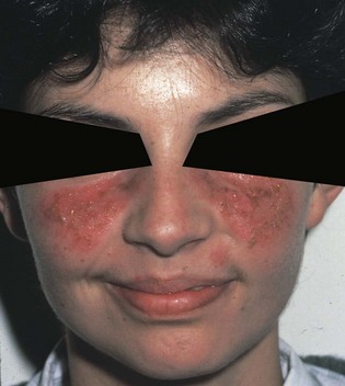
The triad of fever, joint pain, and rash in a woman of childbearing age suggests SLE. The most well-recognized cutaneous finding is the red, raised butterfly rash (Fig. 108.1), but malaise, fatigue, aches, fever, and weight loss are the most common symptoms. The rash, which does not cross the nasolabial fold, may be painful or pruritic. It may be precipitated by sunlight and can last from days to weeks.

Fig. 108.1 Erythematous malar rash of systemic lupus erythematosus.
Note that the rash does not cross the nasolabial fold.
(From Gladman DD, Urowitz MB. Clinical features. In: Hochberg MC, Silman AJ, Smolen JS, editors. Rheumatology. Philadelphia:, Mosby; 2003. pp. 1359–79.)
More than two thirds of patients have vague constitutional symptoms. A thorough evaluation is required before attributing such symptoms to lupus alone. Patients can have kidney failure, infections, adrenal failure, and other complications with similar symptoms (Box 108.1).
Box 108.1
Criteria for the Classification of Systemic Lupus Erythematosus*
Immunologic Disorder
Positive ANA Test
Abnormal ANA titers by immunofluorescence or an equivalent assay at any time in the absence of drugs
ANA, Antinuclear antibody; anti-Sm, anti-Smith; IgG, immunoglobulin G; IgM, immunoglobulin M.
Modified from Hochberg MC, Silman AJ, Smolen JS, editors. Rheumatology, vol 2. 3rd ed. London: Mosby; 2003, Chapter 122.
* Based on the American Rheumatism Association revised criteria for the classification of lupus. The criteria consist of conditions associated with systemic lupus erythematosus, including clinical symptoms, systemic complications, and diagnostic and laboratory test findings (see text for discussion).
See Box 108.1, Criteria for the Classification of Systemic Lupus Erythematosus, online at www.expertconsult.com
Differential Diagnosis and Medical Decision Making
Kidneys
Patients with active urine sediment may benefit from aggressive steroid or other immunosuppressive therapy. Indications for treatment include worsening renal failure, decreasing serum complement levels, increasing anti–double-stranded DNA levels, and nephritic urinary sediment, especially when accompanied by increasing or nephrotic-range proteinuria.3
Cardiovascular System
Pulmonary System4
Pleuritis
Pleurisy and pleural effusions occur in more than half of patients with SLE.5 The pleural effusions are usually small and bilateral but can occasionally be very large. Pleural fluid is generally exudative, with glucose levels similar to serum glucose levels. In contrast, the pleural fluid of patients with rheumatoid arthritis has very low glucose levels.
Drug-induced Lupus Erythematosus
Procainamide has been known for more than 40 years to induce a lupus reaction. Since then, a large number of agents have been implicated, with hydralazine and procainamide being the most common (Table 108.1). The clinical manifestations vary, with most patients experiencing arthralgias and occasional pleuropericardial pain. The full manifestations are present in less than 1% of patients taking high-risk drugs, although a positive antinuclear antibody titer can be found in more than 50%. Patients are generally women, middle-aged or older, and rarely African American, but this may be representative of the group of patients. The condition is usually reversible when drug therapy is stopped, with resolution occurring within days or weeks. Manifestations lasting for years have been reported. In patients with significant pleuropericardial disease, a short course of tapered steroids has been used successfully once use of the implicated medication has been discontinued.
| SYSTEM | DRUG | RISK |
|---|---|---|
| Cardiovascular | Procainamide, quinidine, practolol* | High |
| Antihypertensive | Hydralazine, methyldopa, reserpine | High |
| Antimicrobial | Isoniazid, nitrofurantoin, penicillin, sulfonamides, streptomycin, tetracycline | Moderate |
| Anticonvulsant | Ethosuximide, mephenytoin, phenytoin, primidone | Moderate |
| Antithyroid | Methylthiouracil, propylthiouracil | Low |
| Psychotropic | Chlorpromazine, lithium carbonate | Low |
| Miscellaneous | AllopurinolAminoglutethimide, gold salts, D-penicillamine, phenylbutazone, methysergide | HighLow |
* Removed from the market because of lupuslike syndrome.
Adapted from Marx J. Rosen’s emergency medicine: concepts and clinical practice. 5th ed. St. Louis: Mosby; 2001.
Treatment
Medical therapy attempts to reduce inflammation, suppress the immune system, and control pain. Nonsteroidal antiinflammatory drugs (NSAIDs), corticosteroids, and immunosuppressive agents are the mainstays of treatment. Aspirin and other NSAIDs are the primary treatment of arthralgias, pleurisy, and pericarditis. The maximum standard recommended doses of these agents are usually needed. These agents should be avoided in patients with severe gastrointestinal complications, renal insufficiency, nephritis, or thrombocytopenia. Treatment with NSAIDs can worsen lupus nephritis, either by causing interstitial nephritis or by inhibiting prostaglandins.5
Immunosuppressive agents (azathioprine, methotrexate, cyclophosphamide) are reserved for patients with severe renal or cerebral disease in whom other therapies have failed and for patients who cannot tolerate corticosteroids. Studies of the use of immunosuppressants have shown decreased chronic renal scarring and a reduced likelihood of end-stage renal disease without an increase in mortality. The toxic effects of such drugs are numerous and include myelosuppression, risk for neoplasms, and infections,6 especially with gram-negative organisms, encapsulated gram-positive organisms, herpes zoster, and opportunistic organisms. Febrile patients who are taking azathioprine, methotrexate, or cyclophosphamide should be admitted regardless of whether a source is evident because gram-negative or streptococcal sepsis occurs in this population. Immunosuppressed patients with localized herpes zoster should be admitted for intravenous acyclovir treatment to prevent viral dissemination.
follow-up, next steps in care, and patient education
Patients with evidence of lupus nephritis and worsening renal failure should be admitted for therapy with steroids and, frequently, immunosuppressive agents.6 The serum creatinine level may be elevated, but serious disease may be present even with normal creatinine levels. Proteinuria may be present, or red blood cell casts in urine may be the only sign of severe renal involvement.
1 Moldovan I. Systemic lupus erythematosus: current state of diagnosis and treatment. Compr Ther. 2006;32:158–162.
2 Ward MM. Prevalence of physician-diagnosed systemic lupus erythematosus in the United States: results from the Third National Health and Nutrition Examination Survey. J Womens Health. 2004;13:713–718.
3 O’Callaghan CA. Renal manifestations of systemic lupus erythematosus. Nephrol Ther. 2006;2:140–151.
4 Ion DA, Chivu RD, Chivu LI. Aspects of pleural/pulmonary involvement in systemic lupus erythematosus. Pneumologia. 2006;55:151–155.
5 Sercombe CT. Systemic lupus erythematosus and the vasculidites. In: Marx J, Hockberger R, Walls R. Rosen’s emergency medicine: concepts and practice. 6th ed. Philadelphia: Mosby; 2006:1805–1818.
6 Boumpas DT, Sidiropoulos P, Bertsias G. Optimum therapeutic approaches for lupus nephritis: what therapy and for whom? Nat Clin Pract Rheumatol. 2005;1:22–30.


