Surgical Treatment for Athletic Pubalgia (“Sports Hernia”)

Published on 11/04/2015 by admin

Filed under Orthopaedics

Last modified 22/04/2025

Print this page

rate 1 star rate 2 star rate 3 star rate 4 star rate 5 star
Your rating: none, Average: 0 (0 votes)

This article have been viewed 3208 times

CHAPTER 38 Surgical Treatment for Athletic Pubalgia (“Sports Hernia”)

Basic science

When treating these injuries, a detailed understanding of the musculoskeletal and visceral anatomy of the pelvis is necessary. One must also appreciate the physiologic concept that the pubic symphysis is a functional joint. The pubis is the center of normal symmetric motion—flexion, extension, abduction, adduction, and rotation—with naturally opposing groups of muscles and other soft tissues. We have described the musculoskeletal anatomy and the concept of the pubic joint in several publications.

Briefly, the musculoskeletal anatomic considerations are as follows. Consider mainly the anterior pelvis, excluding the spine, and then consider the anatomy inside and outside of the hip joint. It is easiest to think of the hip joint as a ball-and-socket joint that is relatively independent of the musculature outside of the socket. From the standpoint of anatomic proximity, it also seems logical to think that some interplay must occur between the hip joint and the pelvic musculature around it. If so, then injury to the adjacent musculature might also negatively affect the hip joint or vice versa, and primary and compensatory forces likely play important roles in the pathogeneses of these two types of injuries.

The hip is a synovial joint that is comprised of the femoral head and the acetabulum of the pelvis. The hip, which is also known as the acetabulofemoral joint, connects the lower limb to the axial skeleton. Both joint surfaces are covered in hyaline cartilage, and the acetabulum also has a fibrocartilaginous rim called the labrum that firmly holds the femoral head in place. In addition, the joint is encased in a fibrous capsule and stabilized by three ligaments: the iliofemoral ligament, the pubofemoral ligament, and the ischiofemoral ligament. The ligamentum teres, which is located at the femoral head, also serves to support joint integrity.

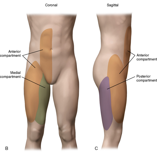
The pubic musculature outside of the hip joint consists of a vast set of muscles and soft tissues. The anterior pelvis includes multiple structures (excluding the hip, sacrum, and spine) such as the lower abdominal soft tissues; both sides of the pubic symphysis; and multiple thigh and pelvic adductors, abductors, flexors, extendors, and rotators. We think in terms of three compartments of muscles or other attachments that provide ligamentous- type support (Figure 38-1). The anterior compartment consists mainly of the abdominal muscles, including the sartorius, the anterior attachment of the psoas, portions of the quadriceps, and some complex interdigitations with fibers from the thighs and the medial and posterior pelvis. The posterior compartment consists primarily of the hamstrings, a portion of the adductor magnus, several key nerves, and an artery. The medial compartment consists of the most important thigh components, which include the three adductors that attach to the symphysis, the gracilis, the obturator externus, and several other structures.

image image

Figure 38–1 A, Anterior view of muscle origins and insertions. Note that all adductors originate in the pubic ramus, and also note the relatively anterior location of insertion of the psoas tendon onto the lesser trochanter. B, Anterior view. C, Lateral view.

From: Meyers WC, Yoo E, Devon O, et al.,: Understanding “sports hernia” (athletic pubalgia): the anatomic and pathophysiologic basis for abdominal and groin pain in athletes. Oper Tech Sport Med. Elsevier, 15:4, pp 165-177, October 2007.

The muscular attachments provide different types of either central or strap supports, depending on their medial or lateral locations, insertions, or origins. For example, a combination of the rectus femoris and the obturator externus is particularly important for place kicking, and the adductor longus and magnus are particularly important as push-off muscles for pitching. We can think in terms of four groups of muscles: adductors, abdominal flexors, thigh flexors, and internal or external rotators. The most important adductors are the adductor longus, the adductor brevis, and the pectineus. The adductor magnus and gracilis usually play minor roles in pelvic stabilization. The rectus abdominis and, to a lesser degree, the obliques and the transversalis comprise the more superior or anterior flexors, and the psoas major and minor combine with other thigh flexors as key inferior or posterior flexors of the pubic joint. The rotators consist primarily of the obturator externus and internus and the quadrator femoris, although other muscles also play roles in rotation. One should not forget the importance of some of the back muscles, particularly the large transversus, which play important roles in both rehabilitation after injury and in performance in general.

Lastly, in addition to musculoskeletal tissue, one must take into account the solid and hollow viscera organs in the pelvis. These include the small and large intestine, the rectum, the genitourinary system, the gynecologic system, and some important blood vessels. Any of these systems can be involved in patients who present with sports hernia. The causes of pelvic pain can be many, and accurate diagnosis can be confusing. To make matters even more complex, visceral diseases can produce symptoms during exertion. Therefore, during the evaluation of the athlete or nonathlete with any type of pelvic pain, one needs to strongly consider possible visceral causes. Thus, one has three main things to consider as part of the differential diagnosis: 1) hip problems; 2) nonhip musculoskeletal problems; and 3) visceral problems. Remember, we are excluding the spine as part of this anatomy, because it rarely causes problems in this area.

History and physical examination

A careful history must take into account all of the previously mentioned systems and anatomy. One must remember that pelvic pain has a wide differential diagnosis and that these patients, who are for the most part young, can have pain from a wide variety of sources. For example, Crohn’s disease and endometriosis have mimicked musculoskeletal disorders, and they have initially presented with exertion-related pelvic pain. In addition, we have picked up a wide variety of malignancies, benign tumors, and unusual other problems involving the gastrointestinal, genitourinary, and gynecologic systems. More recently, now that trainers and team physicians are recognizing these problems earlier, we have also seen appendicitis as a primary cause of acute exertion-related pelvic pain in athletes.

One must also consider the historic differentiators between intra-articular hip problems and problems that involve the soft tissues outside of the hip. These can be tricky, particularly considering that there is considerable overlap among these symptoms. For the hip, one usually looks for pain with simple changes in posture that often involve minimal exertion. There can be a continuous nature to the pain, particularly after activity. Any inflammatory process (e.g., pubic osteitis) can cause the pain to have a continuous nature, but injuries to the soft-tissue structures outside of the hip joint usually cause pain with extremes of activity that is often reproducible with the same maneuvers.

During the physical examination, one looks primarily for pain with resistive maneuvers versus pain with passive range of motion of the hip. Each of the soft-tissue structures outside of the hip can be tested with specific physical tests. For example, one can easily differentiate among the three adductors that insert on the pubic symphysis, and one can also test individually for the adductor magnus, the rectus femoris, the sartorius, the iliopsoas, the gracilis, and other structures. Tenderness should immediately alert the observer to the possibility of an intraperitoneal problem. Hip examination is often particularly important for these patients. The examination should be thorough and consider all of the potential musculoskeletal and nonmusculoskeletal causes of pain; it definitely includes the examination of the abdomen and the chest as well as the scrutiny of the regional lymph nodes and the vascular system. This examination may even include rectal and gynecologic examinations.

Imaging and other diagnostic tools

During the past 2 to 3 years, imaging and the proper interpretation of that imaging have gained increasing importance. The most important advances have been in magnetic resonance imaging and magnetic resonance arthrography with Sensorcaine. A magnetic resonance image of the pelvis details the soft-tissue structures that attach to and cross the pubic symphysis. We can now identify with about 91% specificity the soft-tissue injuries that occur outside of the hip. This accuracy of interpretation is directly related to improvement in the techniques and an improved understanding of the anatomy and pathophysiology. Things that were documented in these patients as “nonspecific findings” in the past have turned out, in fact, to have specific correlations with the pathology.

With magnetic resonance arthrography of the hip with an anesthetic, a combination of a gadolinium injection and a Sensorcaine/lidocaine mixture is injected into the hip joint to determine whether signs or symptoms go away after the injection. When performed properly, this test can be very important for differentiating between hip and nonhip musculoskeletal problems. One must also appreciate that hip and nonhip problems can occur together. We see simultaneous injury (i.e., intra-articular hip injury and athletic pubalgia) in as many as 10% of athletes.

Other anesthetic blocks are also used to help determine the site of the pain. Psoas injections can be useful. Although one must understand that simply blocking certain nerves usually leads to a relatively nonspecific diagnosis, nerve injections can be useful for differentiating lateral pelvic musculature problems from intrinsic hip injury. We see many patients with persistent or new pain after an attempt at conventional hernia repair for athletic pubalgia. The differential diagnosis often includes pain related to mesh or nerve entrapment. In these cases, ilioinguinal, iliohypogastric, and genitofemoral blocks may be considered.

Other imaging that we commonly use is abdominal and pelvic computed tomography. This can be particularly helpful for identifying certain visceral problems, abscesses, and tumors. For many gastrointestinal problems, endoscopy and contrast imaging are often more helpful. However, these are rarely helpful for the diagnosis of most musculoskeletal disorders of the pelvis. Abdominal, pelvic, or lower-extremity computed tomography scanning may be better tests than magnetic resonance imaging for suspected soft-tissue neoplasms. In addition, bone scans may be helpful for identifying osteitis that involves the pubic bone or other bones of the pelvis, and these scans may occasionally be useful for addressing the possibility of metastatic disease as the cause of the pelvic pain. For example, we have had several cases of patients with prostate cancer for which this test was useful. In general, magnetic resonance imaging of the pelvis is more useful than bone scan for osteitis as well as for most of the other considerations in the differential diagnosis.

We rarely use ultrasound as a modality for diagnosing most of the problems of athletic pubalgia. Ultrasound can sometimes be helpful for identifying hematomas or soft-tissue avulsions. This modality can occasionally be helpful for women to identify other sources of pathology. If one is using this test routinely for identifying abdominal or pelvic hernias, then one misunderstands the basic considerations involved in these injuries.

Surgical technique

Because of the complexity of the anatomy of this region of the body, one should readily appreciate that many injuries may occur here. Therefore, a wide variety of operations and combinations of procedures apply, depending on the proper identification of the primary injury and any compensatory injuries. In a recent review, we identified 19 clear categories of injuries, and we applied 26 different procedures and 121 different combinations of procedures.

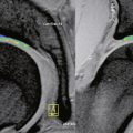
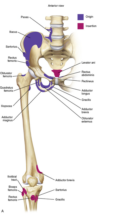
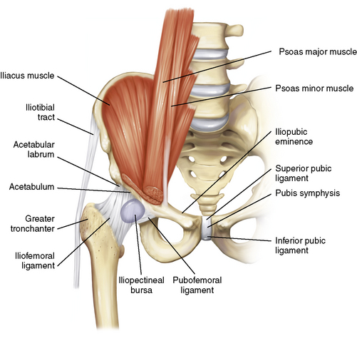
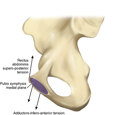
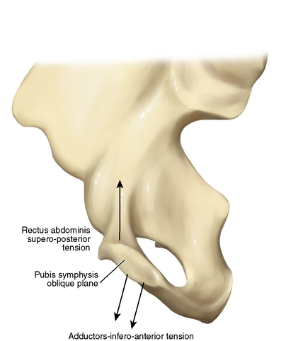
The specific identification of the primary and secondary injuries as well as of the possibility of an associated hip injury provides a guideline for the correction of the problem. First, consider the principal soft-tissue structures that attach to and help stabilize the pelvis. Table 38-1 lists many of these. In general, for males, the more medial structures play greater roles. For women, the lateral structures come more into play. This may be the result of differences in anatomic structure (Figure 38-2). The forces applied to the pubis by these structures are important to consider, and they vary considerably. Note the subtle differences that are schematically represented in the anterior and medial views in Figures 38-3 and 38-4. For each force, there are structures that apply parallel counterforces. In the fresh cadaver laboratory, one can cut certain structures and cause dramatic increases in pressure within other structures or compartments. Such changes in forces likely account for much of what we see clinically in terms of primary and compensatory injuries. The compensatory problems likely result from the “over pulling” of the attachments related to the loss of counterforces and increased pressures within muscles or compartments. Clinically and on a magnetic resonance image, one often finds acute or chronic reactions in the various tissues. These reactions probably represent avulsion, scarring, or tightness within the various compartments involved. Figures 38-5 through 38-8 provide more anatomic direction.

image

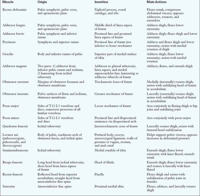
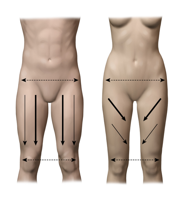
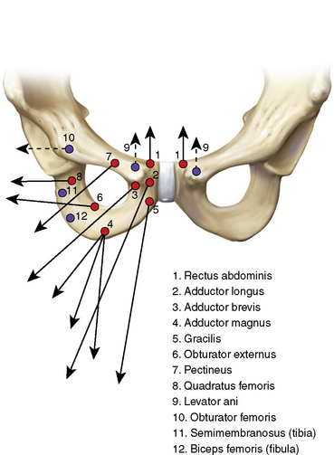
Figure 38–3 Anterior view of the pubic ramus with a schematic depiction of the many forces that act on the pubic joint.

From: Meyers WC, Greenleaf R, Saad A: Anatomic basis for evaluation of abdominal and groin pain in athletes. Oper Tech Sports Med. Elsevier, 13:1, pp 55-61, 2005.

image

Figure 38–4 Medial view of the pelvis that depicts the direction of the forces that act on the pelvis and that influence pelvic tilt.

From: Meyers WC, Greenleaf R, Saad A: Anatomic basis for evaluation of abdominal and groin pain in athletes. Oper Tech Sports Med. Elsevier, 13:1, pp 55-61, 2005.

image

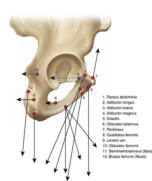
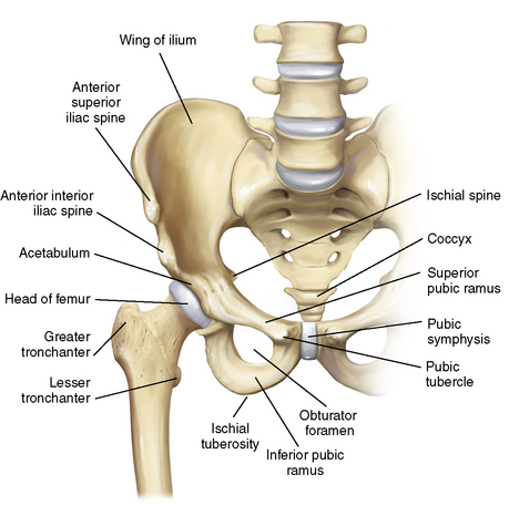
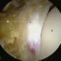
Figure 38–5 Anterior view of the bony anatomy and the proximal femur.

From: Meyers WC, Greenleaf R, Saad A: Anatomic basis for evaluation of abdominal and groin pain in athletes. Oper Tech Sports Med. Elsevier, 13:1, pp 55-61, 2005.

image

Figure 38–6 The ligamentous anatomy of the pelvis and the proximal femur. The iliopsoas muscle tendon, which inserts on the lesser trochanter, is cut.

From: Meyers WC, Greenleaf R, Saad A: Anatomic basis for evaluation of abdominal and groin pain in athletes. Oper Tech Sports Med. Elsevier, 13:1, pp 55-61, 2005.

image

Figure 38–7 Cross-sectional view through the pubis symphysis that shows the main opposing forces that act on the pubic joint.

From: Meyers WC, Greenleaf R, Saad A: Anatomic basis for evaluation of abdominal and groin pain in athletes. Oper Tech Sports Med. Elsevier, 13:1, pp 55-61, 2005.

image

Figure 38–8 Oblique view of the pubis symphysis that shows the direction of the opposing forces exerted on the pubic joint by the rectus abdominis and the adductors.

From: Meyers WC, Greenleaf R, Saad A: Anatomic basis for evaluation of abdominal and groin pain in athletes. Oper Tech Sports Med. Elsevier, 13:1, pp 55-61, 2005.

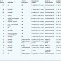
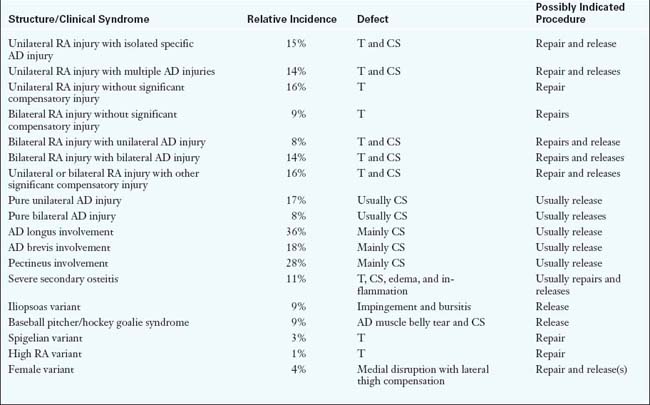
Next, consider the wide variety of musculoskeletal problems that can occur as a result of the disruption of these forces. Table 38-2 lists a number of them that were determined by a recent review of our data. There are still a variety of other problems that are not listed here. The table also summarizes the general approach for the correction of these problems. Initial treatment is usually nonsurgical, and some of these problems can be adequately treated this way. However, when the nonoperative approach does not work, surgery is indicated.

It is important to recognize that these procedures require a combination of the tightening and loosening of various structures, depending on the primary and secondary problems. Mesh for the most part is not necessary. “Tightening” usually means that some specifically placed sutures are used to create more stability in the region where the primary injury occurred. “Loosening” or “releasing” usually refers to decreasing the pressure in a compartment, usually via a specific epis- iotomy. For the most part, all intact muscle fibers are left intact. However, if an intracompartmental scar is identified, this should be released. When we talk in terms of release operations, we are usually talking about a focal decompression of a particular muscle or part of a muscle, like the treatment of a compartment syndrome. The muscle usually remains intact. The muscles that attach to the pubis or the cartilaginous plate usually attach separately from the investing epimysium. In most cases, we aim to keep as much normal muscle intact as possible.

Postoperative rehabilitation

Recovery time varies according to the specific injury and the procedure performed. The repair associated with the shortest time might be called a minimal repair, which involves a specific tightening and a slight reinforcement of the specifically identified afflicted area, possibly in combination with the nerve division of afflicted branches. Athletes with minimal injury and minimal repair may be able to return to activity within days of surgery. However, the downside of the minimal repair of a minimal injury is the possibility of early recurrence as a result of the undertreatment of pelvic instability. We have had to perform revision surgery on a number of such patients. The patient needs to understand the increased risk of revision surgery if he or she chooses this in-season approach. However, in selected patients, return to full play by 3 weeks after the procedure is possible. So far, early return in general has not been associated with an early recurrence of injury. It is interesting to note that such an aggressive protocol does seem to work despite the relatively limited time allowed for wound healing.

Alternatively, more extensive repairs require the longest recovery times. An example of one of these is the reattachment of multiple avulsions, such as the entire rectus abdominis in combination with the entire pubic–adductor complex, which is common among bull riders. In such cases, a full return to performance may not occur for several months. Patients with severe osteitis pubis are less predictable in terms of returning to full play without experiencing a recurrence. Other variables that affect both the timing of surgery and the duration of postoperative rehabilitation include the following: the timing of the injury (i.e., within a season or between seasons); contractual considerations; injury complexity; concomitant hip injury; performance with the injury; the specific sport and the player’s position; and team standings.

Briefly, the most important parts of postoperative rehabilitation are an aggressive return to activity, the avoidance of movements that cause pain, and massage and other mechanical means of reducing scar bands. We have some specific sport rehabilitation programs, and we modify these protocols on the basis of the specific injury and repair. For long-term rehabilitation and the prevention of further pelvic injury, we strongly recommend certain core stability programs.

Results

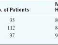
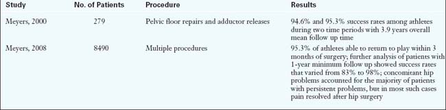
In general, 95% to 96% of athletes can expect to return to sports participation (Table 38-3). The most important endpoints are at 1 and 2 years after surgery. Success rates vary according to multiple factors, such as the site and severity of the injury, concomitant hip injury, and whether the patient is an athlete or a nonathlete. Recurrence rates are about 0.4%, but new involvement of the contralateral side can be as high as 4%. Return to play by 3 months should occur in approximately 90% of patients. Nonathletes and patients without a clear primary injury going into surgery will have a success rate of between 50% and 90%, depending on the specific problem. Workers compensation injuries are less predictable than other injuries with regard to the reporting of successful operations.

Annotated references and suggested readings

Albers S.L., Spritzer C.E., Garrett W.E., Meyers W.C. MRI findings in athletes with pubalgia. Skeletal Radiol. 2006;30:270-277.

Gilmore O.J.A. Gilmore’s groin: ten years of experience of groin disruption—a previously unsolved problem in sportsmen. Sport Med Soft Tissue Trauma.. 1991;3:12-14.

About the same time that we did, Dr. Gilmore recognized that there were nonhernia problems that afflicted athletes. He provided a new name for the set of syndromes..

McKechnie McKechnie A., Celebrini R. Hard core strength. Vancouver, BC. Available at http://www.p2soccer.com/Content/Main%20pages/Resource%20Centre.asp

Meyers WC, Foley DP, Garrett WE, et al. Management of severe lower abdominal or inguinal pain in high-performance athletes. Am J Sports Med. 200;28:2–8.

Meyers W.C., Greenleaf R., Saad A. Anatomic basis for evaluation of abdominal and groin pain in athletes. Oper Tech Sports Med. 2005;13(1):55-61.

Meyers W.C., McKechnie A., Philippon M.J., et al. Experience with “sports hernia” spanning two decades. Presented at the 2008 meeting of the American Surgical Association. Ann Surg. Oct 2008;248(4):656-665.

Meyers W.C., Szalai L., Potter N., et al Extraarticular sources of hip pain. Byrd J.W.T., editor. Operative hip arthroscopy, 2nd ed, Vol 5. New York: Springer, 2005;86-97.

Meyers W.C., Yoo E.Y., Devon O.N., et al. Understanding “sports hernia” (athletic pubalgia): the anatomic and pathophysiologic basis for abdominal and groin pain in athletes. Oper Tech Sports Med. 2007;15:165-177.

Nesovic Nesovic, Treatise on maladies of the pubic symphysis (privately published monograph).

Omar I.M., Zoga A.C., Meyers W.C., et al Athletic pubalgia and the “sports hernia”: optimal MR imaging technique and pictorial review of MR findings. Scientific exhibit. Proceedings of the Radiologic Society of North America, Cum Laude Award winner 2006. Radiographics, 28. 2008:1415-1438.

Swan K.G., Wolcott M. The athletic hernia; a systematic review. Clin Orthop Rel Res.. 2006;455:78-87.

This review describes well the tremendous confusion in the literature regarding this set of problems, the large number of different descriptions of these entities, and the numerous reports that do not include adequate follow up..

Taylor D.C., Meyers W.C., Moylan J.A., et al. Abdominal musculature abnormalities as a cause of groin pain in athletes. Am J Sports Med.. 1991;19:239-242.

This was our first article about these entities. We saw that these were not hernias, and we were initially very selective with regard to the patients on which we chose to operate. The term athletic pubalgia originated with this paper..

Zoga A.C., Kavanaugh E.C., Meyers W.C., et al MRI of the rectus abdominis/adductor aponeurosis: findings in the “sports hernia.”. Scientific paper presentation, Proceedings of the American Roentgen Ray Society; 2007.

Zoga A.C., Kavanaugh E.C., Meyers W.C., et al. MRI findings in athletic pubalgia and the “sports hernia.”. Radiol. 2008;247(3):797-807.